
Bacterial Endocarditis
The Common Vein Copyright 2008
Definition
Bacterial endocarditis is a microbial infection of the endothelial surface of the heart.
Structurally the lesions consist of circulating bacteria and inflammatory cells adhering to and growing on thrombi formed on damaged or denuded endothelium.
Functionally these vegetations may affect valvular closure or opening and also embolize to virtually any organ in the body.
Clinically patients present with fever , new onset murmur , signs of systemic embolization ( janeway lesions, strokes , splinter hemorrhages ) and immunological phenomena ( glomerulonephritis , oslers nodes , roth spots ) .
Diagnosis is made by the Dukes criteria which includes clinical, laboratory and echocardiographic criteria. The treatment of bacterial endocarditis is primarily medical ? with antimicrobial agents. Surgery may be used in rare circumstances.
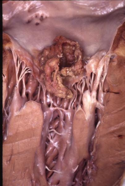
Heavy vegetation on the Mitral Valve |
| 13414 heart + cardiac mitral valve + bacterial endocarditis papillary muscles chordae fx vegetation dx SBE grosspathology Courtesy Henri Cuenoud MD |
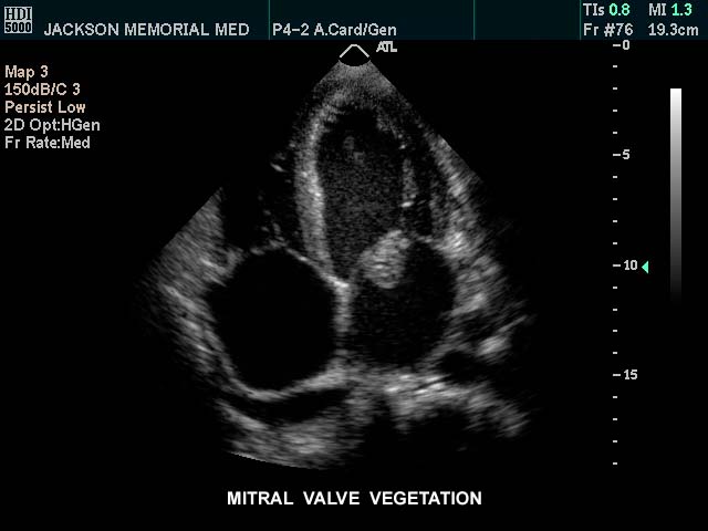
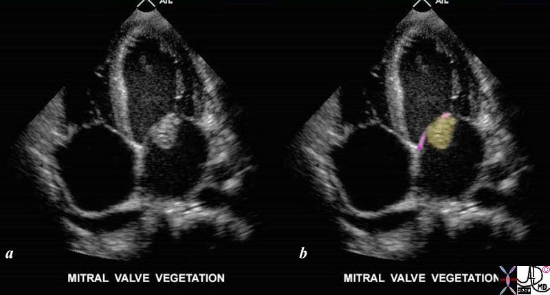
Mitral Valve Vegetation (yellow) |
| This gray scale echo of the heart using a left apical 4-chamber view, and demonstrating an echogenic mass on the atrial side of the mitral valve. This is a vegetation in a patient with bacterial endocarditis of the mitral valve.
Courtesy Philips Medical Systems 33139 code cardiac heart echo MV mass vegetation SBE LA LV imaging cardiac echo infection 33139c01.8s |
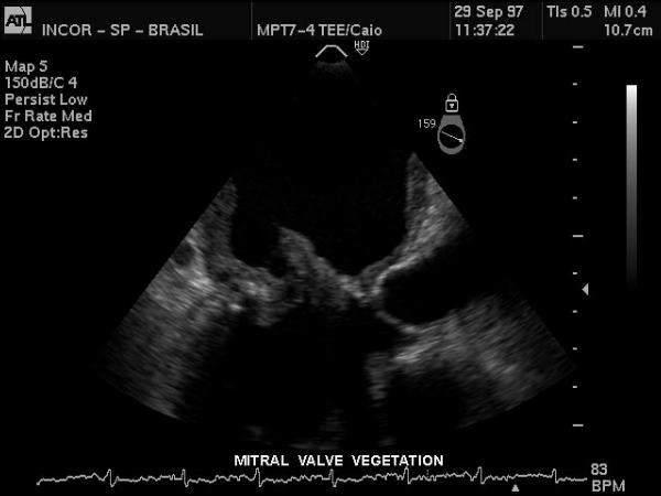
33172 |
| This gray scale echo of the heart showing a 4 chambered view with a focus on the mitral valve, reveals a thickened and irregular valve consistent with a vegetation on the valve. The patient has a diagnosis of bacterial endocarditis. Courtesy Philips Medical Systems 33172 code cardiac heart echo gray scale MV vegetation thick bacterial endocarditis infection imaging cardiac echo |
33133c02.8s |
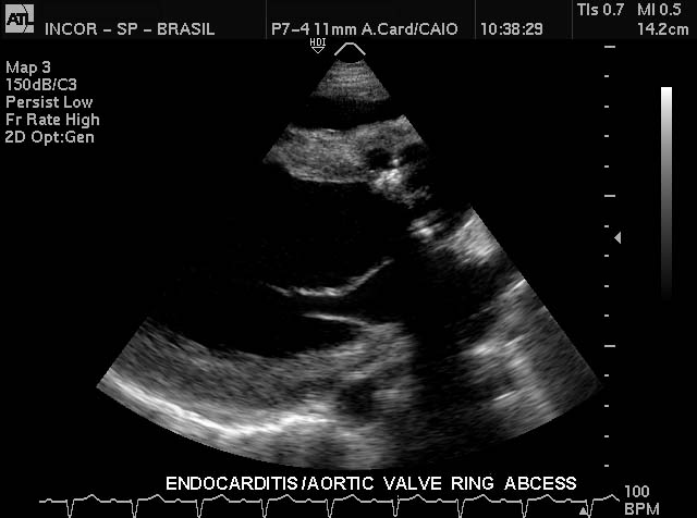
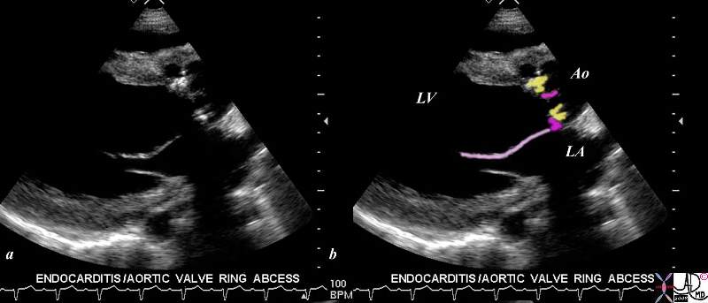
| This gray scale echo of the heart shows the left ventricle, anterior and posterior leaflets of the mitral valve, the aortic valve and the base of the aorta. There is a rounded echogenic focus on the aortic valve ring. In the setting of a febrile illness this represents a ring abscess complicating bacterial endocarditis.
Courtesy Philips Medical Systems 33133 code cardiac heart echo MV aortic valve, echogenic, accumulation SBE ring abscess infection imaging cardiac echo 33133c02.8s |
33125 |
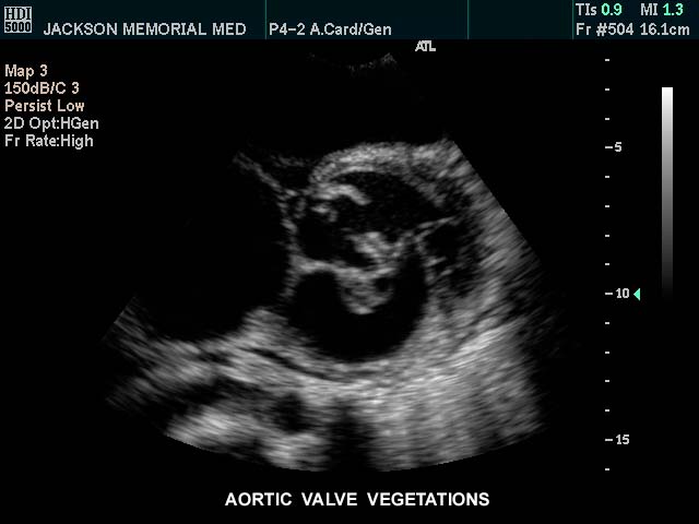
| This gray scale echo of the heart showing a short-axis aorta left atrial view, and demonstrating echogenic vegetations on the aortic valve. The patient has a diagnosis of bacterial endocarditis. Courtesy Philips Medical Systems 33125 code cardiac heart echo AOV vegetation bacterial endocarditis infection imaging cardiac echo |
33127 |
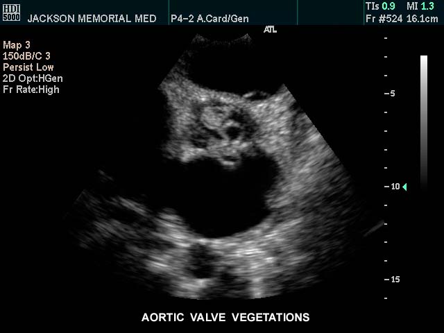
| This gray scale echo of the heart showing a short-axis aorta left atrial view, and demonstrating echogenic vegetations on the deformed valve. The patient has a diagnosis of bacterial endocarditis Courtesy Philips Medical Systems 33127 code cardiac heart echo aorta artic valve BE bacterial endocarditis infection imaging cardiac echo |
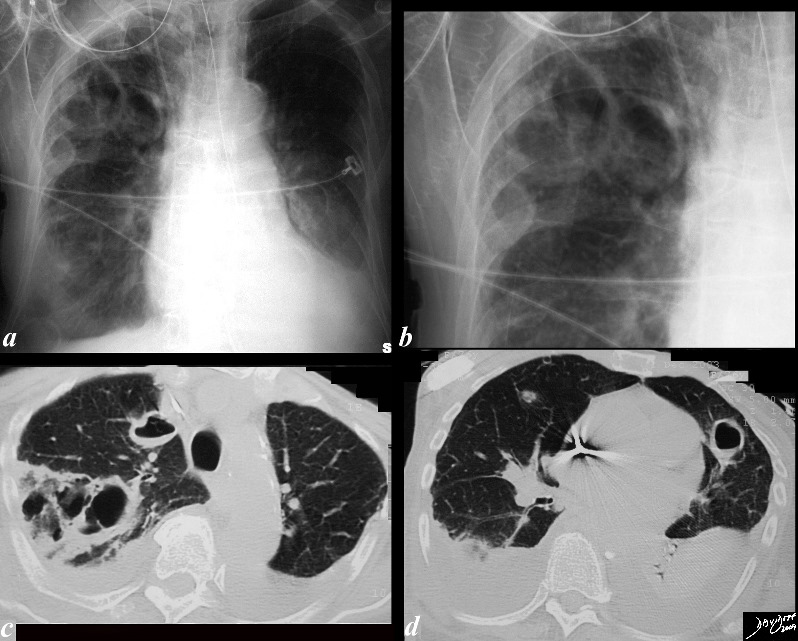
33015c01.8s |
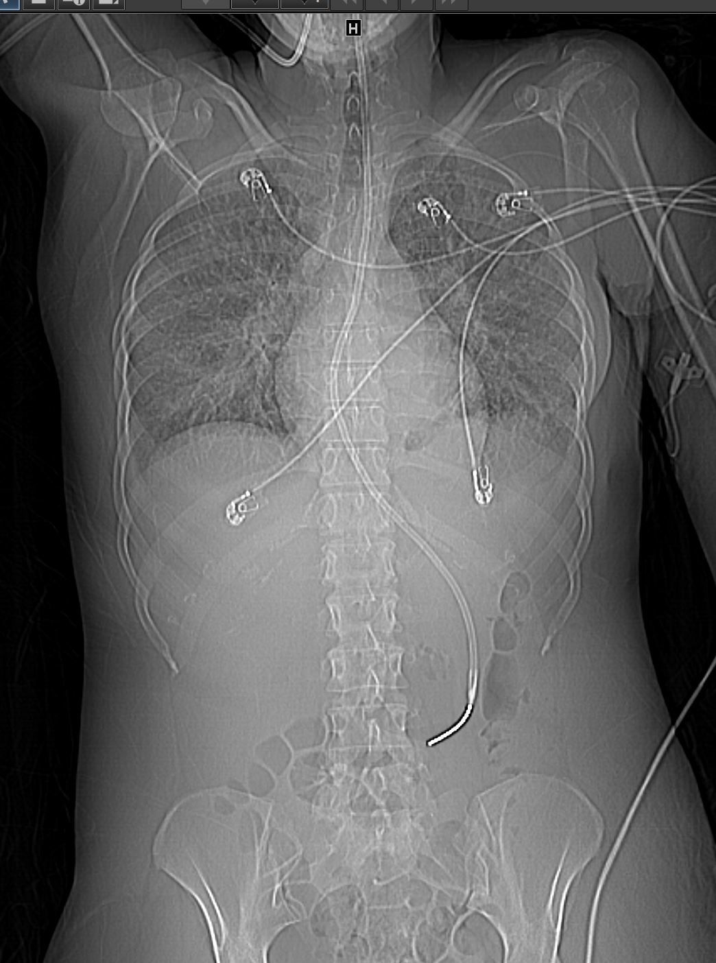
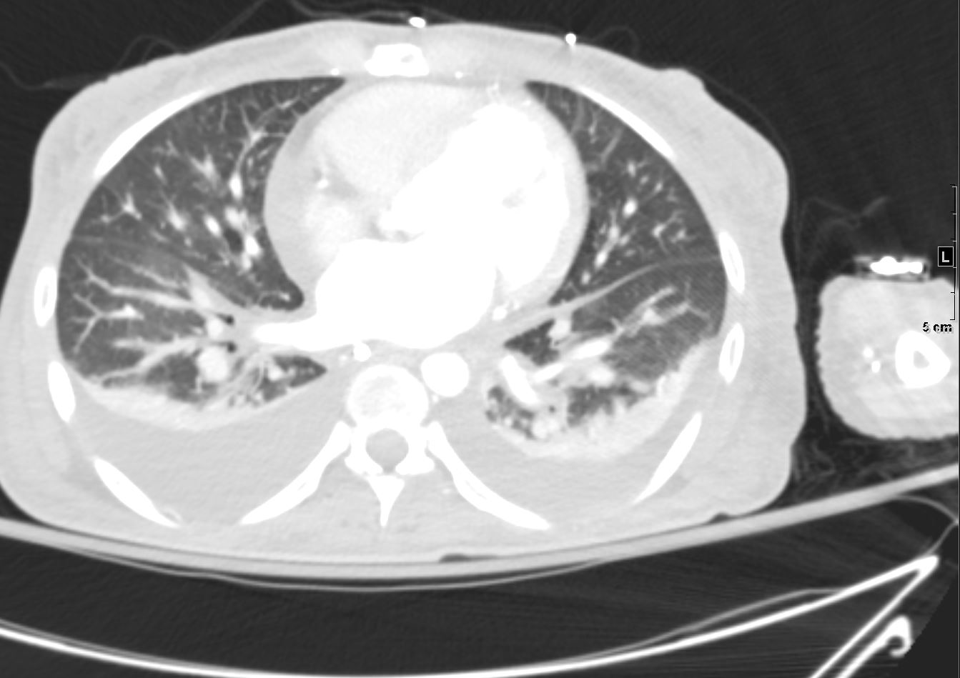
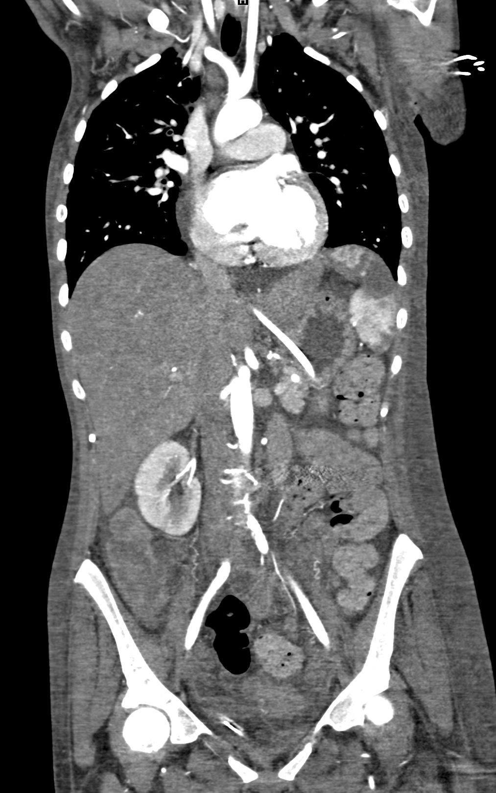
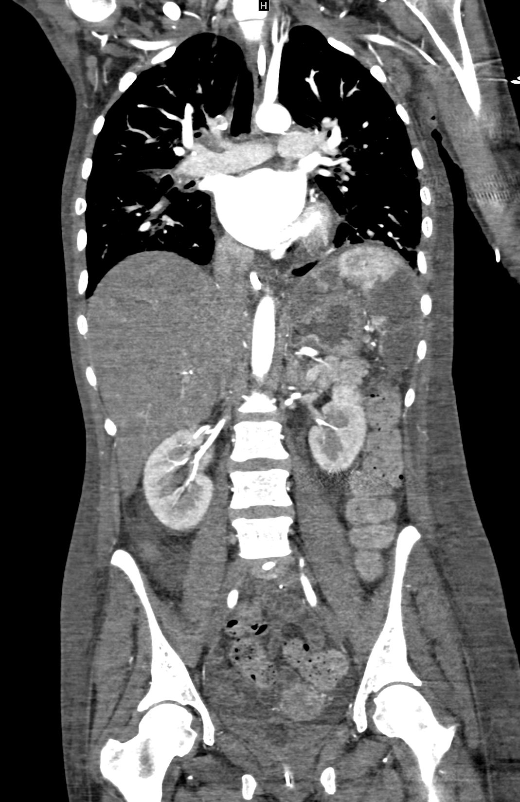
| 33015c01.8s The CXR and CT scan is from a 76 year patient who had a complicated history of septicemia and bacterial endocarditis of the mitral valve associated with a septic arthritis and an infected ICD wire. She has a PMH of MI, ischemic cardiomyopathyEF of 20%, PTCA of LAD, RCA stent, chronic renal failure and COPD. Her wcc was 33 thousand Staphylocosccus was isolated from her sputum. The CXR shows multiple large cavitating lesions with 3 large lesions in the right upper lobe (a,b,c), one in the right lower lobe (a), and one large lesion alongside the heart in the lingula. (b,d) These findings are consistent with septic emboli with cavitation. lung infection cavitation septic emboli SBE BE bacterial endocarditis pleural effusions CXR chest X-ray CTscan Courtesy Ashley DAvidoff MD copyright 2009 all rights reserved |
|
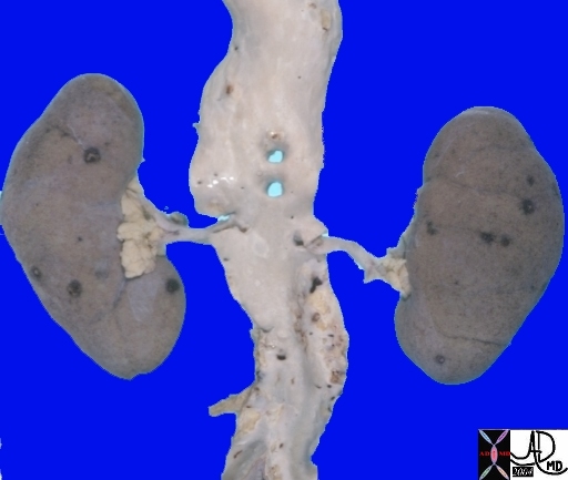
| 05447c aorta SMA celiax axis renal artery origin fx normal anatomy kidney flea bitten endocarditis vs aspergillosis grosspathology Courtesy Ashley Davidoff MD DB |
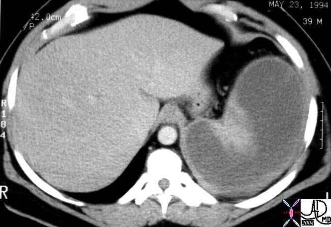
20776 |
| History staphylococcal bacterial endocarditis May 23rd 1994 with lack of abnormality on April 25th 1994. Patient was found to have a staphylococcal splenic abscess at surgery. 20776 spleen + non perfusion + dx infarct + imaging radiology CTscan C+ spleen + hypodense abscess staphylococcus + dx infarct + SBE bacterial endocarditis mycotic embolus infection |
Endocarditis With Splenic Infarcts-
DOMElement Object
(
    [schemaTypeInfo] =>
    [tagName] => table
    [firstElementChild] => (object value omitted)
    [lastElementChild] => (object value omitted)
    [childElementCount] => 1
    [previousElementSibling] => (object value omitted)
    [nextElementSibling] => (object value omitted)
    [nodeName] => table
    [nodeValue] =>


20776


History staphylococcal bacterial endocarditis May 23rd 1994 with lack of abnormality on April 25th 1994. Patient was found to have a staphylococcal splenic abscess at surgery. 20776 spleen + non perfusion + dx infarct + imaging radiology CTscan C+ spleen + hypodense abscess staphylococcus + dx infarct + SBE bacterial endocarditis mycotic embolus infection


    [nodeType] => 1
    [parentNode] => (object value omitted)
    [childNodes] => (object value omitted)
    [firstChild] => (object value omitted)
    [lastChild] => (object value omitted)
    [previousSibling] => (object value omitted)
    [nextSibling] => (object value omitted)
    [attributes] => (object value omitted)
    [ownerDocument] => (object value omitted)
    [namespaceURI] =>
    [prefix] =>
    [localName] => table
    [baseURI] =>
    [textContent] =>


20776


History staphylococcal bacterial endocarditis May 23rd 1994 with lack of abnormality on April 25th 1994. Patient was found to have a staphylococcal splenic abscess at surgery. 20776 spleen + non perfusion + dx infarct + imaging radiology CTscan C+ spleen + hypodense abscess staphylococcus + dx infarct + SBE bacterial endocarditis mycotic embolus infection


)
DOMElement Object
(
    [schemaTypeInfo] =>
    [tagName] => td
    [firstElementChild] =>
    [lastElementChild] =>
    [childElementCount] => 0
    [previousElementSibling] =>
    [nextElementSibling] =>
    [nodeName] => td
    [nodeValue] => History staphylococcal bacterial endocarditis May 23rd 1994 with lack of abnormality on April 25th 1994. Patient was found to have a staphylococcal splenic abscess at surgery. 20776 spleen + non perfusion + dx infarct + imaging radiology CTscan C+ spleen + hypodense abscess staphylococcus + dx infarct + SBE bacterial endocarditis mycotic embolus infection
    [nodeType] => 1
    [parentNode] => (object value omitted)
    [childNodes] => (object value omitted)
    [firstChild] => (object value omitted)
    [lastChild] => (object value omitted)
    [previousSibling] => (object value omitted)
    [nextSibling] => (object value omitted)
    [attributes] => (object value omitted)
    [ownerDocument] => (object value omitted)
    [namespaceURI] =>
    [prefix] =>
    [localName] => td
    [baseURI] =>
    [textContent] => History staphylococcal bacterial endocarditis May 23rd 1994 with lack of abnormality on April 25th 1994. Patient was found to have a staphylococcal splenic abscess at surgery. 20776 spleen + non perfusion + dx infarct + imaging radiology CTscan C+ spleen + hypodense abscess staphylococcus + dx infarct + SBE bacterial endocarditis mycotic embolus infection
)
DOMElement Object
(
    [schemaTypeInfo] =>
    [tagName] => td
    [firstElementChild] => (object value omitted)
    [lastElementChild] => (object value omitted)
    [childElementCount] => 2
    [previousElementSibling] =>
    [nextElementSibling] =>
    [nodeName] => td
    [nodeValue] =>
20776
    [nodeType] => 1
    [parentNode] => (object value omitted)
    [childNodes] => (object value omitted)
    [firstChild] => (object value omitted)
    [lastChild] => (object value omitted)
    [previousSibling] => (object value omitted)
    [nextSibling] => (object value omitted)
    [attributes] => (object value omitted)
    [ownerDocument] => (object value omitted)
    [namespaceURI] =>
    [prefix] =>
    [localName] => td
    [baseURI] =>
    [textContent] =>
20776
)
DOMElement Object
(
    [schemaTypeInfo] =>
    [tagName] => table
    [firstElementChild] => (object value omitted)
    [lastElementChild] => (object value omitted)
    [childElementCount] => 1
    [previousElementSibling] => (object value omitted)
    [nextElementSibling] => (object value omitted)
    [nodeName] => table
    [nodeValue] =>


05447c aorta SMA celiax axis renal artery origin fx normal anatomy kidney flea bitten endocarditis vs aspergillosis grosspathology Courtesy Ashley Davidoff MD DB


    [nodeType] => 1
    [parentNode] => (object value omitted)
    [childNodes] => (object value omitted)
    [firstChild] => (object value omitted)
    [lastChild] => (object value omitted)
    [previousSibling] => (object value omitted)
    [nextSibling] => (object value omitted)
    [attributes] => (object value omitted)
    [ownerDocument] => (object value omitted)
    [namespaceURI] =>
    [prefix] =>
    [localName] => table
    [baseURI] =>
    [textContent] =>


05447c aorta SMA celiax axis renal artery origin fx normal anatomy kidney flea bitten endocarditis vs aspergillosis grosspathology Courtesy Ashley Davidoff MD DB


)
DOMElement Object
(
    [schemaTypeInfo] =>
    [tagName] => td
    [firstElementChild] =>
    [lastElementChild] =>
    [childElementCount] => 0
    [previousElementSibling] =>
    [nextElementSibling] =>
    [nodeName] => td
    [nodeValue] => 05447c aorta SMA celiax axis renal artery origin fx normal anatomy kidney flea bitten endocarditis vs aspergillosis grosspathology Courtesy Ashley Davidoff MD DB
    [nodeType] => 1
    [parentNode] => (object value omitted)
    [childNodes] => (object value omitted)
    [firstChild] => (object value omitted)
    [lastChild] => (object value omitted)
    [previousSibling] => (object value omitted)
    [nextSibling] => (object value omitted)
    [attributes] => (object value omitted)
    [ownerDocument] => (object value omitted)
    [namespaceURI] =>
    [prefix] =>
    [localName] => td
    [baseURI] =>
    [textContent] => 05447c aorta SMA celiax axis renal artery origin fx normal anatomy kidney flea bitten endocarditis vs aspergillosis grosspathology Courtesy Ashley Davidoff MD DB
)
DOMElement Object
(
    [schemaTypeInfo] =>
    [tagName] => td
    [firstElementChild] => (object value omitted)
    [lastElementChild] => (object value omitted)
    [childElementCount] => 2
    [previousElementSibling] =>
    [nextElementSibling] =>
    [nodeName] => td
    [nodeValue] =>

    [nodeType] => 1
    [parentNode] => (object value omitted)
    [childNodes] => (object value omitted)
    [firstChild] => (object value omitted)
    [lastChild] => (object value omitted)
    [previousSibling] => (object value omitted)
    [nextSibling] => (object value omitted)
    [attributes] => (object value omitted)
    [ownerDocument] => (object value omitted)
    [namespaceURI] =>
    [prefix] =>
    [localName] => td
    [baseURI] =>
    [textContent] =>

)
DOMElement Object
(
    [schemaTypeInfo] =>
    [tagName] => table
    [firstElementChild] => (object value omitted)
    [lastElementChild] => (object value omitted)
    [childElementCount] => 1
    [previousElementSibling] => (object value omitted)
    [nextElementSibling] => (object value omitted)
    [nodeName] => table
    [nodeValue] =>


33015c01.8s


33015c01.8s The CXR and CT scan is from a 76 year patient who had a complicated history of septicemia and bacterial endocarditis of the mitral valve associated with a septic arthritis and an infected ICD wire. She has a PMH of MI, ischemic cardiomyopathyEF of 20%, PTCA of LAD, RCA stent, chronic renal failure and COPD. Her wcc was 33 thousand Staphylocosccus was isolated from her sputum. The CXR shows multiple large cavitating lesions with 3 large lesions in the right upper lobe (a,b,c), one in the right lower lobe (a), and one large lesion alongside the heart in the lingula. (b,d) These findings are consistent with septic emboli with cavitation. lung infection cavitation septic emboli SBE BE bacterial endocarditis pleural effusions CXR chest X-ray CTscan Courtesy Ashley DAvidoff MD copyright 2009 all rights reserved


    [nodeType] => 1
    [parentNode] => (object value omitted)
    [childNodes] => (object value omitted)
    [firstChild] => (object value omitted)
    [lastChild] => (object value omitted)
    [previousSibling] => (object value omitted)
    [nextSibling] => (object value omitted)
    [attributes] => (object value omitted)
    [ownerDocument] => (object value omitted)
    [namespaceURI] =>
    [prefix] =>
    [localName] => table
    [baseURI] =>
    [textContent] =>


33015c01.8s


33015c01.8s The CXR and CT scan is from a 76 year patient who had a complicated history of septicemia and bacterial endocarditis of the mitral valve associated with a septic arthritis and an infected ICD wire. She has a PMH of MI, ischemic cardiomyopathyEF of 20%, PTCA of LAD, RCA stent, chronic renal failure and COPD. Her wcc was 33 thousand Staphylocosccus was isolated from her sputum. The CXR shows multiple large cavitating lesions with 3 large lesions in the right upper lobe (a,b,c), one in the right lower lobe (a), and one large lesion alongside the heart in the lingula. (b,d) These findings are consistent with septic emboli with cavitation. lung infection cavitation septic emboli SBE BE bacterial endocarditis pleural effusions CXR chest X-ray CTscan Courtesy Ashley DAvidoff MD copyright 2009 all rights reserved


)
DOMElement Object
(
    [schemaTypeInfo] =>
    [tagName] => td
    [firstElementChild] =>
    [lastElementChild] =>
    [childElementCount] => 0
    [previousElementSibling] =>
    [nextElementSibling] =>
    [nodeName] => td
    [nodeValue] => 33015c01.8s The CXR and CT scan is from a 76 year patient who had a complicated history of septicemia and bacterial endocarditis of the mitral valve associated with a septic arthritis and an infected ICD wire. She has a PMH of MI, ischemic cardiomyopathyEF of 20%, PTCA of LAD, RCA stent, chronic renal failure and COPD. Her wcc was 33 thousand Staphylocosccus was isolated from her sputum. The CXR shows multiple large cavitating lesions with 3 large lesions in the right upper lobe (a,b,c), one in the right lower lobe (a), and one large lesion alongside the heart in the lingula. (b,d) These findings are consistent with septic emboli with cavitation. lung infection cavitation septic emboli SBE BE bacterial endocarditis pleural effusions CXR chest X-ray CTscan Courtesy Ashley DAvidoff MD copyright 2009 all rights reserved
    [nodeType] => 1
    [parentNode] => (object value omitted)
    [childNodes] => (object value omitted)
    [firstChild] => (object value omitted)
    [lastChild] => (object value omitted)
    [previousSibling] => (object value omitted)
    [nextSibling] => (object value omitted)
    [attributes] => (object value omitted)
    [ownerDocument] => (object value omitted)
    [namespaceURI] =>
    [prefix] =>
    [localName] => td
    [baseURI] =>
    [textContent] => 33015c01.8s The CXR and CT scan is from a 76 year patient who had a complicated history of septicemia and bacterial endocarditis of the mitral valve associated with a septic arthritis and an infected ICD wire. She has a PMH of MI, ischemic cardiomyopathyEF of 20%, PTCA of LAD, RCA stent, chronic renal failure and COPD. Her wcc was 33 thousand Staphylocosccus was isolated from her sputum. The CXR shows multiple large cavitating lesions with 3 large lesions in the right upper lobe (a,b,c), one in the right lower lobe (a), and one large lesion alongside the heart in the lingula. (b,d) These findings are consistent with septic emboli with cavitation. lung infection cavitation septic emboli SBE BE bacterial endocarditis pleural effusions CXR chest X-ray CTscan Courtesy Ashley DAvidoff MD copyright 2009 all rights reserved
)
DOMElement Object
(
    [schemaTypeInfo] =>
    [tagName] => td
    [firstElementChild] => (object value omitted)
    [lastElementChild] => (object value omitted)
    [childElementCount] => 2
    [previousElementSibling] =>
    [nextElementSibling] =>
    [nodeName] => td
    [nodeValue] =>
33015c01.8s
    [nodeType] => 1
    [parentNode] => (object value omitted)
    [childNodes] => (object value omitted)
    [firstChild] => (object value omitted)
    [lastChild] => (object value omitted)
    [previousSibling] => (object value omitted)
    [nextSibling] => (object value omitted)
    [attributes] => (object value omitted)
    [ownerDocument] => (object value omitted)
    [namespaceURI] =>
    [prefix] =>
    [localName] => td
    [baseURI] =>
    [textContent] =>
33015c01.8s
)
DOMElement Object
(
    [schemaTypeInfo] =>
    [tagName] => table
    [firstElementChild] => (object value omitted)
    [lastElementChild] => (object value omitted)
    [childElementCount] => 1
    [previousElementSibling] => (object value omitted)
    [nextElementSibling] => (object value omitted)
    [nodeName] => table
    [nodeValue] =>


33127


This gray scale echo of the heart showing a short-axis aorta left atrial view, and demonstrating echogenic vegetations on the deformed valve. The patient has a diagnosis of bacterial endocarditis Courtesy Philips Medical Systems 33127 code cardiac heart echo aorta artic valve BE bacterial endocarditis infection imaging cardiac echo


    [nodeType] => 1
    [parentNode] => (object value omitted)
    [childNodes] => (object value omitted)
    [firstChild] => (object value omitted)
    [lastChild] => (object value omitted)
    [previousSibling] => (object value omitted)
    [nextSibling] => (object value omitted)
    [attributes] => (object value omitted)
    [ownerDocument] => (object value omitted)
    [namespaceURI] =>
    [prefix] =>
    [localName] => table
    [baseURI] =>
    [textContent] =>


33127


This gray scale echo of the heart showing a short-axis aorta left atrial view, and demonstrating echogenic vegetations on the deformed valve. The patient has a diagnosis of bacterial endocarditis Courtesy Philips Medical Systems 33127 code cardiac heart echo aorta artic valve BE bacterial endocarditis infection imaging cardiac echo


)
DOMElement Object
(
    [schemaTypeInfo] =>
    [tagName] => td
    [firstElementChild] =>
    [lastElementChild] =>
    [childElementCount] => 0
    [previousElementSibling] =>
    [nextElementSibling] =>
    [nodeName] => td
    [nodeValue] => This gray scale echo of the heart showing a short-axis aorta left atrial view, and demonstrating echogenic vegetations on the deformed valve. The patient has a diagnosis of bacterial endocarditis Courtesy Philips Medical Systems 33127 code cardiac heart echo aorta artic valve BE bacterial endocarditis infection imaging cardiac echo
    [nodeType] => 1
    [parentNode] => (object value omitted)
    [childNodes] => (object value omitted)
    [firstChild] => (object value omitted)
    [lastChild] => (object value omitted)
    [previousSibling] => (object value omitted)
    [nextSibling] => (object value omitted)
    [attributes] => (object value omitted)
    [ownerDocument] => (object value omitted)
    [namespaceURI] =>
    [prefix] =>
    [localName] => td
    [baseURI] =>
    [textContent] => This gray scale echo of the heart showing a short-axis aorta left atrial view, and demonstrating echogenic vegetations on the deformed valve. The patient has a diagnosis of bacterial endocarditis Courtesy Philips Medical Systems 33127 code cardiac heart echo aorta artic valve BE bacterial endocarditis infection imaging cardiac echo
)
DOMElement Object
(
    [schemaTypeInfo] =>
    [tagName] => td
    [firstElementChild] => (object value omitted)
    [lastElementChild] => (object value omitted)
    [childElementCount] => 2
    [previousElementSibling] =>
    [nextElementSibling] =>
    [nodeName] => td
    [nodeValue] =>
33127
    [nodeType] => 1
    [parentNode] => (object value omitted)
    [childNodes] => (object value omitted)
    [firstChild] => (object value omitted)
    [lastChild] => (object value omitted)
    [previousSibling] => (object value omitted)
    [nextSibling] => (object value omitted)
    [attributes] => (object value omitted)
    [ownerDocument] => (object value omitted)
    [namespaceURI] =>
    [prefix] =>
    [localName] => td
    [baseURI] =>
    [textContent] =>
33127
)
DOMElement Object
(
    [schemaTypeInfo] =>
    [tagName] => table
    [firstElementChild] => (object value omitted)
    [lastElementChild] => (object value omitted)
    [childElementCount] => 1
    [previousElementSibling] => (object value omitted)
    [nextElementSibling] => (object value omitted)
    [nodeName] => table
    [nodeValue] =>


33125


This gray scale echo of the heart showing a short-axis aorta left atrial view, and demonstrating echogenic vegetations on the aortic valve. The patient has a diagnosis of bacterial endocarditis. Courtesy Philips Medical Systems 33125 code cardiac heart echo AOV vegetation bacterial endocarditis infection imaging cardiac echo


    [nodeType] => 1
    [parentNode] => (object value omitted)
    [childNodes] => (object value omitted)
    [firstChild] => (object value omitted)
    [lastChild] => (object value omitted)
    [previousSibling] => (object value omitted)
    [nextSibling] => (object value omitted)
    [attributes] => (object value omitted)
    [ownerDocument] => (object value omitted)
    [namespaceURI] =>
    [prefix] =>
    [localName] => table
    [baseURI] =>
    [textContent] =>


33125


This gray scale echo of the heart showing a short-axis aorta left atrial view, and demonstrating echogenic vegetations on the aortic valve. The patient has a diagnosis of bacterial endocarditis. Courtesy Philips Medical Systems 33125 code cardiac heart echo AOV vegetation bacterial endocarditis infection imaging cardiac echo


)
DOMElement Object
(
    [schemaTypeInfo] =>
    [tagName] => td
    [firstElementChild] =>
    [lastElementChild] =>
    [childElementCount] => 0
    [previousElementSibling] =>
    [nextElementSibling] =>
    [nodeName] => td
    [nodeValue] => This gray scale echo of the heart showing a short-axis aorta left atrial view, and demonstrating echogenic vegetations on the aortic valve. The patient has a diagnosis of bacterial endocarditis. Courtesy Philips Medical Systems 33125 code cardiac heart echo AOV vegetation bacterial endocarditis infection imaging cardiac echo
    [nodeType] => 1
    [parentNode] => (object value omitted)
    [childNodes] => (object value omitted)
    [firstChild] => (object value omitted)
    [lastChild] => (object value omitted)
    [previousSibling] => (object value omitted)
    [nextSibling] => (object value omitted)
    [attributes] => (object value omitted)
    [ownerDocument] => (object value omitted)
    [namespaceURI] =>
    [prefix] =>
    [localName] => td
    [baseURI] =>
    [textContent] => This gray scale echo of the heart showing a short-axis aorta left atrial view, and demonstrating echogenic vegetations on the aortic valve. The patient has a diagnosis of bacterial endocarditis. Courtesy Philips Medical Systems 33125 code cardiac heart echo AOV vegetation bacterial endocarditis infection imaging cardiac echo
)
DOMElement Object
(
    [schemaTypeInfo] =>
    [tagName] => td
    [firstElementChild] => (object value omitted)
    [lastElementChild] => (object value omitted)
    [childElementCount] => 2
    [previousElementSibling] =>
    [nextElementSibling] =>
    [nodeName] => td
    [nodeValue] =>
33125
    [nodeType] => 1
    [parentNode] => (object value omitted)
    [childNodes] => (object value omitted)
    [firstChild] => (object value omitted)
    [lastChild] => (object value omitted)
    [previousSibling] => (object value omitted)
    [nextSibling] => (object value omitted)
    [attributes] => (object value omitted)
    [ownerDocument] => (object value omitted)
    [namespaceURI] =>
    [prefix] =>
    [localName] => td
    [baseURI] =>
    [textContent] =>
33125
)
DOMElement Object
(
    [schemaTypeInfo] =>
    [tagName] => table
    [firstElementChild] => (object value omitted)
    [lastElementChild] => (object value omitted)
    [childElementCount] => 1
    [previousElementSibling] => (object value omitted)
    [nextElementSibling] => (object value omitted)
    [nodeName] => table
    [nodeValue] =>


33133c02.8s


This gray scale echo of the heart shows the left ventricle, anterior and posterior leaflets of the mitral valve, the aortic valve and the base of the aorta. There is a rounded echogenic focus on the aortic valve ring. In the setting of a febrile illness this represents a ring abscess complicating bacterial endocarditis.
Courtesy Philips Medical Systems 33133 code cardiac heart echo MV aortic valve, echogenic, accumulation SBE ring abscess infection imaging cardiac echo 33133c02.8s


    [nodeType] => 1
    [parentNode] => (object value omitted)
    [childNodes] => (object value omitted)
    [firstChild] => (object value omitted)
    [lastChild] => (object value omitted)
    [previousSibling] => (object value omitted)
    [nextSibling] => (object value omitted)
    [attributes] => (object value omitted)
    [ownerDocument] => (object value omitted)
    [namespaceURI] =>
    [prefix] =>
    [localName] => table
    [baseURI] =>
    [textContent] =>


33133c02.8s


This gray scale echo of the heart shows the left ventricle, anterior and posterior leaflets of the mitral valve, the aortic valve and the base of the aorta. There is a rounded echogenic focus on the aortic valve ring. In the setting of a febrile illness this represents a ring abscess complicating bacterial endocarditis.
Courtesy Philips Medical Systems 33133 code cardiac heart echo MV aortic valve, echogenic, accumulation SBE ring abscess infection imaging cardiac echo 33133c02.8s


)
DOMElement Object
(
    [schemaTypeInfo] =>
    [tagName] => td
    [firstElementChild] => (object value omitted)
    [lastElementChild] => (object value omitted)
    [childElementCount] => 2
    [previousElementSibling] =>
    [nextElementSibling] =>
    [nodeName] => td
    [nodeValue] => This gray scale echo of the heart shows the left ventricle, anterior and posterior leaflets of the mitral valve, the aortic valve and the base of the aorta. There is a rounded echogenic focus on the aortic valve ring. In the setting of a febrile illness this represents a ring abscess complicating bacterial endocarditis.
Courtesy Philips Medical Systems 33133 code cardiac heart echo MV aortic valve, echogenic, accumulation SBE ring abscess infection imaging cardiac echo 33133c02.8s
    [nodeType] => 1
    [parentNode] => (object value omitted)
    [childNodes] => (object value omitted)
    [firstChild] => (object value omitted)
    [lastChild] => (object value omitted)
    [previousSibling] => (object value omitted)
    [nextSibling] => (object value omitted)
    [attributes] => (object value omitted)
    [ownerDocument] => (object value omitted)
    [namespaceURI] =>
    [prefix] =>
    [localName] => td
    [baseURI] =>
    [textContent] => This gray scale echo of the heart shows the left ventricle, anterior and posterior leaflets of the mitral valve, the aortic valve and the base of the aorta. There is a rounded echogenic focus on the aortic valve ring. In the setting of a febrile illness this represents a ring abscess complicating bacterial endocarditis.
Courtesy Philips Medical Systems 33133 code cardiac heart echo MV aortic valve, echogenic, accumulation SBE ring abscess infection imaging cardiac echo 33133c02.8s
)
DOMElement Object
(
    [schemaTypeInfo] =>
    [tagName] => td
    [firstElementChild] => (object value omitted)
    [lastElementChild] => (object value omitted)
    [childElementCount] => 2
    [previousElementSibling] =>
    [nextElementSibling] =>
    [nodeName] => td
    [nodeValue] =>
33133c02.8s
    [nodeType] => 1
    [parentNode] => (object value omitted)
    [childNodes] => (object value omitted)
    [firstChild] => (object value omitted)
    [lastChild] => (object value omitted)
    [previousSibling] => (object value omitted)
    [nextSibling] => (object value omitted)
    [attributes] => (object value omitted)
    [ownerDocument] => (object value omitted)
    [namespaceURI] =>
    [prefix] =>
    [localName] => td
    [baseURI] =>
    [textContent] =>
33133c02.8s
)
DOMElement Object
(
    [schemaTypeInfo] =>
    [tagName] => table
    [firstElementChild] => (object value omitted)
    [lastElementChild] => (object value omitted)
    [childElementCount] => 1
    [previousElementSibling] => (object value omitted)
    [nextElementSibling] => (object value omitted)
    [nodeName] => table
    [nodeValue] =>


33172


This gray scale echo of the heart showing a 4 chambered view with a focus on the mitral valve, reveals a thickened and irregular valve consistent with a vegetation on the valve. The patient has a diagnosis of bacterial endocarditis. Courtesy Philips Medical Systems 33172 code cardiac heart echo gray scale MV vegetation thick bacterial endocarditis infection imaging cardiac echo


    [nodeType] => 1
    [parentNode] => (object value omitted)
    [childNodes] => (object value omitted)
    [firstChild] => (object value omitted)
    [lastChild] => (object value omitted)
    [previousSibling] => (object value omitted)
    [nextSibling] => (object value omitted)
    [attributes] => (object value omitted)
    [ownerDocument] => (object value omitted)
    [namespaceURI] =>
    [prefix] =>
    [localName] => table
    [baseURI] =>
    [textContent] =>


33172


This gray scale echo of the heart showing a 4 chambered view with a focus on the mitral valve, reveals a thickened and irregular valve consistent with a vegetation on the valve. The patient has a diagnosis of bacterial endocarditis. Courtesy Philips Medical Systems 33172 code cardiac heart echo gray scale MV vegetation thick bacterial endocarditis infection imaging cardiac echo


)
DOMElement Object
(
    [schemaTypeInfo] =>
    [tagName] => td
    [firstElementChild] =>
    [lastElementChild] =>
    [childElementCount] => 0
    [previousElementSibling] =>
    [nextElementSibling] =>
    [nodeName] => td
    [nodeValue] => This gray scale echo of the heart showing a 4 chambered view with a focus on the mitral valve, reveals a thickened and irregular valve consistent with a vegetation on the valve. The patient has a diagnosis of bacterial endocarditis. Courtesy Philips Medical Systems 33172 code cardiac heart echo gray scale MV vegetation thick bacterial endocarditis infection imaging cardiac echo
    [nodeType] => 1
    [parentNode] => (object value omitted)
    [childNodes] => (object value omitted)
    [firstChild] => (object value omitted)
    [lastChild] => (object value omitted)
    [previousSibling] => (object value omitted)
    [nextSibling] => (object value omitted)
    [attributes] => (object value omitted)
    [ownerDocument] => (object value omitted)
    [namespaceURI] =>
    [prefix] =>
    [localName] => td
    [baseURI] =>
    [textContent] => This gray scale echo of the heart showing a 4 chambered view with a focus on the mitral valve, reveals a thickened and irregular valve consistent with a vegetation on the valve. The patient has a diagnosis of bacterial endocarditis. Courtesy Philips Medical Systems 33172 code cardiac heart echo gray scale MV vegetation thick bacterial endocarditis infection imaging cardiac echo
)
DOMElement Object
(
    [schemaTypeInfo] =>
    [tagName] => td
    [firstElementChild] => (object value omitted)
    [lastElementChild] => (object value omitted)
    [childElementCount] => 2
    [previousElementSibling] =>
    [nextElementSibling] =>
    [nodeName] => td
    [nodeValue] =>
33172
    [nodeType] => 1
    [parentNode] => (object value omitted)
    [childNodes] => (object value omitted)
    [firstChild] => (object value omitted)
    [lastChild] => (object value omitted)
    [previousSibling] => (object value omitted)
    [nextSibling] => (object value omitted)
    [attributes] => (object value omitted)
    [ownerDocument] => (object value omitted)
    [namespaceURI] =>
    [prefix] =>
    [localName] => td
    [baseURI] =>
    [textContent] =>
33172
)
DOMElement Object
(
    [schemaTypeInfo] =>
    [tagName] => table
    [firstElementChild] => (object value omitted)
    [lastElementChild] => (object value omitted)
    [childElementCount] => 1
    [previousElementSibling] => (object value omitted)
    [nextElementSibling] => (object value omitted)
    [nodeName] => table
    [nodeValue] =>


Mitral Valve Vegetation (yellow)


This gray scale echo of the heart using a left apical 4-chamber view, and demonstrating an echogenic mass on the atrial side of the mitral valve. This is a vegetation in a patient with bacterial endocarditis of the mitral valve.
Courtesy Philips Medical Systems 33139 code cardiac heart echo MV mass vegetation SBE LA LV imaging cardiac echo infection 33139c01.8s


    [nodeType] => 1
    [parentNode] => (object value omitted)
    [childNodes] => (object value omitted)
    [firstChild] => (object value omitted)
    [lastChild] => (object value omitted)
    [previousSibling] => (object value omitted)
    [nextSibling] => (object value omitted)
    [attributes] => (object value omitted)
    [ownerDocument] => (object value omitted)
    [namespaceURI] =>
    [prefix] =>
    [localName] => table
    [baseURI] =>
    [textContent] =>


Mitral Valve Vegetation (yellow)


This gray scale echo of the heart using a left apical 4-chamber view, and demonstrating an echogenic mass on the atrial side of the mitral valve. This is a vegetation in a patient with bacterial endocarditis of the mitral valve.
Courtesy Philips Medical Systems 33139 code cardiac heart echo MV mass vegetation SBE LA LV imaging cardiac echo infection 33139c01.8s


)
DOMElement Object
(
    [schemaTypeInfo] =>
    [tagName] => td
    [firstElementChild] => (object value omitted)
    [lastElementChild] => (object value omitted)
    [childElementCount] => 2
    [previousElementSibling] =>
    [nextElementSibling] =>
    [nodeName] => td
    [nodeValue] => This gray scale echo of the heart using a left apical 4-chamber view, and demonstrating an echogenic mass on the atrial side of the mitral valve. This is a vegetation in a patient with bacterial endocarditis of the mitral valve.
Courtesy Philips Medical Systems 33139 code cardiac heart echo MV mass vegetation SBE LA LV imaging cardiac echo infection 33139c01.8s
    [nodeType] => 1
    [parentNode] => (object value omitted)
    [childNodes] => (object value omitted)
    [firstChild] => (object value omitted)
    [lastChild] => (object value omitted)
    [previousSibling] => (object value omitted)
    [nextSibling] => (object value omitted)
    [attributes] => (object value omitted)
    [ownerDocument] => (object value omitted)
    [namespaceURI] =>
    [prefix] =>
    [localName] => td
    [baseURI] =>
    [textContent] => This gray scale echo of the heart using a left apical 4-chamber view, and demonstrating an echogenic mass on the atrial side of the mitral valve. This is a vegetation in a patient with bacterial endocarditis of the mitral valve.
Courtesy Philips Medical Systems 33139 code cardiac heart echo MV mass vegetation SBE LA LV imaging cardiac echo infection 33139c01.8s
)
DOMElement Object
(
    [schemaTypeInfo] =>
    [tagName] => td
    [firstElementChild] => (object value omitted)
    [lastElementChild] => (object value omitted)
    [childElementCount] => 2
    [previousElementSibling] =>
    [nextElementSibling] =>
    [nodeName] => td
    [nodeValue] =>

Mitral Valve Vegetation (yellow)

    [nodeType] => 1
    [parentNode] => (object value omitted)
    [childNodes] => (object value omitted)
    [firstChild] => (object value omitted)
    [lastChild] => (object value omitted)
    [previousSibling] => (object value omitted)
    [nextSibling] => (object value omitted)
    [attributes] => (object value omitted)
    [ownerDocument] => (object value omitted)
    [namespaceURI] =>
    [prefix] =>
    [localName] => td
    [baseURI] =>
    [textContent] =>

Mitral Valve Vegetation (yellow)

)
DOMElement Object
(
    [schemaTypeInfo] =>
    [tagName] => table
    [firstElementChild] => (object value omitted)
    [lastElementChild] => (object value omitted)
    [childElementCount] => 1
    [previousElementSibling] => (object value omitted)
    [nextElementSibling] => (object value omitted)
    [nodeName] => table
    [nodeValue] =>


Heavy vegetation on the Mitral Valve


13414 heart + cardiac mitral valve + bacterial endocarditis papillary muscles chordae fx vegetation dx SBE grosspathology Courtesy Henri Cuenoud MD


    [nodeType] => 1
    [parentNode] => (object value omitted)
    [childNodes] => (object value omitted)
    [firstChild] => (object value omitted)
    [lastChild] => (object value omitted)
    [previousSibling] => (object value omitted)
    [nextSibling] => (object value omitted)
    [attributes] => (object value omitted)
    [ownerDocument] => (object value omitted)
    [namespaceURI] =>
    [prefix] =>
    [localName] => table
    [baseURI] =>
    [textContent] =>


Heavy vegetation on the Mitral Valve


13414 heart + cardiac mitral valve + bacterial endocarditis papillary muscles chordae fx vegetation dx SBE grosspathology Courtesy Henri Cuenoud MD


)
DOMElement Object
(
    [schemaTypeInfo] =>
    [tagName] => td
    [firstElementChild] => (object value omitted)
    [lastElementChild] => (object value omitted)
    [childElementCount] => 1
    [previousElementSibling] =>
    [nextElementSibling] =>
    [nodeName] => td
    [nodeValue] => 13414 heart + cardiac mitral valve + bacterial endocarditis papillary muscles chordae fx vegetation dx SBE grosspathology Courtesy Henri Cuenoud MD
    [nodeType] => 1
    [parentNode] => (object value omitted)
    [childNodes] => (object value omitted)
    [firstChild] => (object value omitted)
    [lastChild] => (object value omitted)
    [previousSibling] => (object value omitted)
    [nextSibling] => (object value omitted)
    [attributes] => (object value omitted)
    [ownerDocument] => (object value omitted)
    [namespaceURI] =>
    [prefix] =>
    [localName] => td
    [baseURI] =>
    [textContent] => 13414 heart + cardiac mitral valve + bacterial endocarditis papillary muscles chordae fx vegetation dx SBE grosspathology Courtesy Henri Cuenoud MD
)
DOMElement Object
(
    [schemaTypeInfo] =>
    [tagName] => td
    [firstElementChild] => (object value omitted)
    [lastElementChild] => (object value omitted)
    [childElementCount] => 2
    [previousElementSibling] =>
    [nextElementSibling] =>
    [nodeName] => td
    [nodeValue] =>
Heavy vegetation on the Mitral Valve

    [nodeType] => 1
    [parentNode] => (object value omitted)
    [childNodes] => (object value omitted)
    [firstChild] => (object value omitted)
    [lastChild] => (object value omitted)
    [previousSibling] => (object value omitted)
    [nextSibling] => (object value omitted)
    [attributes] => (object value omitted)
    [ownerDocument] => (object value omitted)
    [namespaceURI] =>
    [prefix] =>
    [localName] => td
    [baseURI] =>
    [textContent] =>
Heavy vegetation on the Mitral Valve

)