
81 YEAR OLD FEMALE with history of nicotine dependance presented for lung screen evaluation. CXR and CT were negative. In retrospect small focal fat noted in the RV which also demonstrates an elongated RV apex.
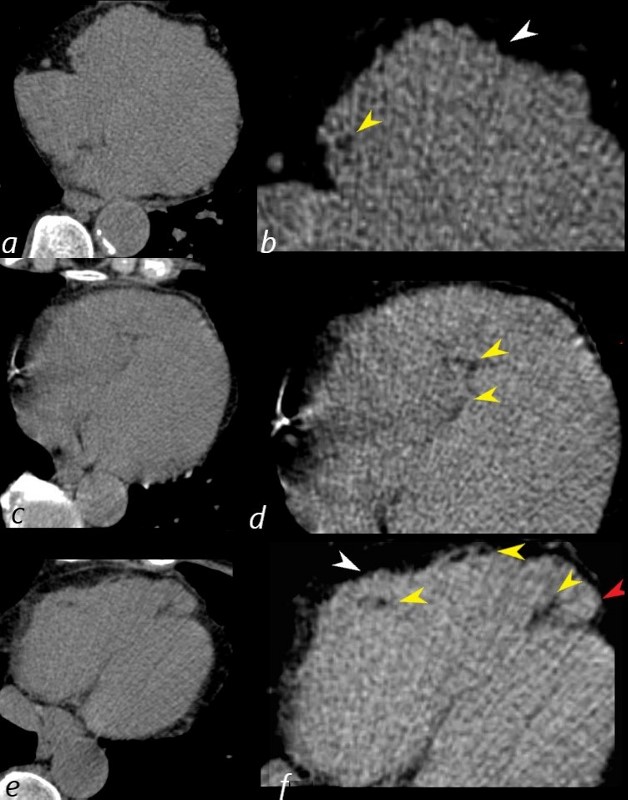
81 year old female presents 6 years earlier with non specific symptoms and abdominal CT without contrast was negative. Review of the inferior portions of the heart (images a,c,e are correspondingly magnified b,d,f) however show an unusually prominent and pointed RV apex (red arrowhead) , an irregular contour of the anterior wall of the RV (white arrowhead, f) and small focal regions of fat (yellow arrowheads b,d,f).
Ashley Davidoff MD Hristina Natcheva MD
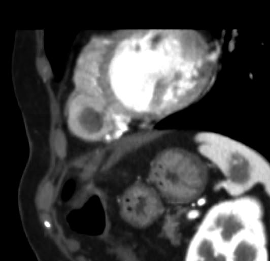
CT for rectal bleeding from 3 months prior to the MRI showed active bleeding Also noted on the lower cuts was an unusual RV wall and apex and evidence of thrombus in the apex of the RV
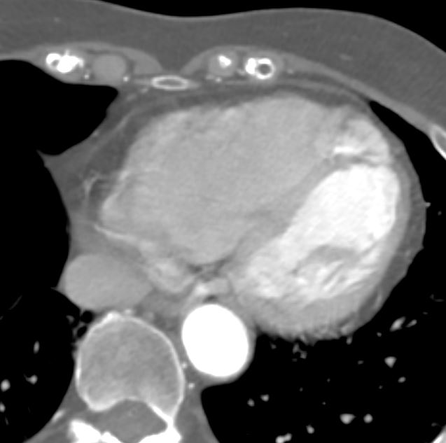
81 year old female presents 6 years earlier with non specific symptoms and abdominal CT without contrast was negative. Review of the inferior portion of the shows an enlarged RV and an unusually prominent and pointed RV apex with trapped contrast ,
Ashley Davidoff MD Hristina Natcheva MD
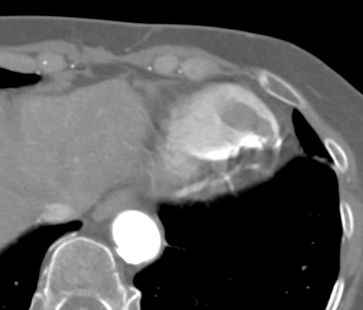
81 year old female presents 6 years earlier with nonspecific symptoms and abdominal CT without contrast was negative. Review of the inferior portion of the shows an enlarged RV and an unusually prominent and pointed RV apex with trapped contrast snf contained within the apex is a filling defect consistent the apical thrombus in the right ventricle. This image is from a dual energy CT and shows no enhancement of the mass.
Ashley Davidoff MD Hristina Natcheva MD
In the interim 2 years prior to the cardiac MRI she had a CT for rectal bleeding. The lower cuts demonstrate an unusual shape of the RV apex and a fairly large non enhancing RV apical filling defect reminiscent of thrombus.
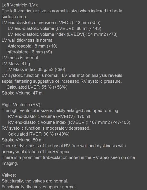
At the time of the cardiac MRI, she was having non sustained VT
Ashley Davidoff MD Hristina Natcheva MD
81 year old female presents 6 years earlier with nonspecific symptoms and abdominal CT without contrast was negative. Review of the inferior portion of the shows an enlarged RV and an unusually prominent and pointed RV apex with trapped contrast snf contained within the apex is a filling defect consistent the apical thrombus in the right ventricle. This image is from a dual energy CT and shows no enhancement of the mass.
Ashley Davidoff MD Hristina Natcheva MD
In the interim 2 years prior to the cardiac MRI she had a CT for rectal bleeding. The lower cuts demonstrate an unusual shape of the RV apex and a fairly large non enhancing RV apical filling defect reminiscent of thrombus.
At the time of the cardiac MRI, she was having non sustained VT and echo raised the possibility of ARVD and an intracavitary mass.
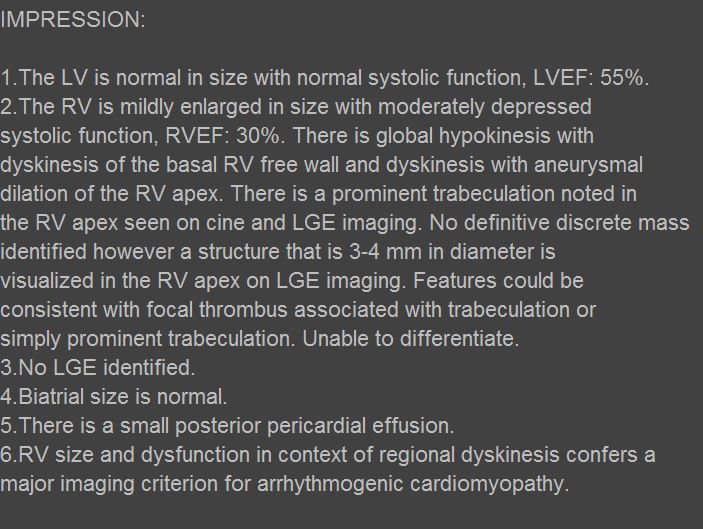
The MRI shows a large abnormal appearing apex of the RV with accentuated trabeculations and cine images that show regions of dyskinesis. These finding are consistent with ARVD.
Ashley Davidoff MD Hristina Natcheva MD
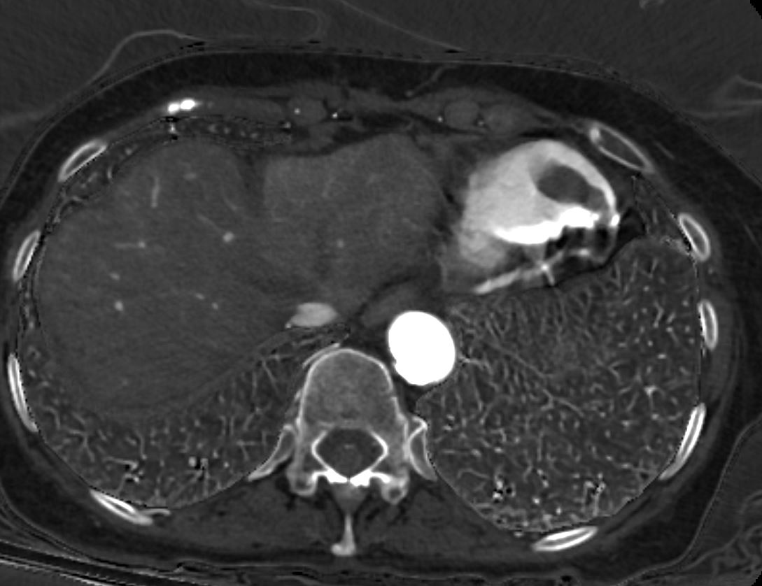
81 year old female presents 6 years earlier with nonspecific symptoms and abdominal CT without contrast was negative. Review of the inferior portion of the shows an enlarged RV and an unusually prominent and pointed RV apex with trapped contrast snf contained within the apex is a filling defect consistent the apical thrombus in the right ventricle. This image is from a sagittal reconstruction of the chest and shows the thrombus as a non-enhancing mass in the RV.
Ashley Davidoff MD Hristina Natcheva MD
ULTRASOUND REPORT
81 year old female presents 6 years earlier with nonspecific symptoms and abdominal CT without contrast was negative. Review of prior CT scans from 3 months prior showed a large filling defect in the apex of the RV consistent with a clot
At the time of the cardiac MRI, she was having non sustained VT and echo raised the possibility of ARVD and an intracavitary mass.
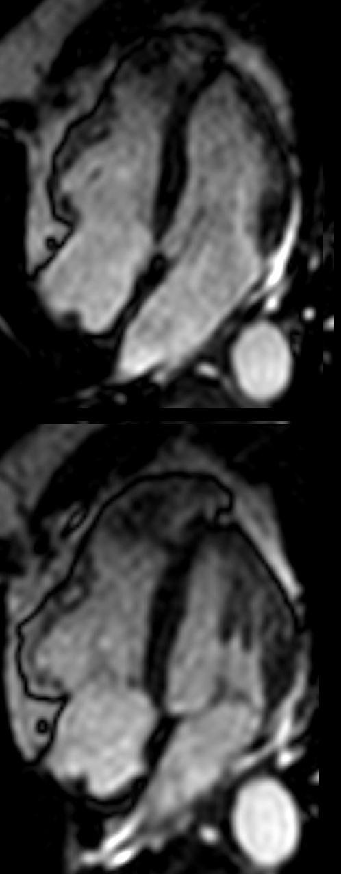
The white blood sequence in diastole (above) and systole (below shows) an unusual shape to the apex of the RV with prominent trabeculation and dyskinesia
The white blood sequence (SSFP) shows a large abnormal appearing apex of the RV with accentuated trabeculations and cine images that show regions of dyskinesis. These finding are consistent with ARVD.
Ashley Davidoff MD Hristina Natcheva MD
81 year old female presents 6 years earlier with non specific symptoms and abdominal CT without contrast was negative. Review of the inferior portions of the heart however show a pointed RV apex, an irregular contour of the anterior wall of the RV and small focal regions of fat.
In the interim 2 years prior to the cardiac MRI she had a CT for rectal bleeding. The lower cuts demonstrate an unusual shape of the RV apex and a fairly large non enhancing RV apical filling defect reminiscent of thrombus.
At the time of the cardiac MRI, she was having non sustained VT and echo raised the possibility of ARVD and an intracavitary mass.
The MRI shows a large abnormal appearing apex of the RV with accentuated trabeculations and cine images that show regions of dyskinesis. These finding are consistent with ARVD.
Ashley Davidoff MD Hristina Natcheva MD
References and Links
- TCV
- Cardiomyopathy Map
- TCVEmbryologicallyAssociated Diseases