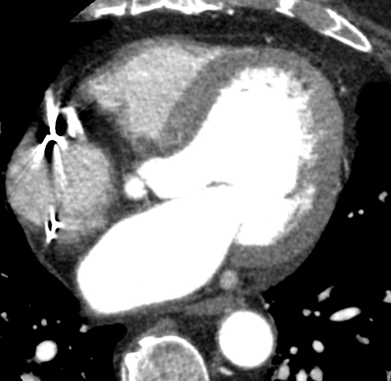
58M Ventricular tachycardia,
Post-contrast images were timed for maximum of the left ventricular cavity. In addition, delayed images were acquired 8 minutes after acquisition of contrast enhanced images.
he left ventricle is dilated with visually globally severely reduced systolic function. There is thinning and akinesis of the basal to apical inferior, basal to mid inferolateral and basal inferoseptal segments
CORONARY ARTERIES: This study was not optimized for the assessment of the coronary arteries. There are severe coronary artery calcifications. The RCA appears totally occluded in the mid RCA.
The left ventricular myocardium is thinned and akinetic in the basal to apical inferior, basal to mid inferolateral and basal inferoseptal segments. All other segments have normal myocardial wall thickness. The myocardium is hypointense in the basal to apical inferior, basal to mid inferolateral and basal inferoseptal walls on the contrast enhanced images. In addition, on delayed imaging, there is sub-endocardial to transmural hyperenhancement of the myocardium in the basal to mid inferolateral and basal inferoseptal segments. Due to artifact from the RV ICD lead, unable to assess the inferior wall on delayed imaging. Collectively, areas of hypointensity on contrast enhanced imaging and hyperenhancement on delayed imaging are consistent scar due to a prior myocardial infarction in the RCA/PDA territory.


