40 F
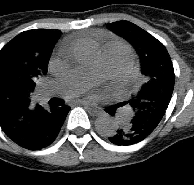
Sinus venosus defect with PAPVR complicated by Pulmonary Hypertension, and Systemic Hypertension
5 Years Ago

Ashley Davidoff MD TheCommonVein.net

Ashley Davidoff MD TheCommonVein.net
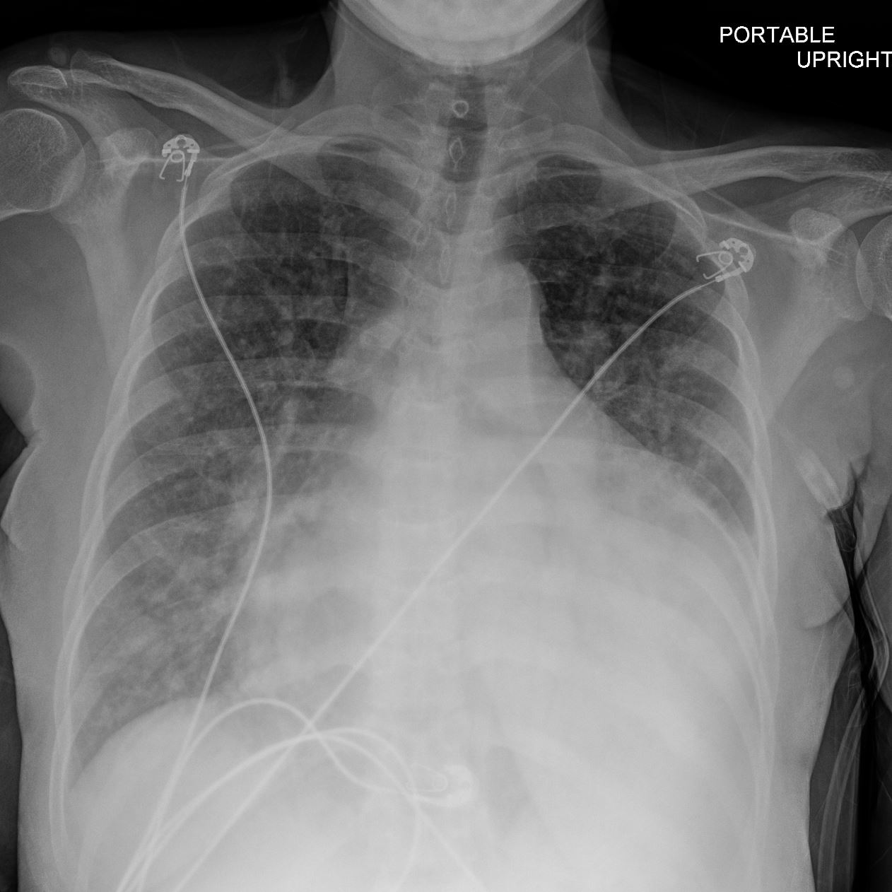
Current
Ashley Davidoff MD TheCommonVein.net
Left Atrial Enlargement
Ashley Davidoff MD TheCommonVein.net
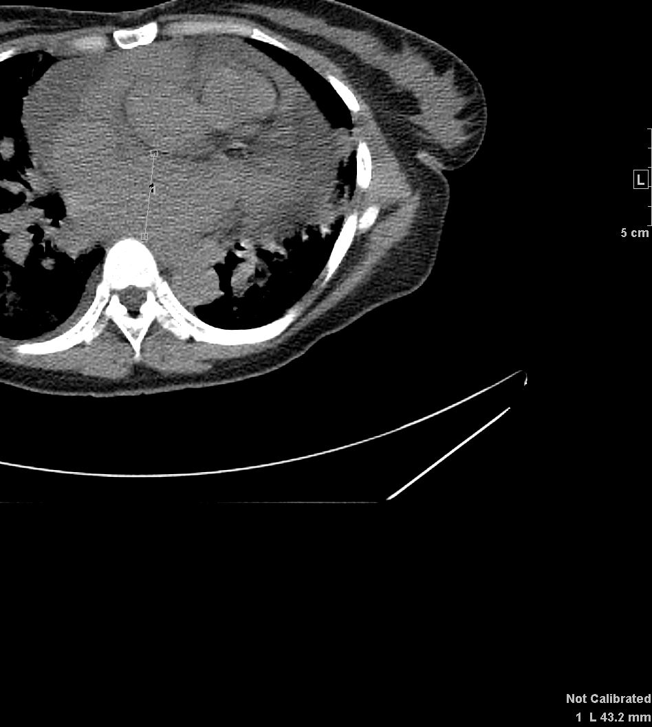
Right Atrial Enlargement and Pericardial Effusion

Ashley Davidoff MD TheCommonVein.net
Enlarged IVC and Pericardial Effusion

Ashley Davidoff MD TheCommonVein.net
Pulmonary Hypertension
Ashley Davidoff MD TheCommonVein.net
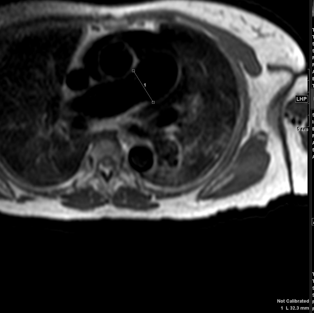
Anomalous Right Upper Lobe Pulmonary Vein to SVC

Ashley Davidoff MD TheCommonVein.net
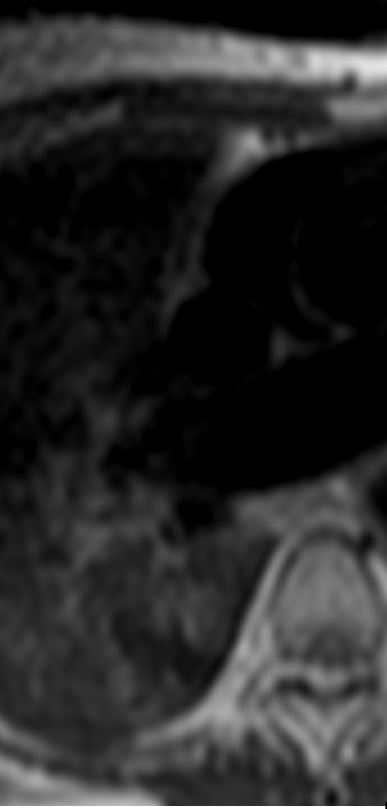
Ashley Davidoff MD TheCommonVein.net
Enlarged Right Atrium IVC and Hepatic Veins

Ashley Davidoff MD TheCommonVein.net

Ashley Davidoff MD TheCommonVein.net
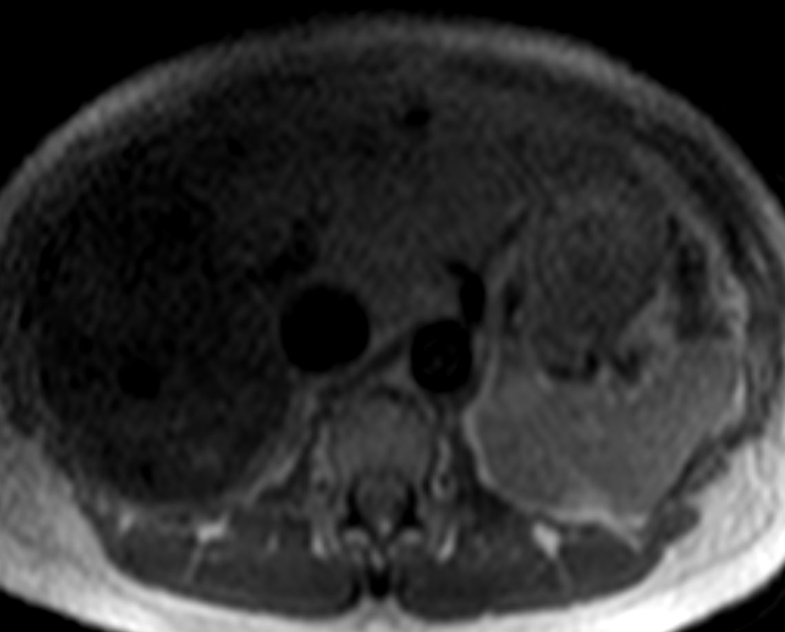
Ashley Davidoff MD TheCommonVein.net

Ashley Davidoff MD TheCommonVein.net
Sinus Venosus ASD

Ashley Davidoff MD TheCommonVein.net
Right Ventricular Enlargement

Ashley Davidoff MD TheCommonVein.net

Ashley Davidoff MD TheCommonVein.net
Current
Echo
Left ventricular wall thickness is severely increased.
Left ventricular systolic function is normal.
The ejection fraction based on measurements and visual assessment is estimated
to be 55-60%.
No regional wall motion abnormalities noted.
The left atrium is mildly enlarged.
There is a known large sinus venosus ASD which was not well seen in this
study.
The right atrium is enlarged.
The right ventricular systolic function is mildly reduced.
The right ventricle is mild to moderately dilated.
There is trace mitral regurgitation.
There is moderate tricuspid regurgitation.
Estimated PA systolic pressure is 74 mmHg.
The inferior vena cava is > 2.1 cm and exhibits normal respiratory variability
consistent with a right atrial pressure of ~ 8 mm Hg.
There is a small to moderate pericardial effusion that appears largest
posteriorly measuring 1.4 cm. There is no chamber collapse, no tamponade
findings noted.
Compared to prior study of 5years ago, LVH and pericardial effusion are noted. h/o
ASD noted.
MRI
1. The LV is normal in size with normal systolic function, LVEF: 54%. LV wall thickness is mildly increased.
2. The RV is moderately dilated with mildly reduced systolic function, RVEF: 37%.
3. There is a large sinus venosus atrial septal defect, with an associated anomalous right upper pulmonary vein, with evidence of significant left to right shunting (Qp:Qs = 3.1). An additional anomalous right pulmonary vein drains directly into the SVC, just superior to the sinus venosus defect.
5. Valves are structurally normal. There is visually mild mitral regurgitation and moderate tricuspid regurgitation.
6. The right atrium is mildly dilated.
7. The main pulmonary artery is dilated.
8. There is a moderate-large circumferential pericardial effusion
PCW 18, RA14

