66-year-old female presents with chest pain
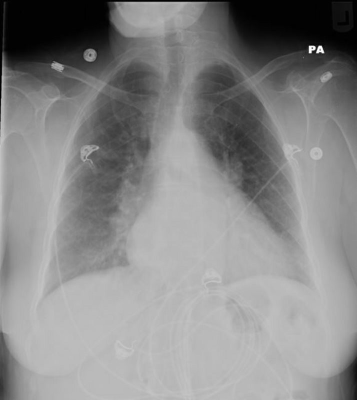
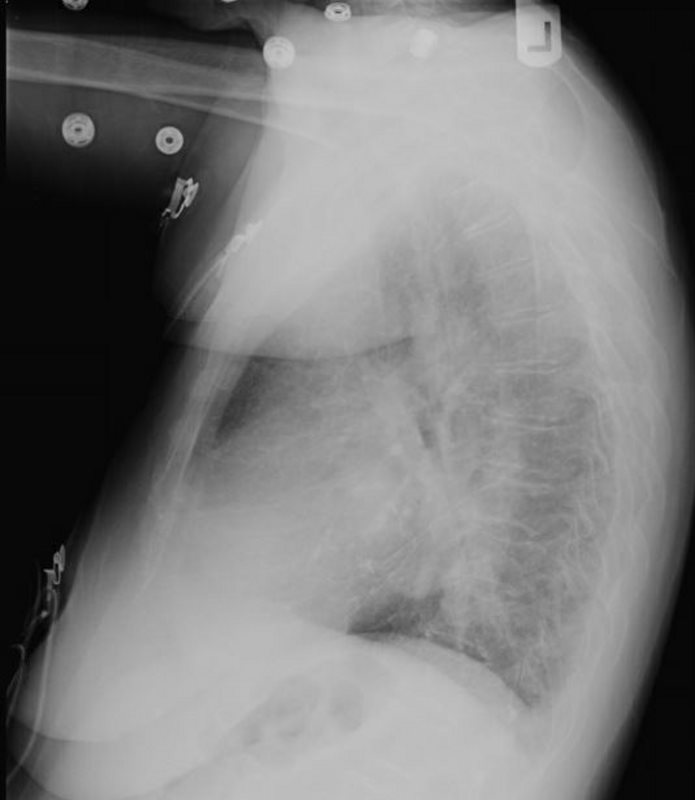
The frontal CXR shows left atrial enlargement characterized by elevated L main stem bronchus, and in retrospect some calcifications overlying the left atrium on the lateral
Ashley Davidoff MD
Ashley Davidoff MD

CALCIFIED LEFT ATRIAL MYXOMA
Ashley Davidoff MD
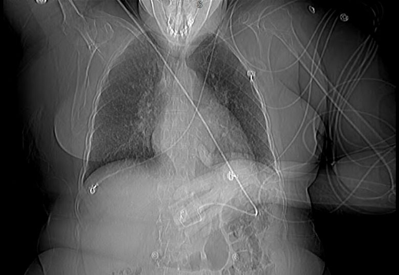
The scout film prior to the CT confirms the elevated left main stem bronchus
Ashley Davidoff MD
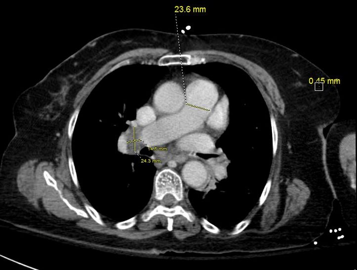
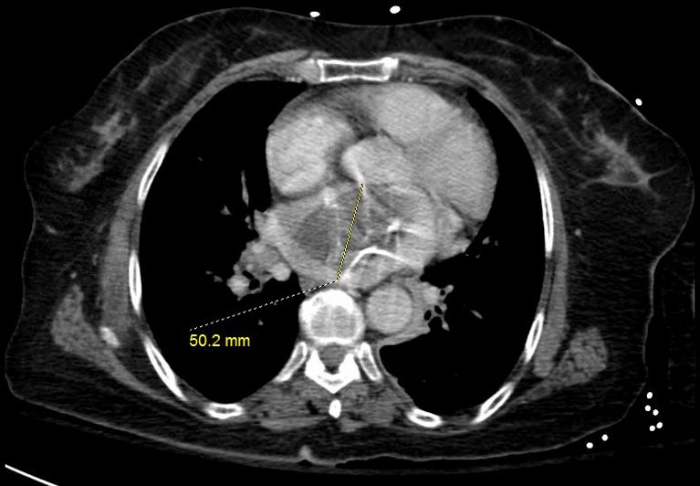
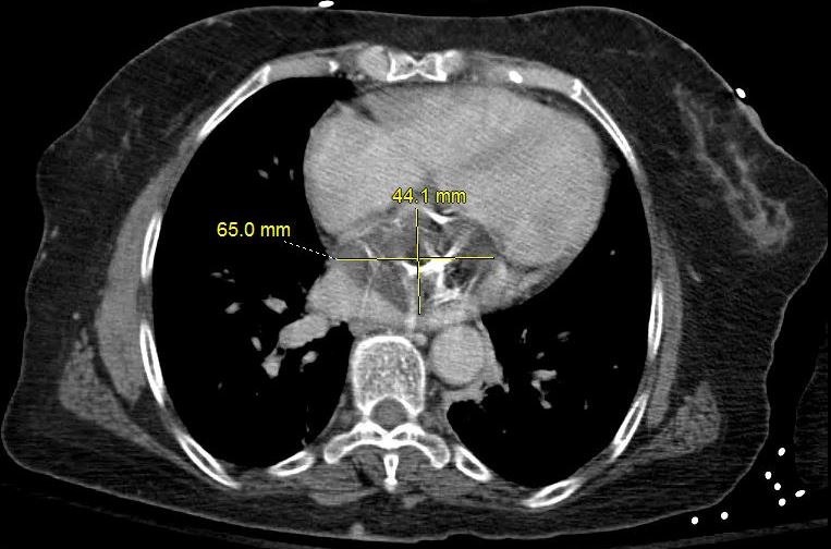
The dominant finding is a calcified left atrial mass that is attached to the atrial septum. It measures 6.5 X 4.4 X 4.6cms and shows mild enhancement.
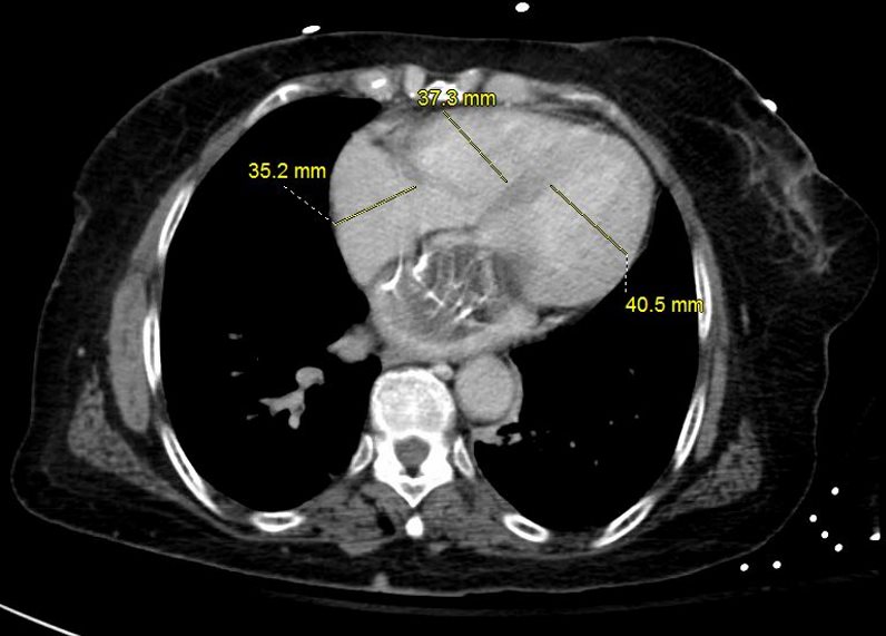
The other chambers are normal in size

66-year-old female presents with chest pain
The frontal CXR shows left atrial enlargement characterized by elevated L main stem bronchus, and in retrospect some calcifications overlying the left atrium on the lateral
The scout film prior to the CT confirms the elevated left main stem bronchus, but the dominant finding is a calcified left atrial mass that is attached to the atrial septum. It measures 6.5 X 4.4 X 4.6cms and shows mild enhancement.
The other chambers are normal in size
The mass was resected and pathology findings were consistent with an atrial myxoma
Ashley Davidoff MD

Ashley Davidoff MD
Ashley Davidoff MD
Ashley Davidoff MD
CALCIFIED LEFT ATRIAL MYXOMA
Ashley Davidoff MD
66-year-old female presents with chest pain
The frontal CXR shows left atrial enlargement characterized by elevated L main stem bronchus, and in retrospect some calcifications overlying the left atrium on the lateral
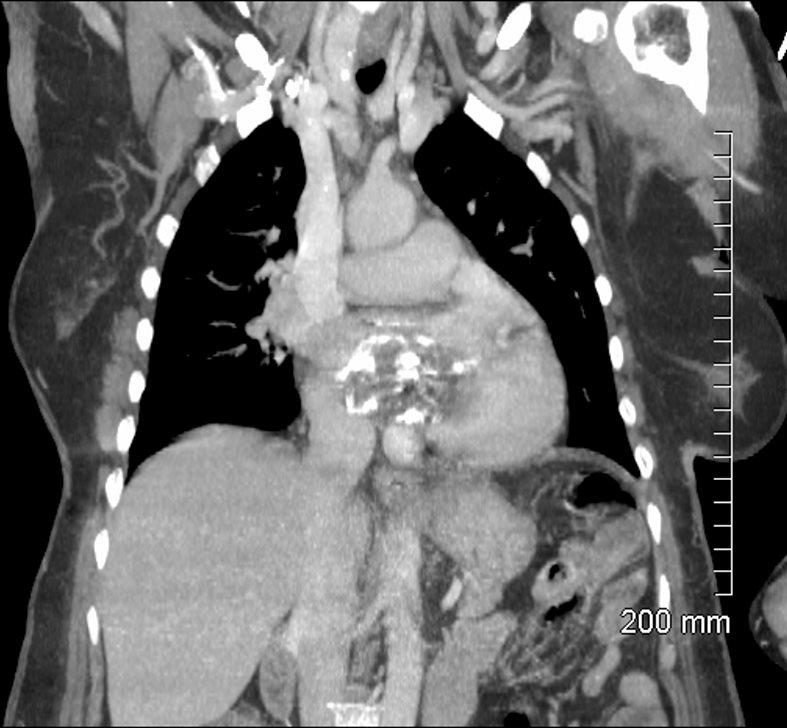
The scout film prior to the CT confirms the elevated left main stem bronchus, but the dominant finding is a calcified left atrial mass that is attached to the atrial septum. It measures 6.5 X 4.4 X 4.6cms and shows mild enhancement.
The other chambers are normal in size
The mass was resected and pathology findings were consistent with an atrial myxoma
Ashley Davidoff MD
Ashley Davidoff MD
Pre Contrast
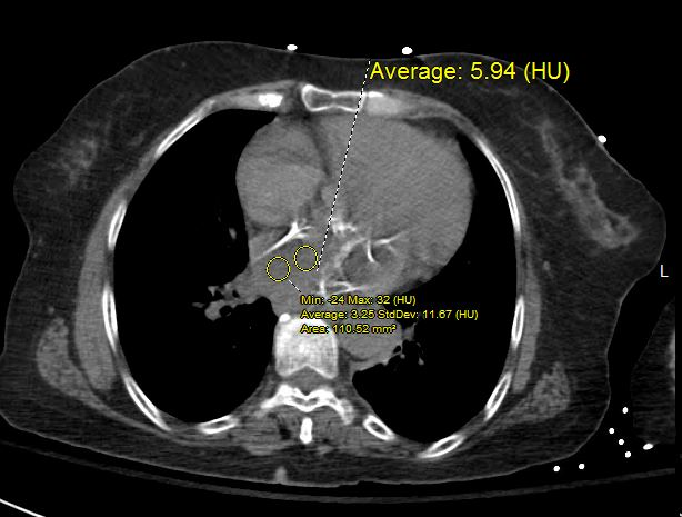
Ashley Davidoff MD
During Contrast
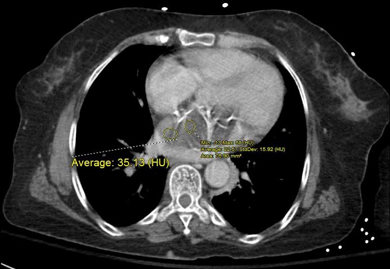
CALCIFIED LEFT ATRIAL MYXOMA
Ashley Davidoff MD
Post Contrast

12 HU
Ashley Davidoff MD
The mass was resected and pathology findings were consistent with an atrial myxoma
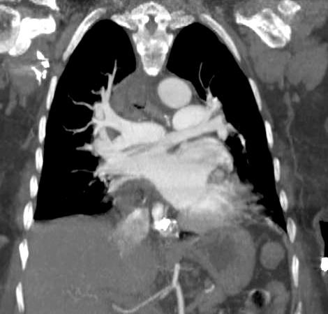
WIDELY PATENT LA
Ashley Davidoff MD

POST OP REMOVAL OF CALCIFIED LEFT ATRIAL MYXOMA IVC REPAIR
Ashley Davidoff MD
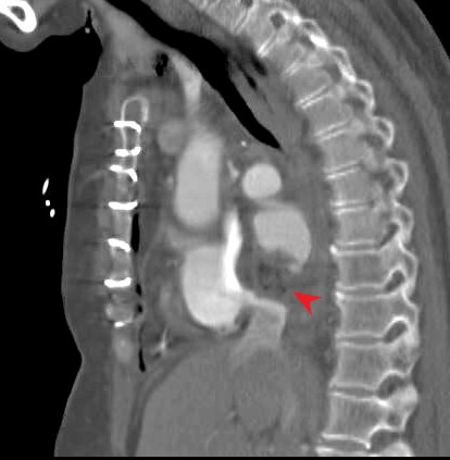
POST OP REPAIR OF CALCIFIED LEFT ATRIAL MYXOMA
Ashley Davidoff MD
