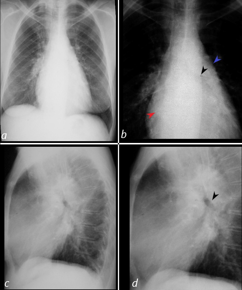
58-year-old female presents with a hacking non productive cough
The frontal CXR shows probable left atrial enlargement characterized by elevated L main stem bronchus (b black arrow), double density (red arrow, b) and a straight heart border caused mostly by a large PA segment (blue arrow b) and evidence of redistribution (a). The lateral confirms left atrial enlargement with posterior displacement and elevation of the left main stem bronchus (d, black arrow). The left ventricle is not enlarged. These findings suggest mitral stenosis.

58-year-old female presents with cough
The frontal CXR shows probable left atrial enlargement characterized by elevated L main stem bronchus (b black arrow), double density (red arrow, b) and a straight heart border caused mostly by a large PA segment (blue arrow b) and evidence of redistribution (a). The lateral confirms left atrial enlargement with posterior displacement and elevation of the left main stem bronchus (d, black arrow). The left ventricle is not enlarged. These findings suggest mitral stenosis.
Ashley Davidoff MD
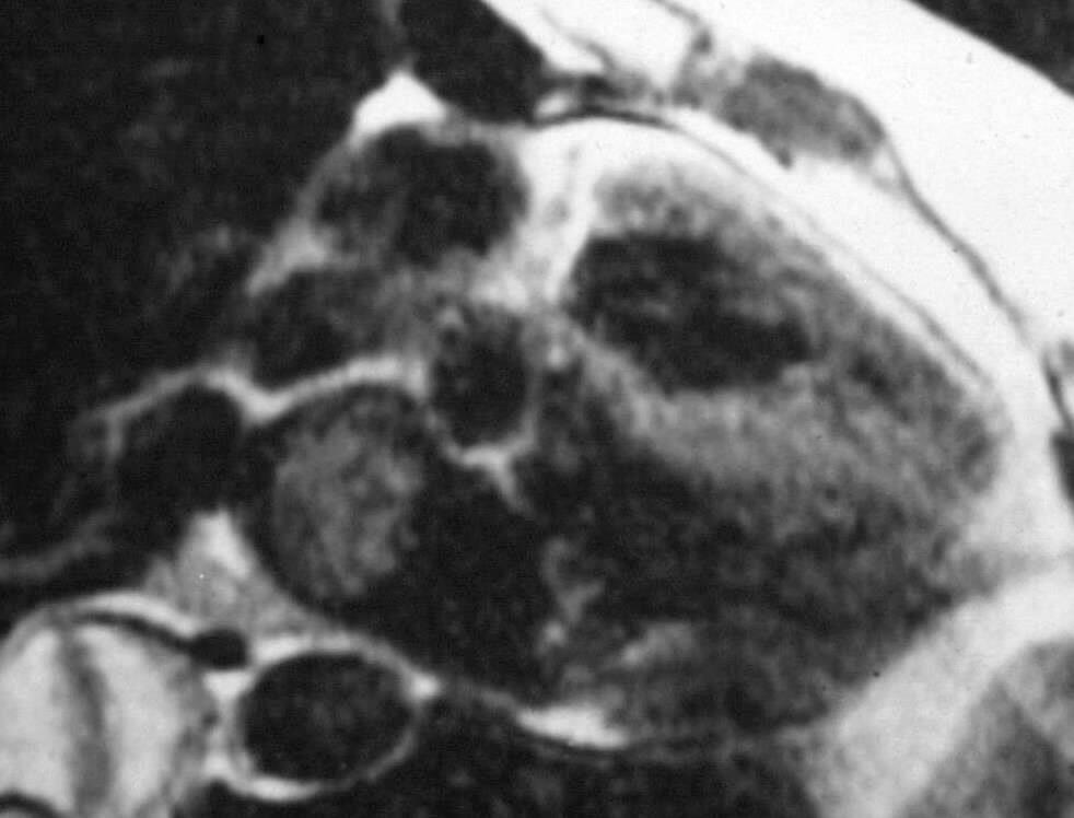
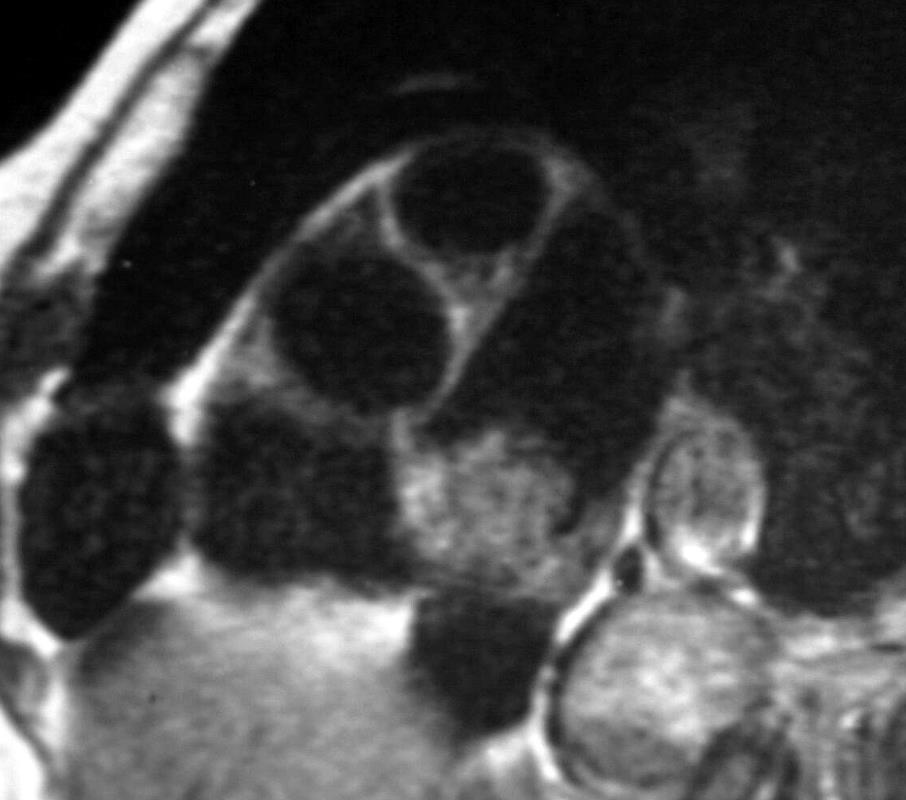
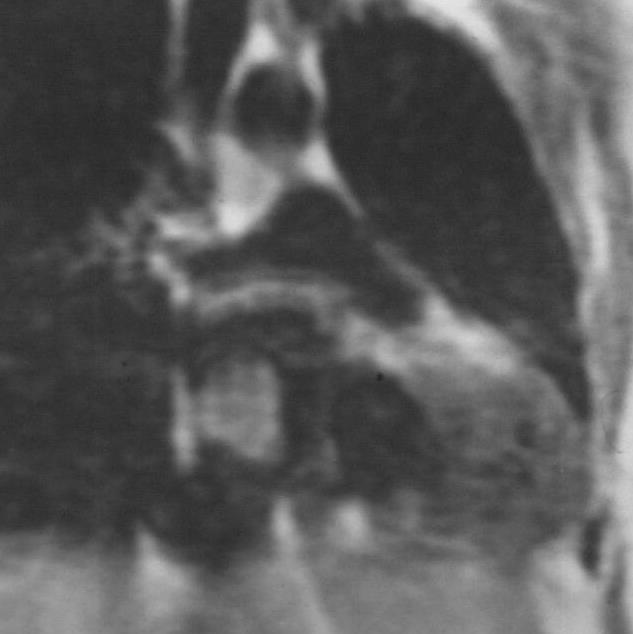
Black blood sequence shows a heterogeneous mass in the left atrium (LA) attached to the septum with signal brighter than the myocardium and less than fat. These findings are characteristic of an atrial myxoma. The mass was removed and pathology confirmed an atrial myxoma

ATRIAL MYXOMA
Ashley Davidoff MD

Ashley Davidoff MD

Ashley Davidoff MD
