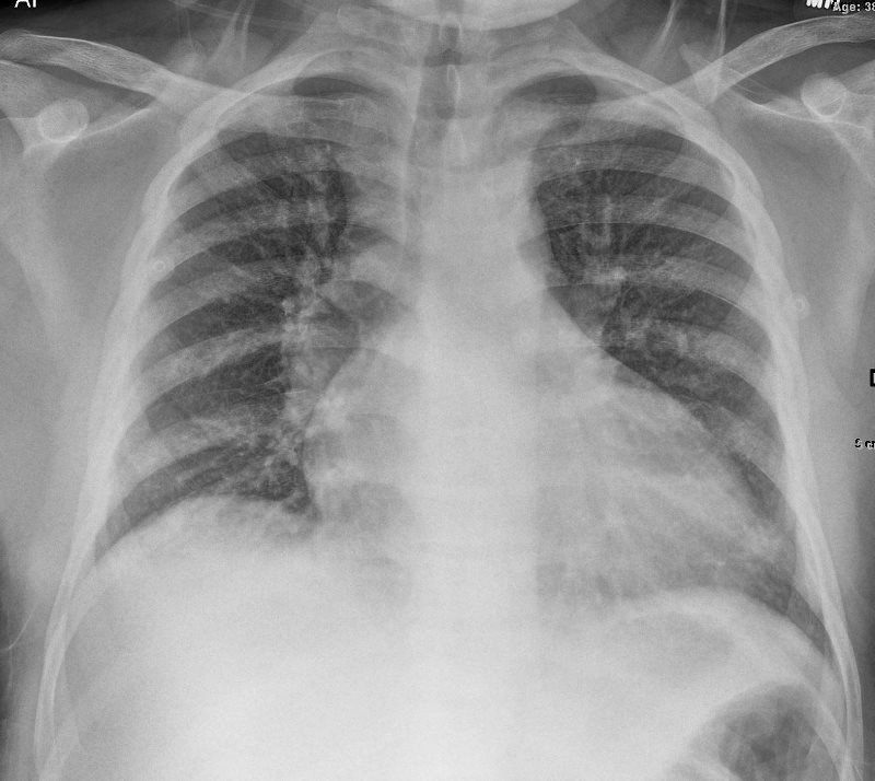
38-year-old female with history of sickle cell disease presents with chest pain.
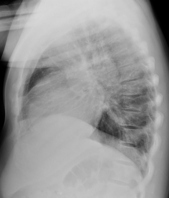
Chest X-ray shows cardiomegaly with left ventricular and left atrial enlargement and mild elevation of the end diastolic pressure characterized by cephalization of the vasculature,
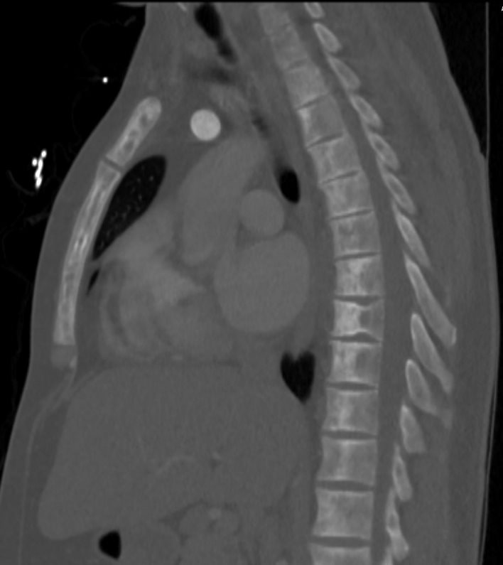
CHF LEFT ATRIAL ENLARGEMENT AND LV ENLARGEMENT
Ashley Davidoff MD
Ashley Davidoff MD
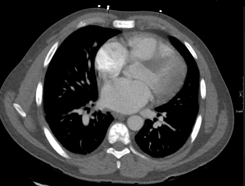
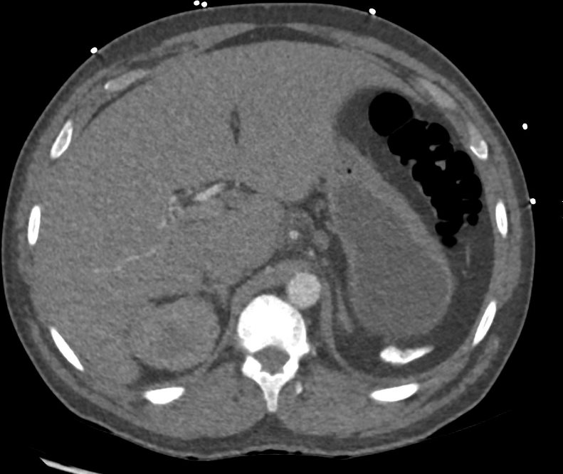
The CT scan confirms left atrial enlargement, auto splenectomy with a small calcified spleen, sclerotic changes in the vertebrae and sternum, and H shaped vertebra characteristic of sequelae of bone infarction.
Ashley Davidoff MD
Ashley Davidoff MD
Ashley Davidoff MD

