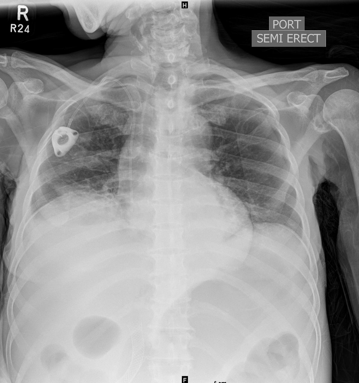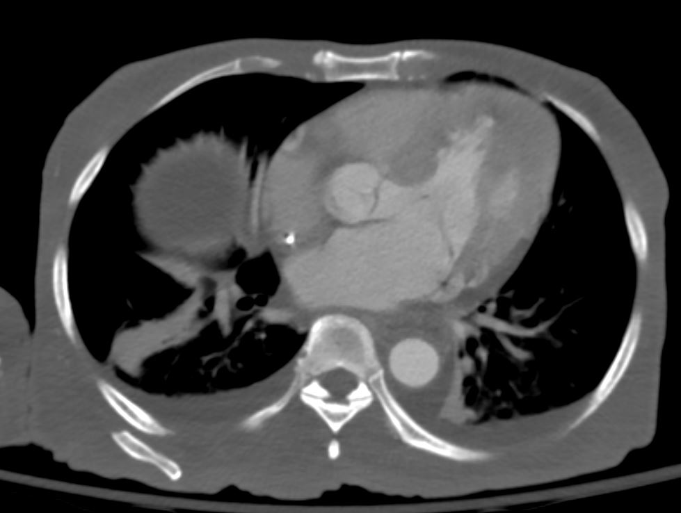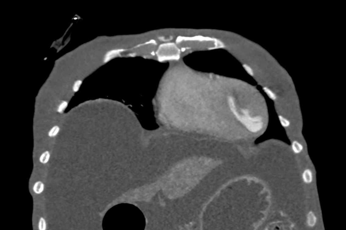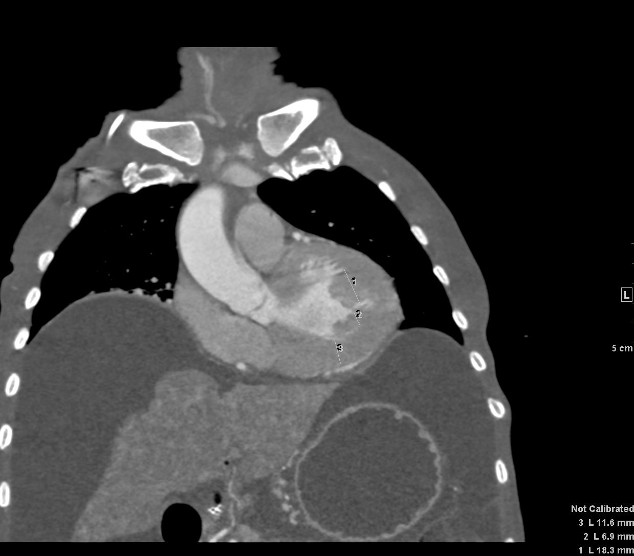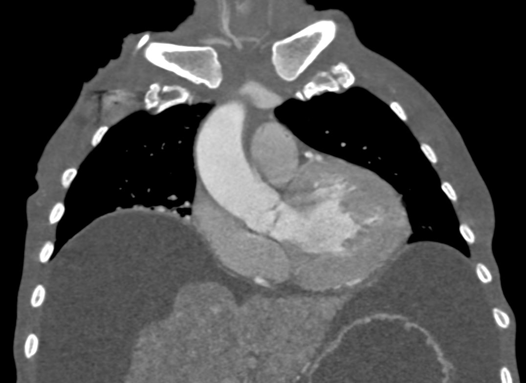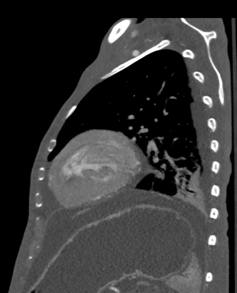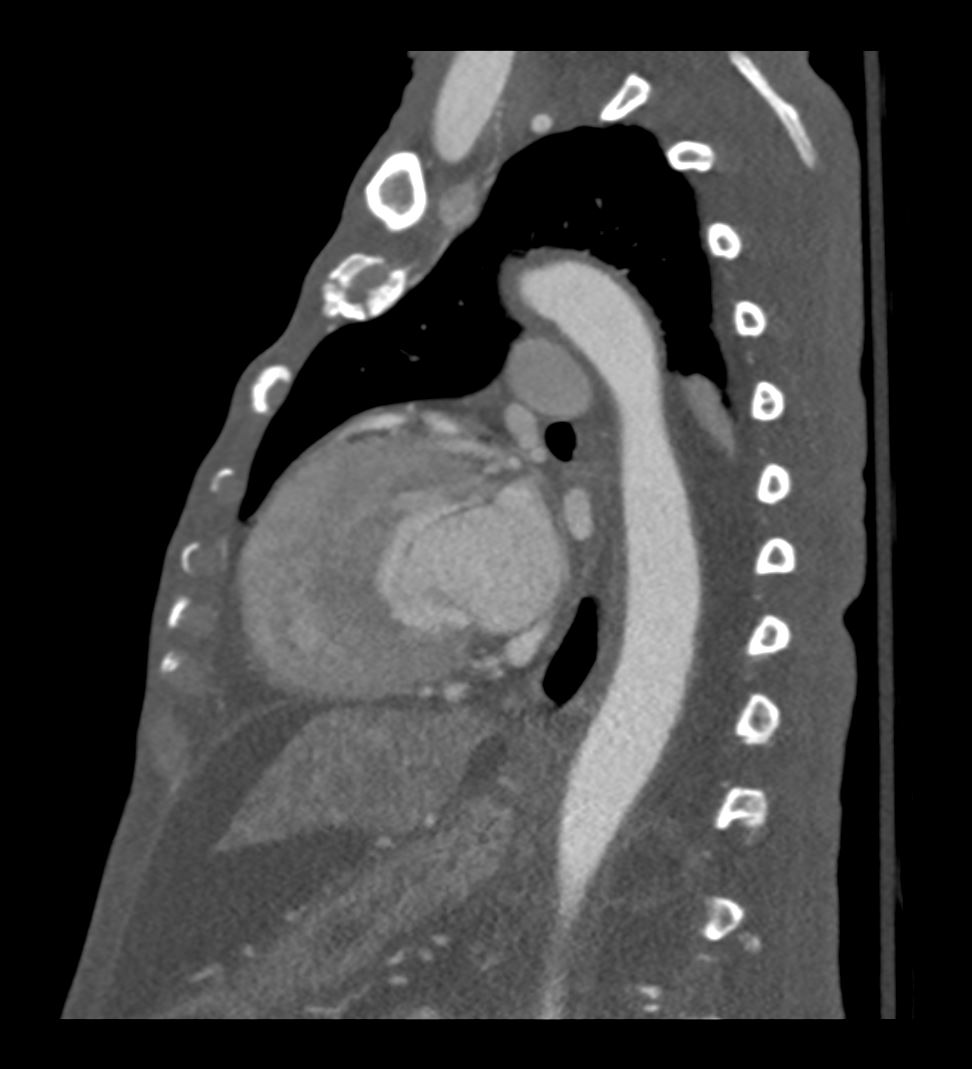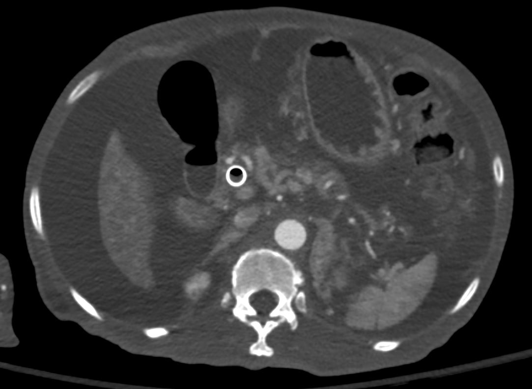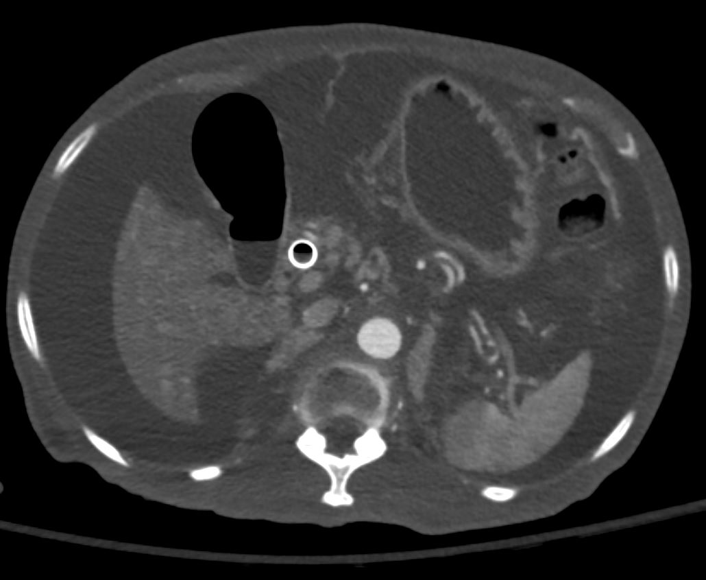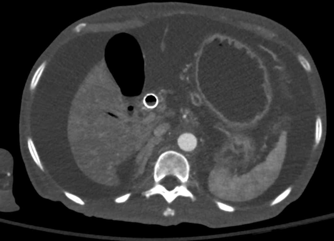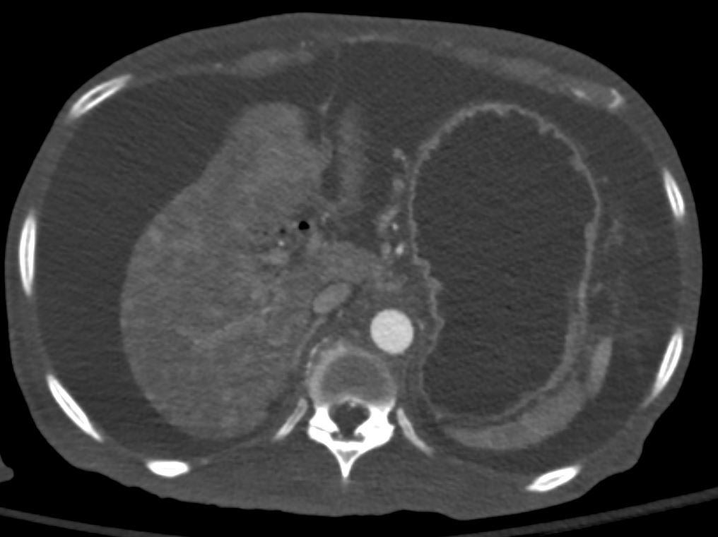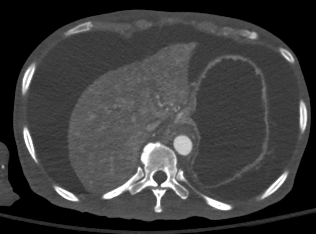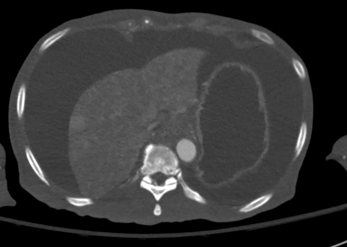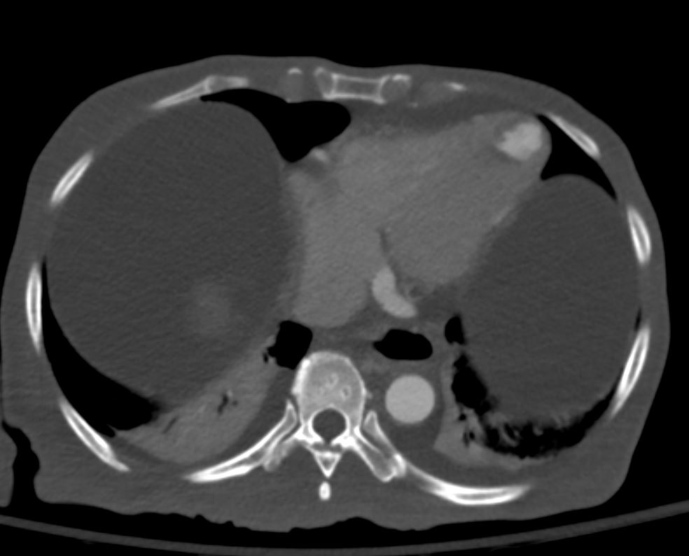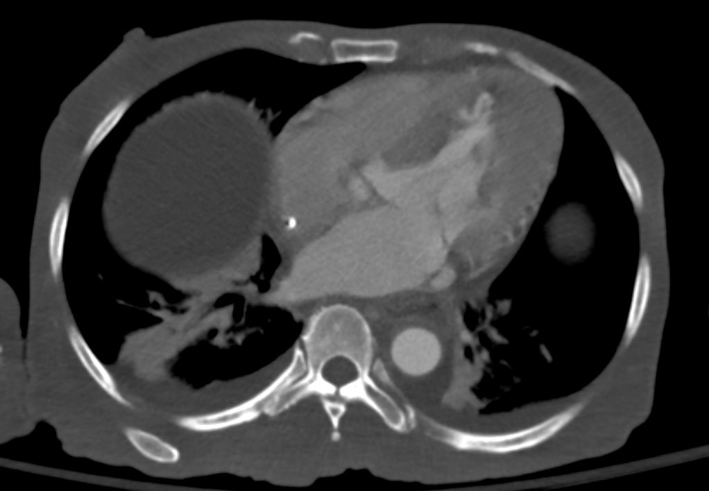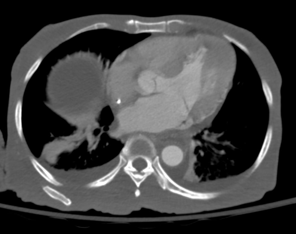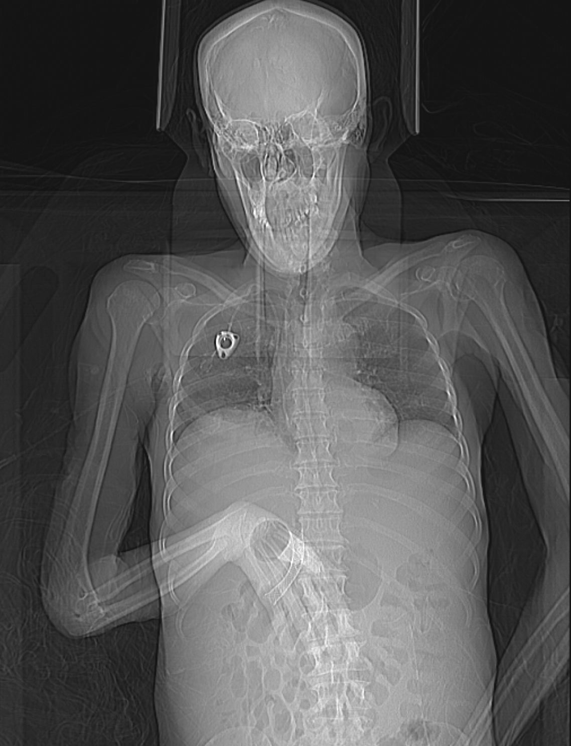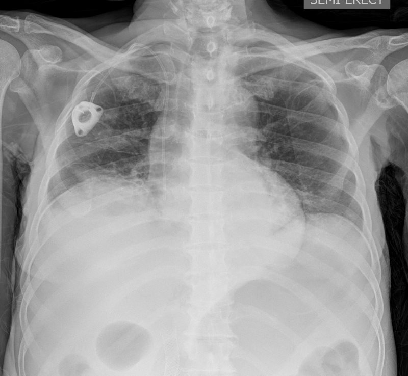
64-year-old male with pancreatic carcinoma and apical hypertrophic cardiomyopathy.
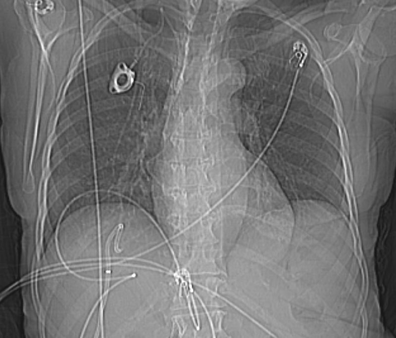
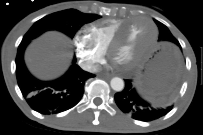
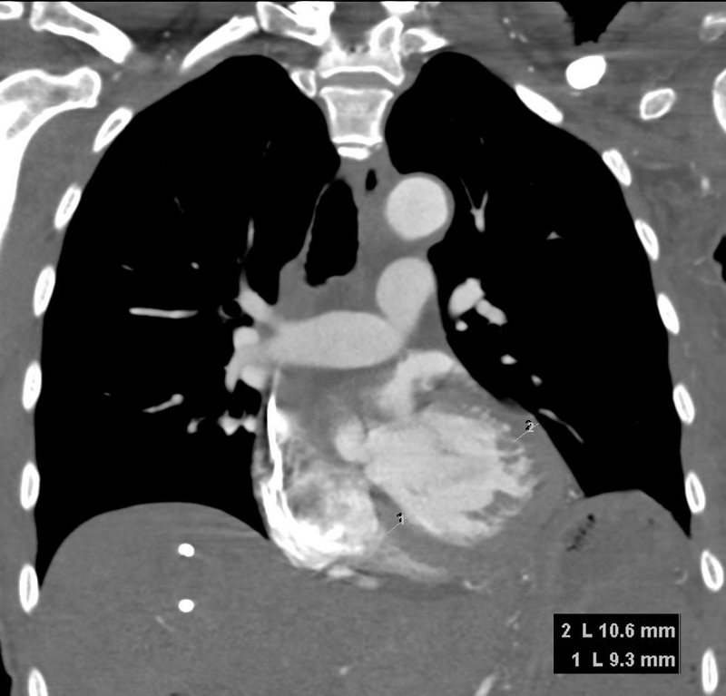
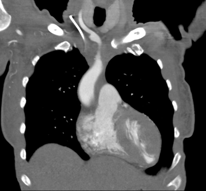
CXR shows left ventricular configuration with an unusually prominent left ventricle. CT shows dominant apical hypertrophy of the LV.
Echocardiogram confirms the presence of apical hypertrophy with mi systolic gradient within the LV cavity of 38mmHg.
Ashley Davidoff MD
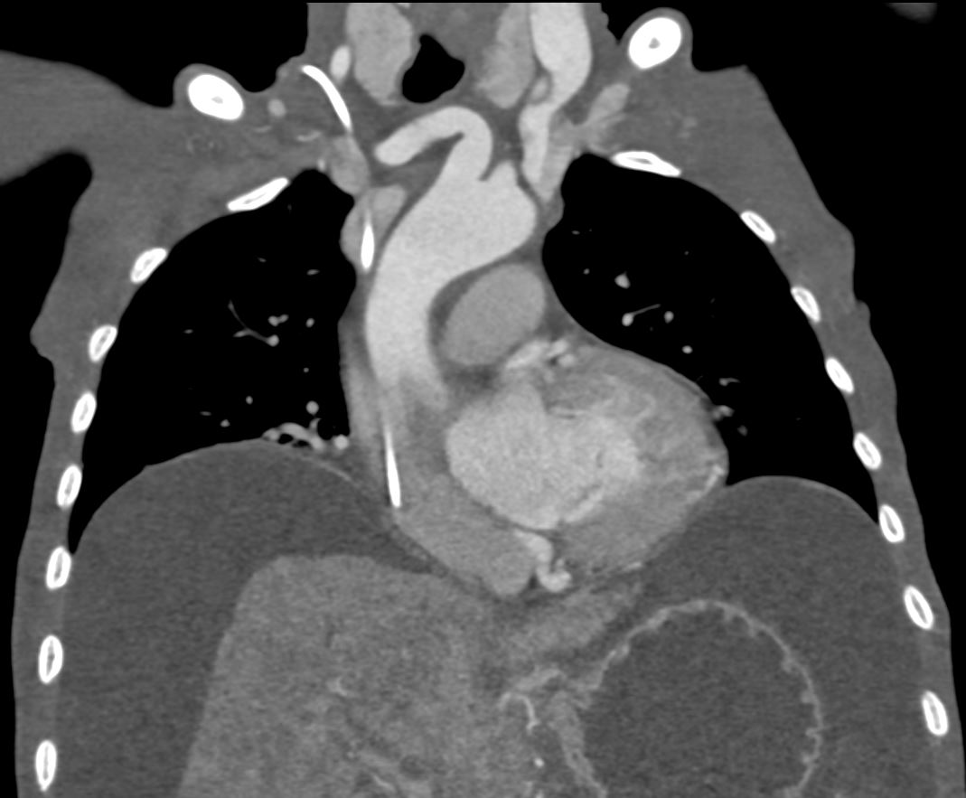
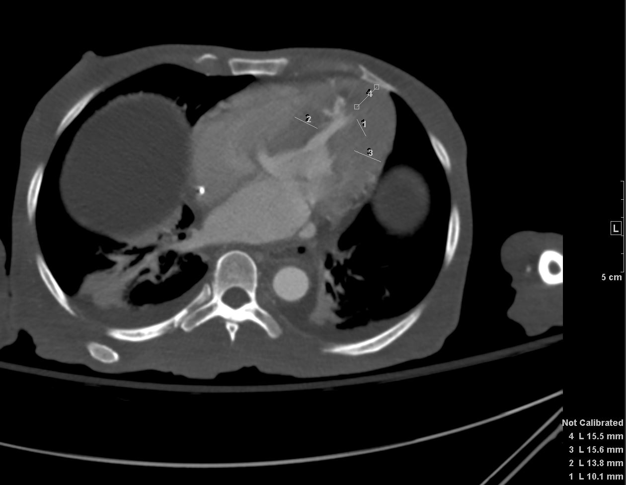
64-year-old male with pancreatic carcinoma and apical hypertrophic cardiomyopathy.
CXR shows left ventricular configuration with an unusually prominent left ventricle. CT shows dominant apical hypertrophy of the LV.
Echocardiogram confirms the presence of apical hypertrophy with mi systolic gradient within the LV cavity of 38 mmHg.
Ashley Davidoff MD
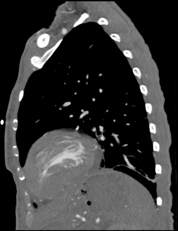
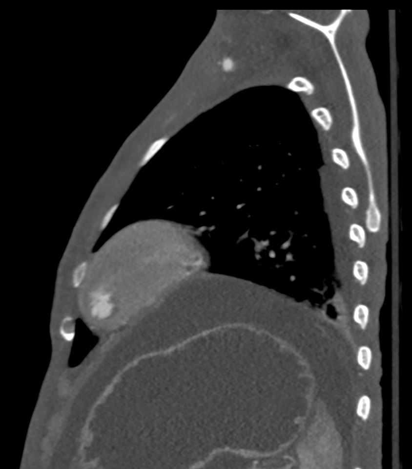
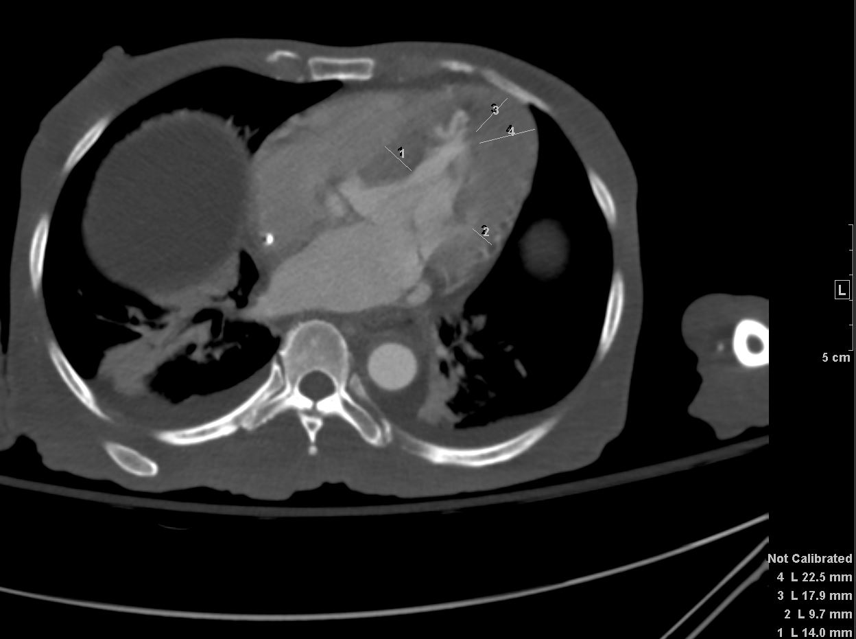
64-year-old male with pancreatic carcinoma and apical hypertrophic cardiomyopathy.
CXR shows left ventricular configuration with an unusually prominent left ventricle. CT shows dominant apical hypertrophy of the LV.
Echocardiogram confirms the presence of apical hypertrophy with mi systolic gradient within the LV cavity of 38 mmHg.
Ashley Davidoff MD
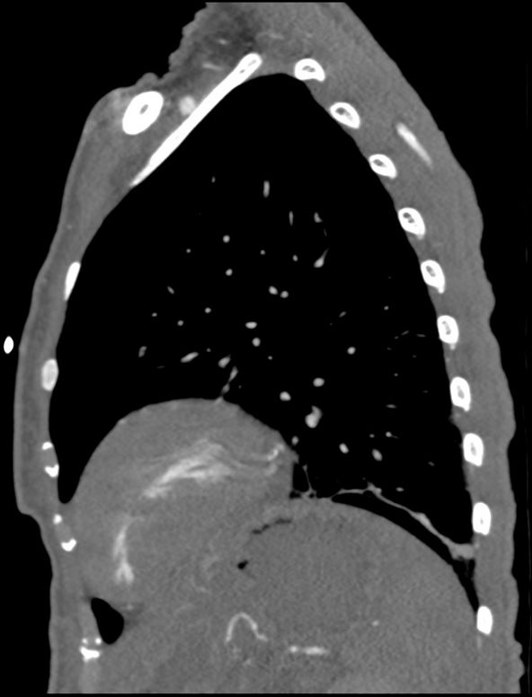
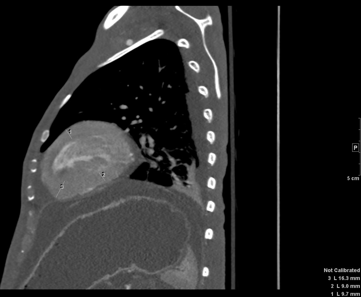
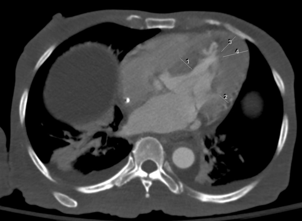
64-year-old male with pancreatic carcinoma and apical hypertrophic cardiomyopathy.
CXR shows left ventricular configuration with an unusually prominent left ventricle. CT shows dominant apical hypertrophy of the LV.
Echocardiogram confirms the presence of apical hypertrophy with mi systolic gradient within the LV cavity of 38mmHg.
Ashley Davidoff MD
Echo showed EF of 70% with mid ventricular cavity obliteration resulting in a gradient of 39mm Hg
References and Links

