Copyright 2008
Definition
The endocardial cushions are embryological precursors of the lower portion of the interatrial septum, the upper portion of the superior end of the interventricular septum, and portions of the mitral and tricuspid valves.
Structurally the epicentre of the endocardial cushions is at the crux of the heart, so that many structures can be affected when there is a disorder of the embryological tissue.
In the extreme of disease, known as a complete A-V canal defect, all the chambers of the heart are potentially connected and blood from any one of the chambers can flow to any one of the others. In addition the conduction system is close by and so it is often affected by endocardial cushion defects. The atrial septum can be affected in both the complete and the partial defects. An isolated primum ASD affects the lowest portion of the atrial septum and usually affects the conduction system and left axis deviation ofn the EKG is characteristic. Sometimes a cleft mitral valve is associated with the primum ASD.
The diagnosis is made clinically with ejection flow murmur, loud fixed P2, and occasionally a murmur of mitral regurgitation. Echocardiography is the study of choice.
Treatment is surgical.

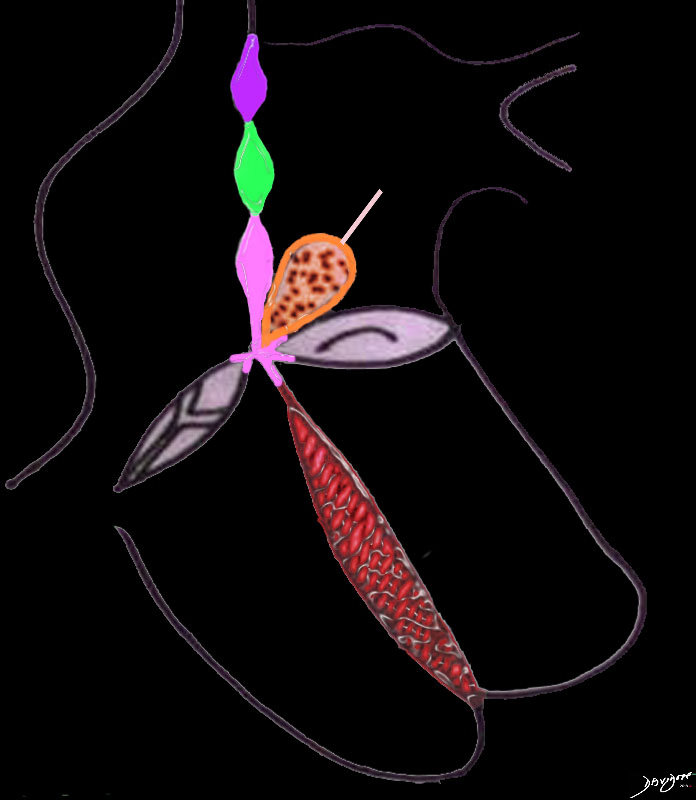
The atrial septum consists of 3 parts; The most superior is the septum derived from the sinus venosus (purple), the middle is the septum primum (green) and the most inferior is derived from the endocardial cushions (pink) The endocardial cushions also contribute to the formation of the of the membranous component of the interventricular septum. The interventricular septum consists of the membranous septum and the muscular septum (red). The great vessels are separated by a muscular conal septum (orange) and the membranous aorticopulmonary septum (light pink).
Ashley Davidoff MD TheCommonVein.net

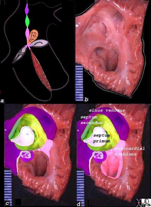
The diagram shows the three portions of the interatrial septum in (a) with the upper portion (purple) deriving from sinus venosus tissue, the middle (grreen) the mesodermal tissue, and the lower (pink) the endocardial cushion tissue. Image b, is an anatomical specimen that is overlaid in reference colors in c, and labelled in d. The middle of the atrial septum is a fibrous membrane called the septum primum, and it is surrounded a rim of muscular tissue called the septum secundum.
The fossa ovalis is the middle portion of the interatrial septum and consists of the central portion called the septum primum (white) and the surrounding muscle called the septum secundum (green).
Ashley Davidoff MD
01653c11b05a04 TheCommonVein.net

The drawing shows the interatrial septum and the defects associated with each of the components. Image a shows a single defect in the septum primum and this is called an ASD secundum, or a secundum ASD. This is the most common ASD. The second image (b) shows an A-V canal defect in green
