Hemangiopericytoma
Elisa Flower MD Asim Mian MD
The Common Vein Copyright 2010
Definition
A hemangiopericytoma is a meningeal tumor which grows from pericytes which are modified smooth muscle cells surrounding capillaries. They typically occur in adults in the fourth or fifth decade. They are similar to meningiomas in that they are extraaxial masses, occur in similar locations (supratentorial location being most common) and have a similar appearance on imaging. Hemangiopericytomas, however, are aggressive and can result in local invasion, high rate of local recurrence after resection and distant metastases.
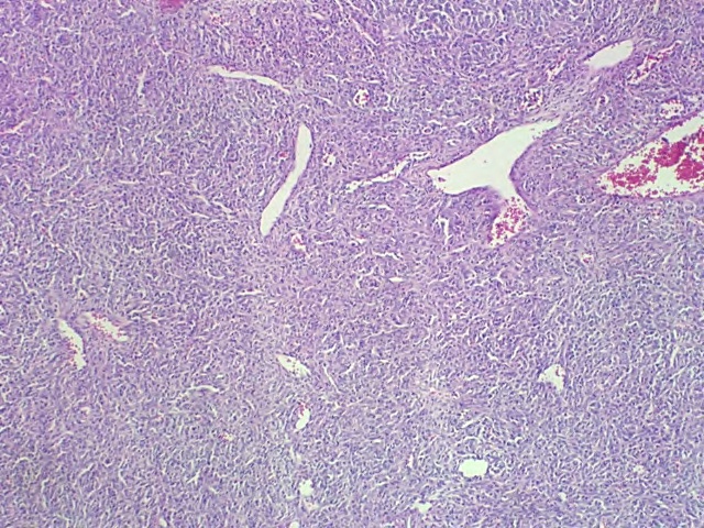
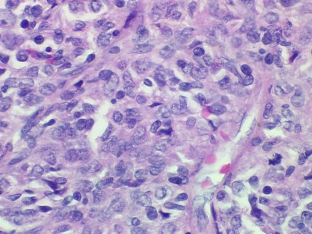
Histologically, hemangiopericytomas are characterized by a highly cellular tumor. There are nests of tumors cells seen to encase a network of branching capillaries. Prominent mitotic activity is seen, correlating with the aggressive nature of this tumor.
Clinical presentation depends on location with a common symptom being headache.
Diagnosis can be suggested by imaging findings but is ultimately made by pathology.
Imaging of hemangiopericytomas includes both CT and MRI, both of which help characterize the lesion which have a similar appearance to meningiomas. They appear as lobulated dural based masses. On CT they are isodense to hyperdense masses without calcifications, which can be seen in meningiomas. CT also helps demonstrate erosion of the skull, which is another suggestive finding as opposed to hyperostosis of the skull which can be found next to meningiomas. On MRI, hemangiopericytoms are T1 isointense and T2 hyperintense. They demonstrate focal areas of low signal corresponding to flow within prominent blood vessels. After contrast administration these masses demonstrate marked enhancement.
Treatment includes primary surgical resection. Preoperative embolization is frequently performed as these are very vascular. Postoperative radiation or radiosurgery may reduce risk of recurrence.

Extraaxial Mass – Eroding through the Skull |
|
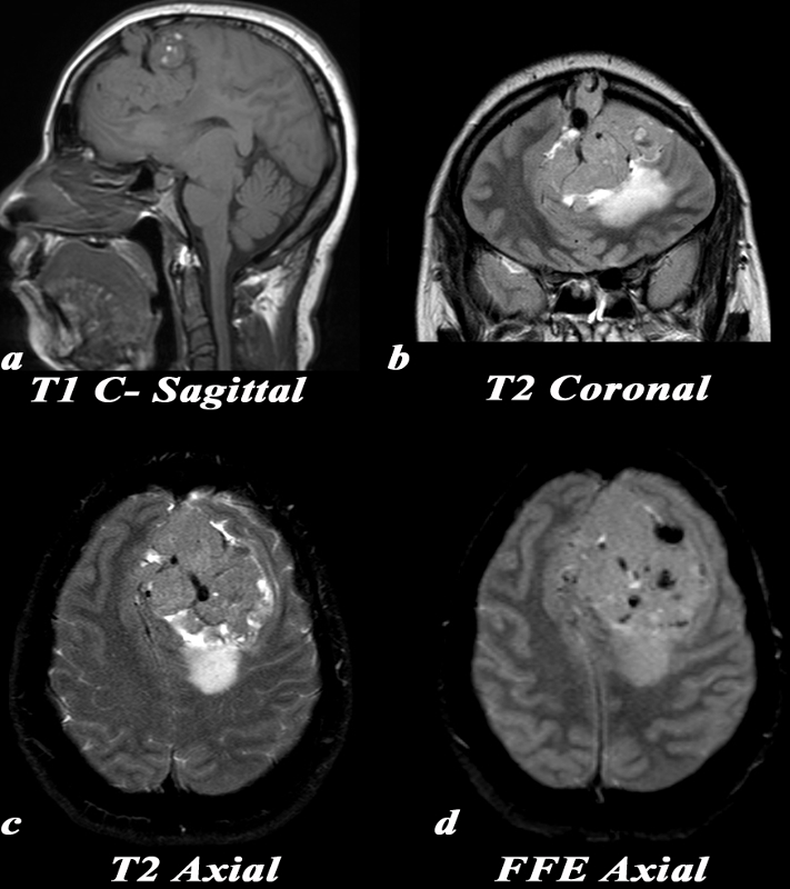
This 30 year old pregnant female presented with a constant headache and an MRI was obtained. Note that MR contrast was not given due to her pregnancy.
MRI: Because this mass is isointense and large, it can be difficult to distinguish if it arises from within the brain parenchyma (intra-axial), or if it is external to it (extra-axial). Note how the brain tissue is buckled around the mass sagittal T1 (a), and coronal T2 (b), and compressed by it, suggesting that it is extra-axial in location.
T1: (a) The mass is predominantly T1 isointense with several areas of high T1 signal which may be related to blood products. A large internal blood vessel is noted.
T2 coronal: (b) Notice the aggressive features of this mass which include erosion through the overlying skull and large internal network of blood vessels which are the linear low intensity structures. Increased T2 signal consistent with edema is seen in the underlying parenchyma. The mass itself is isointense to gray matter with central hypointense foci which are consistent with large blood vessels. T2 axial:(c) Mass effect, shift of the midline, internal network of vessels (linear low intensity structures), and T2 bright posterior peritumoral parenchymal edema.
FFE: (d) This gradient echo MRI sequence is beneficial for picking up substances which alter the local magnetic field, or paramagnetic substances. These substances cause ?blooming? which is a loss of signal, or hypointensity, within and just around them. Note the multiple areas of ?blooming? within the mass which can be caused by blood products or calcification.
Image Courtesy Elisa Flower MD and Asim Mian MD 97656c.8
|
–

H&E Low Power
The low power polymicrograph demonstrates a highly cellular mass with monomorphic cells. There was evidence of necrosis and apoptosis within this hemangiopericytoma.

H&E Higher Power
On the high power polymicrograph, there is a mitotic figure seen at the lower right hand side of the image (the dark blue irregular shaped nucleus).

Hemangiopericytoma |
|
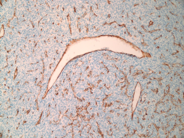
Immunohistochemistry staining for CD31 which highlights vessels demonstrates the characteristic central staghorn vessels seen in hemangiopericytomas.
Image Courtesy of Cheryl Spencer, M.A. and Ivana Delalle, MD, PhD Department of Pathology Boston University School of Medicine 98507/08/09 (S09-14668)
|
—
References
Chieci, MV et al, Intracranial hemangiopericytomas: MR and CT features. AJNR Am J Neuroradiol 17:1365?1371, August 1996
DOMElement Object
(
    [schemaTypeInfo] =>
    [tagName] => td
    [firstElementChild] => (object value omitted)
    [lastElementChild] => (object value omitted)
    [childElementCount] => 2
    [previousElementSibling] =>
    [nextElementSibling] =>
    [nodeName] => td
    [nodeValue] =>
Immunohistochemistry staining for CD31 which highlights vessels demonstrates the characteristic central staghorn vessels seen in hemangiopericytomas.
Image Courtesy of Cheryl Spencer, M.A. and Ivana Delalle, MD, PhD Department of Pathology Boston University School of Medicine 98507/08/09 (S09-14668)

    [nodeType] => 1
    [parentNode] => (object value omitted)
    [childNodes] => (object value omitted)
    [firstChild] => (object value omitted)
    [lastChild] => (object value omitted)
    [previousSibling] => (object value omitted)
    [nextSibling] => (object value omitted)
    [attributes] => (object value omitted)
    [ownerDocument] => (object value omitted)
    [namespaceURI] =>
    [prefix] =>
    [localName] => td
    [baseURI] =>
    [textContent] =>
Immunohistochemistry staining for CD31 which highlights vessels demonstrates the characteristic central staghorn vessels seen in hemangiopericytomas.
Image Courtesy of Cheryl Spencer, M.A. and Ivana Delalle, MD, PhD Department of Pathology Boston University School of Medicine 98507/08/09 (S09-14668)

)
DOMElement Object
(
    [schemaTypeInfo] =>
    [tagName] => table
    [firstElementChild] => (object value omitted)
    [lastElementChild] => (object value omitted)
    [childElementCount] => 1
    [previousElementSibling] => (object value omitted)
    [nextElementSibling] => (object value omitted)
    [nodeName] => table
    [nodeValue] =>


Extraaxial Mass – Eroding through the Skull


This 30 year old pregnant female presented with a constant headache and an MRI was obtained. Note that MR contrast was not given due to her pregnancy.
MRI: Because this mass is isointense and large, it can be difficult to distinguish if it arises from within the brain parenchyma (intra-axial), or if it is external to it (extra-axial). Note how the brain tissue is buckled around the mass sagittal T1 (a), and coronal T2 (b), and compressed by it, suggesting that it is extra-axial in location.
T1: (a) The mass is predominantly T1 isointense with several areas of high T1 signal which may be related to blood products. A large internal blood vessel is noted.
T2 coronal: (b) Notice the aggressive features of this mass which include erosion through the overlying skull and large internal network of blood vessels which are the linear low intensity structures. Increased T2 signal consistent with edema is seen in the underlying parenchyma. The mass itself is isointense to gray matter with central hypointense foci which are consistent with large blood vessels. T2 axial:(c) Mass effect, shift of the midline, internal network of vessels (linear low intensity structures), and T2 bright posterior peritumoral parenchymal edema.
FFE: (d) This gradient echo MRI sequence is beneficial for picking up substances which alter the local magnetic field, or paramagnetic substances. These substances cause ?blooming? which is a loss of signal, or hypointensity, within and just around them. Note the multiple areas of ?blooming? within the mass which can be caused by blood products or calcification.
Image Courtesy Elisa Flower MD and Asim Mian MD 97656c.8


    [nodeType] => 1
    [parentNode] => (object value omitted)
    [childNodes] => (object value omitted)
    [firstChild] => (object value omitted)
    [lastChild] => (object value omitted)
    [previousSibling] => (object value omitted)
    [nextSibling] => (object value omitted)
    [attributes] => (object value omitted)
    [ownerDocument] => (object value omitted)
    [namespaceURI] =>
    [prefix] =>
    [localName] => table
    [baseURI] =>
    [textContent] =>


Extraaxial Mass – Eroding through the Skull


This 30 year old pregnant female presented with a constant headache and an MRI was obtained. Note that MR contrast was not given due to her pregnancy.
MRI: Because this mass is isointense and large, it can be difficult to distinguish if it arises from within the brain parenchyma (intra-axial), or if it is external to it (extra-axial). Note how the brain tissue is buckled around the mass sagittal T1 (a), and coronal T2 (b), and compressed by it, suggesting that it is extra-axial in location.
T1: (a) The mass is predominantly T1 isointense with several areas of high T1 signal which may be related to blood products. A large internal blood vessel is noted.
T2 coronal: (b) Notice the aggressive features of this mass which include erosion through the overlying skull and large internal network of blood vessels which are the linear low intensity structures. Increased T2 signal consistent with edema is seen in the underlying parenchyma. The mass itself is isointense to gray matter with central hypointense foci which are consistent with large blood vessels. T2 axial:(c) Mass effect, shift of the midline, internal network of vessels (linear low intensity structures), and T2 bright posterior peritumoral parenchymal edema.
FFE: (d) This gradient echo MRI sequence is beneficial for picking up substances which alter the local magnetic field, or paramagnetic substances. These substances cause ?blooming? which is a loss of signal, or hypointensity, within and just around them. Note the multiple areas of ?blooming? within the mass which can be caused by blood products or calcification.
Image Courtesy Elisa Flower MD and Asim Mian MD 97656c.8


)
DOMElement Object
(
    [schemaTypeInfo] =>
    [tagName] => td
    [firstElementChild] => (object value omitted)
    [lastElementChild] => (object value omitted)
    [childElementCount] => 6
    [previousElementSibling] =>
    [nextElementSibling] =>
    [nodeName] => td
    [nodeValue] =>
This 30 year old pregnant female presented with a constant headache and an MRI was obtained. Note that MR contrast was not given due to her pregnancy.
MRI: Because this mass is isointense and large, it can be difficult to distinguish if it arises from within the brain parenchyma (intra-axial), or if it is external to it (extra-axial). Note how the brain tissue is buckled around the mass sagittal T1 (a), and coronal T2 (b), and compressed by it, suggesting that it is extra-axial in location.
T1: (a) The mass is predominantly T1 isointense with several areas of high T1 signal which may be related to blood products. A large internal blood vessel is noted.
T2 coronal: (b) Notice the aggressive features of this mass which include erosion through the overlying skull and large internal network of blood vessels which are the linear low intensity structures. Increased T2 signal consistent with edema is seen in the underlying parenchyma. The mass itself is isointense to gray matter with central hypointense foci which are consistent with large blood vessels. T2 axial:(c) Mass effect, shift of the midline, internal network of vessels (linear low intensity structures), and T2 bright posterior peritumoral parenchymal edema.
FFE: (d) This gradient echo MRI sequence is beneficial for picking up substances which alter the local magnetic field, or paramagnetic substances. These substances cause ?blooming? which is a loss of signal, or hypointensity, within and just around them. Note the multiple areas of ?blooming? within the mass which can be caused by blood products or calcification.
Image Courtesy Elisa Flower MD and Asim Mian MD 97656c.8

    [nodeType] => 1
    [parentNode] => (object value omitted)
    [childNodes] => (object value omitted)
    [firstChild] => (object value omitted)
    [lastChild] => (object value omitted)
    [previousSibling] => (object value omitted)
    [nextSibling] => (object value omitted)
    [attributes] => (object value omitted)
    [ownerDocument] => (object value omitted)
    [namespaceURI] =>
    [prefix] =>
    [localName] => td
    [baseURI] =>
    [textContent] =>
This 30 year old pregnant female presented with a constant headache and an MRI was obtained. Note that MR contrast was not given due to her pregnancy.
MRI: Because this mass is isointense and large, it can be difficult to distinguish if it arises from within the brain parenchyma (intra-axial), or if it is external to it (extra-axial). Note how the brain tissue is buckled around the mass sagittal T1 (a), and coronal T2 (b), and compressed by it, suggesting that it is extra-axial in location.
T1: (a) The mass is predominantly T1 isointense with several areas of high T1 signal which may be related to blood products. A large internal blood vessel is noted.
T2 coronal: (b) Notice the aggressive features of this mass which include erosion through the overlying skull and large internal network of blood vessels which are the linear low intensity structures. Increased T2 signal consistent with edema is seen in the underlying parenchyma. The mass itself is isointense to gray matter with central hypointense foci which are consistent with large blood vessels. T2 axial:(c) Mass effect, shift of the midline, internal network of vessels (linear low intensity structures), and T2 bright posterior peritumoral parenchymal edema.
FFE: (d) This gradient echo MRI sequence is beneficial for picking up substances which alter the local magnetic field, or paramagnetic substances. These substances cause ?blooming? which is a loss of signal, or hypointensity, within and just around them. Note the multiple areas of ?blooming? within the mass which can be caused by blood products or calcification.
Image Courtesy Elisa Flower MD and Asim Mian MD 97656c.8

)
DOMElement Object
(
    [schemaTypeInfo] =>
    [tagName] => td
    [firstElementChild] => (object value omitted)
    [lastElementChild] => (object value omitted)
    [childElementCount] => 2
    [previousElementSibling] =>
    [nextElementSibling] =>
    [nodeName] => td
    [nodeValue] =>
Extraaxial Mass – Eroding through the Skull
    [nodeType] => 1
    [parentNode] => (object value omitted)
    [childNodes] => (object value omitted)
    [firstChild] => (object value omitted)
    [lastChild] => (object value omitted)
    [previousSibling] => (object value omitted)
    [nextSibling] => (object value omitted)
    [attributes] => (object value omitted)
    [ownerDocument] => (object value omitted)
    [namespaceURI] =>
    [prefix] =>
    [localName] => td
    [baseURI] =>
    [textContent] =>
Extraaxial Mass – Eroding through the Skull
)


