Pulse Sequences
Sumit Kuria MD
The Common Vein Copyright 2010
Introduction
Spin-echo pulse sequences
Spin-echo pulse sequences yield T1 weighted images, T2 weighted images, and proton density weighted images. T1WIs provide incredible anatomic detail, in which fat, blood and proteinaceous fluids are identifiable. T2WIs are very sensitive to detect edema (water) and presence of potentially pathologic lesions. PDWIs, which convey information about the density of hydrogen in the tissue, are most useful in brain imaging. Spin-echo pulses are adjusted to yield these different images, by selecting the time between radiofrequency pulses (time of repetition), and time allowed for generation and detection of photons secondary to relaxation (time of echo). T1WI?s have both low TR and low TE, whereas T2WI?s have long TR and TE. A long TR with low TE yields PDWI?s.
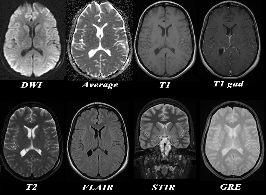
Basic Sequences Used in MRIof the Brain |
|
The MRI sequences used are displayed in the following chart at the level of the lateral ventricles. The CSF is bright on the average diffusion images, the T2 weighted image, the STIR and the GRE sequence. The best differentiation of gray white matter is on the STIR sequences, while the basal ganglia are best seen on the STIR and GRE sequences Acute infarcts are best seen on the DWI images, enhancing tumors on the T1 with gadolinium and iron deposition of subacute or chronic hemorrhage best characterised on the GRE sequences. FLAIR sequences are sensitive to water in lesions and best characterize MS plaques
Courtesy Ashley Davidoff MD Copyright 2010 95233c01.8
|
DOMElement Object
(
    [schemaTypeInfo] =>
    [tagName] => table
    [firstElementChild] => (object value omitted)
    [lastElementChild] => (object value omitted)
    [childElementCount] => 1
    [previousElementSibling] => (object value omitted)
    [nextElementSibling] =>
    [nodeName] => table
    [nodeValue] =>


Basic Sequences Used in MRIof the Brain


The MRI sequences used are displayed in the following chart at the level of the lateral ventricles. The CSF is bright on the average diffusion images, the T2 weighted image, the STIR and the GRE sequence. The best differentiation of gray white matter is on the STIR sequences, while the basal ganglia are best seen on the STIR and GRE sequences Acute infarcts are best seen on the DWI images, enhancing tumors on the T1 with gadolinium and iron deposition of subacute or chronic hemorrhage best characterised on the GRE sequences. FLAIR sequences are sensitive to water in lesions and best characterize MS plaques
Courtesy Ashley Davidoff MD Copyright 2010 95233c01.8


    [nodeType] => 1
    [parentNode] => (object value omitted)
    [childNodes] => (object value omitted)
    [firstChild] => (object value omitted)
    [lastChild] => (object value omitted)
    [previousSibling] => (object value omitted)
    [nextSibling] => (object value omitted)
    [attributes] => (object value omitted)
    [ownerDocument] => (object value omitted)
    [namespaceURI] =>
    [prefix] =>
    [localName] => table
    [baseURI] =>
    [textContent] =>


Basic Sequences Used in MRIof the Brain


The MRI sequences used are displayed in the following chart at the level of the lateral ventricles. The CSF is bright on the average diffusion images, the T2 weighted image, the STIR and the GRE sequence. The best differentiation of gray white matter is on the STIR sequences, while the basal ganglia are best seen on the STIR and GRE sequences Acute infarcts are best seen on the DWI images, enhancing tumors on the T1 with gadolinium and iron deposition of subacute or chronic hemorrhage best characterised on the GRE sequences. FLAIR sequences are sensitive to water in lesions and best characterize MS plaques
Courtesy Ashley Davidoff MD Copyright 2010 95233c01.8


)
DOMElement Object
(
    [schemaTypeInfo] =>
    [tagName] => td
    [firstElementChild] => (object value omitted)
    [lastElementChild] => (object value omitted)
    [childElementCount] => 2
    [previousElementSibling] =>
    [nextElementSibling] =>
    [nodeName] => td
    [nodeValue] =>
The MRI sequences used are displayed in the following chart at the level of the lateral ventricles. The CSF is bright on the average diffusion images, the T2 weighted image, the STIR and the GRE sequence. The best differentiation of gray white matter is on the STIR sequences, while the basal ganglia are best seen on the STIR and GRE sequences Acute infarcts are best seen on the DWI images, enhancing tumors on the T1 with gadolinium and iron deposition of subacute or chronic hemorrhage best characterised on the GRE sequences. FLAIR sequences are sensitive to water in lesions and best characterize MS plaques
Courtesy Ashley Davidoff MD Copyright 2010 95233c01.8

    [nodeType] => 1
    [parentNode] => (object value omitted)
    [childNodes] => (object value omitted)
    [firstChild] => (object value omitted)
    [lastChild] => (object value omitted)
    [previousSibling] => (object value omitted)
    [nextSibling] => (object value omitted)
    [attributes] => (object value omitted)
    [ownerDocument] => (object value omitted)
    [namespaceURI] =>
    [prefix] =>
    [localName] => td
    [baseURI] =>
    [textContent] =>
The MRI sequences used are displayed in the following chart at the level of the lateral ventricles. The CSF is bright on the average diffusion images, the T2 weighted image, the STIR and the GRE sequence. The best differentiation of gray white matter is on the STIR sequences, while the basal ganglia are best seen on the STIR and GRE sequences Acute infarcts are best seen on the DWI images, enhancing tumors on the T1 with gadolinium and iron deposition of subacute or chronic hemorrhage best characterised on the GRE sequences. FLAIR sequences are sensitive to water in lesions and best characterize MS plaques
Courtesy Ashley Davidoff MD Copyright 2010 95233c01.8

)
DOMElement Object
(
    [schemaTypeInfo] =>
    [tagName] => td
    [firstElementChild] => (object value omitted)
    [lastElementChild] => (object value omitted)
    [childElementCount] => 2
    [previousElementSibling] =>
    [nextElementSibling] =>
    [nodeName] => td
    [nodeValue] =>
Basic Sequences Used in MRIof the Brain
    [nodeType] => 1
    [parentNode] => (object value omitted)
    [childNodes] => (object value omitted)
    [firstChild] => (object value omitted)
    [lastChild] => (object value omitted)
    [previousSibling] => (object value omitted)
    [nextSibling] => (object value omitted)
    [attributes] => (object value omitted)
    [ownerDocument] => (object value omitted)
    [namespaceURI] =>
    [prefix] =>
    [localName] => td
    [baseURI] =>
    [textContent] =>
Basic Sequences Used in MRIof the Brain
)
