Insular Cortex
Sumit Karia MD
Copyright 2010
Definition
The insular cortex (aka insula) is one of the lobes of the brain, whose role is still unclear but is associated with limbic functions and emotions. It is also known as the insula of Reil, after the German physician Johann Christian Reil, gave its first anatomical description in 1796.
Structure
The insular cortex is a structure located deep in the brain’s lateral surface, within the Sylvian fissure, separating the temporal and inferior parietal cortices. The overlapping cortical regions – parts of the frontal, parietal and temporal lobes – form an operculum covering the insula, so that it is not visible on the outside surface of the brain.
Function
The insular cortex, especially its anterior portion, is related to the limbic system, having a role in subjective emotional experience, feelings of consciousness.
It is thought to process information received to produce an emotionally relevant context for sensory experience. More specifically, the anterior insula is more linked to emotions related to smell, taste, autonomic nervous system and limbic function, while the posterior insula is more related to somatic motor functions. In essence, it plays an important role in pain and the creation of a large number of basic emotions, such as anger, fear, disgust, happiness and sadness.

Normal Insula |
|
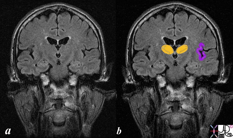
The Coronal T1 weighted image shows a normal brain reflecting the size shape and position of the normal insular cortex. (purple overlay). It is shown in association with the caudate nucleus (orange) with which it is functionally associated in the somatosensory pathway.
Courtesy Ashley Davidoff MD copyright 2008 38610c06b01b01.8s
|

The Insula (purple) in Coronal Section
|
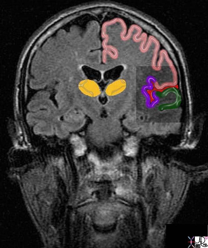
| The coronal T1 weighted image of the brain reveals the insular cortex and its neighbours. The lateral somatosensory cortex is centered around the Sylvian fissure (red) It incorporates the insula (purple) the upper lid of the operculum which is part of the parietal cortex (pink) as well as parts of the frontal cortex, and the lower lid of the operculum (green) which is part of the temporal lobe.
38610c06b07.8s Courtesy Ashley Davidoff MD copyright 2008
|
|

71060c07.8c01.8s
|
|
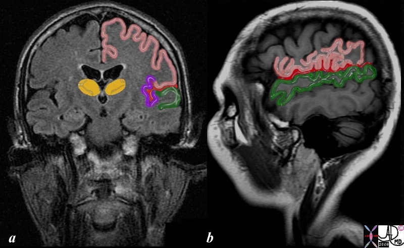
This T1 weighted image is utilized to demonstrate the operculum as seen froma coronal view in the first image (a) and from a sagittal view showing thge the outside in the second (b) Its cortical components consist of frontoparietal regions (pink) and temporal portions (green). The insula cortex is not visible from the outside (b) since it lies deep and medial (purple a). The Sylvian fissure is overlaid in red.
71060c07.8c01.8s MRI T1 weighted Courtesy Ashley Davidoff MD copyright 2008 |

Chronic Infarct Left Insula |
|
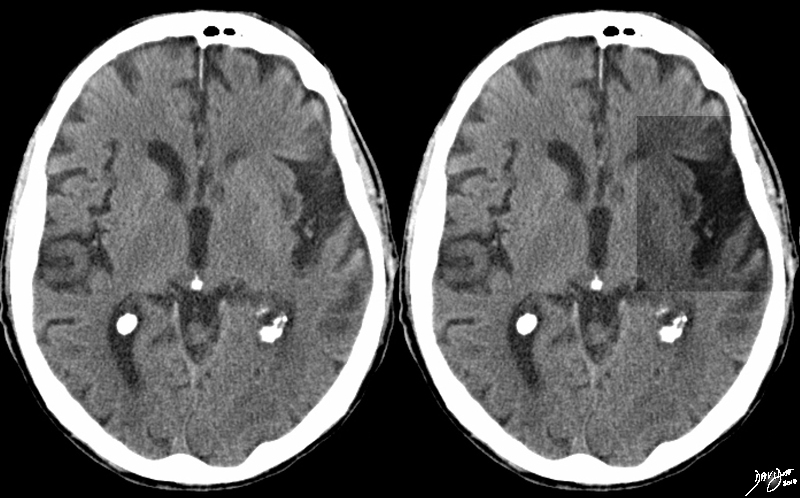
The non contrast CTscan is from elderly patient and reveals volume loss in the region of the insular cortex (insula) with secondary enlargement of the Sylvian fissure as a result of loss of tissue, caused by gliosis and encephalomalacia of the insular cortex. The process may involve the claustrum as well A low density lesion in the left caudate nucleus represents an old infarct. (lacune)
Courtesy Ashley Davidoff MD Copyright 2010 All rights reserved 89384.8s
|
DOMElement Object
(
    [schemaTypeInfo] =>
    [tagName] => table
    [firstElementChild] => (object value omitted)
    [lastElementChild] => (object value omitted)
    [childElementCount] => 1
    [previousElementSibling] => (object value omitted)
    [nextElementSibling] =>
    [nodeName] => table
    [nodeValue] =>


Chronic Infarct Left Insula


The non contrast CTscan is from elderly patient and reveals volume loss in the region of the insular cortex (insula) with secondary enlargement of the Sylvian fissure as a result of loss of tissue, caused by gliosis and encephalomalacia of the insular cortex. The process may involve the claustrum as well A low density lesion in the left caudate nucleus represents an old infarct. (lacune)
Courtesy Ashley Davidoff MD Copyright 2010 All rights reserved 89384.8s


    [nodeType] => 1
    [parentNode] => (object value omitted)
    [childNodes] => (object value omitted)
    [firstChild] => (object value omitted)
    [lastChild] => (object value omitted)
    [previousSibling] => (object value omitted)
    [nextSibling] => (object value omitted)
    [attributes] => (object value omitted)
    [ownerDocument] => (object value omitted)
    [namespaceURI] =>
    [prefix] =>
    [localName] => table
    [baseURI] =>
    [textContent] =>


Chronic Infarct Left Insula


The non contrast CTscan is from elderly patient and reveals volume loss in the region of the insular cortex (insula) with secondary enlargement of the Sylvian fissure as a result of loss of tissue, caused by gliosis and encephalomalacia of the insular cortex. The process may involve the claustrum as well A low density lesion in the left caudate nucleus represents an old infarct. (lacune)
Courtesy Ashley Davidoff MD Copyright 2010 All rights reserved 89384.8s


)
DOMElement Object
(
    [schemaTypeInfo] =>
    [tagName] => td
    [firstElementChild] => (object value omitted)
    [lastElementChild] => (object value omitted)
    [childElementCount] => 2
    [previousElementSibling] =>
    [nextElementSibling] =>
    [nodeName] => td
    [nodeValue] =>
The non contrast CTscan is from elderly patient and reveals volume loss in the region of the insular cortex (insula) with secondary enlargement of the Sylvian fissure as a result of loss of tissue, caused by gliosis and encephalomalacia of the insular cortex. The process may involve the claustrum as well A low density lesion in the left caudate nucleus represents an old infarct. (lacune)
Courtesy Ashley Davidoff MD Copyright 2010 All rights reserved 89384.8s

    [nodeType] => 1
    [parentNode] => (object value omitted)
    [childNodes] => (object value omitted)
    [firstChild] => (object value omitted)
    [lastChild] => (object value omitted)
    [previousSibling] => (object value omitted)
    [nextSibling] => (object value omitted)
    [attributes] => (object value omitted)
    [ownerDocument] => (object value omitted)
    [namespaceURI] =>
    [prefix] =>
    [localName] => td
    [baseURI] =>
    [textContent] =>
The non contrast CTscan is from elderly patient and reveals volume loss in the region of the insular cortex (insula) with secondary enlargement of the Sylvian fissure as a result of loss of tissue, caused by gliosis and encephalomalacia of the insular cortex. The process may involve the claustrum as well A low density lesion in the left caudate nucleus represents an old infarct. (lacune)
Courtesy Ashley Davidoff MD Copyright 2010 All rights reserved 89384.8s

)
DOMElement Object
(
    [schemaTypeInfo] =>
    [tagName] => td
    [firstElementChild] => (object value omitted)
    [lastElementChild] => (object value omitted)
    [childElementCount] => 2
    [previousElementSibling] =>
    [nextElementSibling] =>
    [nodeName] => td
    [nodeValue] =>
Chronic Infarct Left Insula
    [nodeType] => 1
    [parentNode] => (object value omitted)
    [childNodes] => (object value omitted)
    [firstChild] => (object value omitted)
    [lastChild] => (object value omitted)
    [previousSibling] => (object value omitted)
    [nextSibling] => (object value omitted)
    [attributes] => (object value omitted)
    [ownerDocument] => (object value omitted)
    [namespaceURI] =>
    [prefix] =>
    [localName] => td
    [baseURI] =>
    [textContent] =>
Chronic Infarct Left Insula
)
DOMElement Object
(
    [schemaTypeInfo] =>
    [tagName] => table
    [firstElementChild] => (object value omitted)
    [lastElementChild] => (object value omitted)
    [childElementCount] => 1
    [previousElementSibling] => (object value omitted)
    [nextElementSibling] => (object value omitted)
    [nodeName] => table
    [nodeValue] =>


71060c07.8c01.8s


This T1 weighted image is utilized to demonstrate the operculum as seen froma coronal view in the first image (a) and from a sagittal view showing thge the outside in the second (b)  Its cortical components consist of frontoparietal regions (pink) and temporal portions (green). The insula cortex is not visible from the outside (b) since it lies deep and medial (purple a).  The Sylvian fissure is overlaid in red.
71060c07.8c01.8s MRI T1 weighted Courtesy Ashley Davidoff MD copyright 2008


    [nodeType] => 1
    [parentNode] => (object value omitted)
    [childNodes] => (object value omitted)
    [firstChild] => (object value omitted)
    [lastChild] => (object value omitted)
    [previousSibling] => (object value omitted)
    [nextSibling] => (object value omitted)
    [attributes] => (object value omitted)
    [ownerDocument] => (object value omitted)
    [namespaceURI] =>
    [prefix] =>
    [localName] => table
    [baseURI] =>
    [textContent] =>


71060c07.8c01.8s


This T1 weighted image is utilized to demonstrate the operculum as seen froma coronal view in the first image (a) and from a sagittal view showing thge the outside in the second (b)  Its cortical components consist of frontoparietal regions (pink) and temporal portions (green). The insula cortex is not visible from the outside (b) since it lies deep and medial (purple a).  The Sylvian fissure is overlaid in red.
71060c07.8c01.8s MRI T1 weighted Courtesy Ashley Davidoff MD copyright 2008


)
DOMElement Object
(
    [schemaTypeInfo] =>
    [tagName] => td
    [firstElementChild] => (object value omitted)
    [lastElementChild] => (object value omitted)
    [childElementCount] => 2
    [previousElementSibling] =>
    [nextElementSibling] =>
    [nodeName] => td
    [nodeValue] =>
This T1 weighted image is utilized to demonstrate the operculum as seen froma coronal view in the first image (a) and from a sagittal view showing thge the outside in the second (b)  Its cortical components consist of frontoparietal regions (pink) and temporal portions (green). The insula cortex is not visible from the outside (b) since it lies deep and medial (purple a).  The Sylvian fissure is overlaid in red.
71060c07.8c01.8s MRI T1 weighted Courtesy Ashley Davidoff MD copyright 2008
    [nodeType] => 1
    [parentNode] => (object value omitted)
    [childNodes] => (object value omitted)
    [firstChild] => (object value omitted)
    [lastChild] => (object value omitted)
    [previousSibling] => (object value omitted)
    [nextSibling] => (object value omitted)
    [attributes] => (object value omitted)
    [ownerDocument] => (object value omitted)
    [namespaceURI] =>
    [prefix] =>
    [localName] => td
    [baseURI] =>
    [textContent] =>
This T1 weighted image is utilized to demonstrate the operculum as seen froma coronal view in the first image (a) and from a sagittal view showing thge the outside in the second (b)  Its cortical components consist of frontoparietal regions (pink) and temporal portions (green). The insula cortex is not visible from the outside (b) since it lies deep and medial (purple a).  The Sylvian fissure is overlaid in red.
71060c07.8c01.8s MRI T1 weighted Courtesy Ashley Davidoff MD copyright 2008
)
DOMElement Object
(
    [schemaTypeInfo] =>
    [tagName] => td
    [firstElementChild] => (object value omitted)
    [lastElementChild] => (object value omitted)
    [childElementCount] => 2
    [previousElementSibling] =>
    [nextElementSibling] =>
    [nodeName] => td
    [nodeValue] =>

71060c07.8c01.8s

    [nodeType] => 1
    [parentNode] => (object value omitted)
    [childNodes] => (object value omitted)
    [firstChild] => (object value omitted)
    [lastChild] => (object value omitted)
    [previousSibling] => (object value omitted)
    [nextSibling] => (object value omitted)
    [attributes] => (object value omitted)
    [ownerDocument] => (object value omitted)
    [namespaceURI] =>
    [prefix] =>
    [localName] => td
    [baseURI] =>
    [textContent] =>

71060c07.8c01.8s

)
DOMElement Object
(
    [schemaTypeInfo] =>
    [tagName] => table
    [firstElementChild] => (object value omitted)
    [lastElementChild] => (object value omitted)
    [childElementCount] => 1
    [previousElementSibling] => (object value omitted)
    [nextElementSibling] => (object value omitted)
    [nodeName] => table
    [nodeValue] =>


The Insula (purple) in Coronal Section


The coronal T1 weighted image of the brain reveals the insular cortex and its neighbours. The lateral somatosensory cortex is centered around the Sylvian fissure (red) It incorporates the insula (purple) the upper lid of the operculum which is part of the parietal cortex (pink)  as well as parts of the frontal cortex, and the lower lid of the operculum (green) which is part of the temporal lobe.
38610c06b07.8s Courtesy Ashley Davidoff MD copyright 2008


    [nodeType] => 1
    [parentNode] => (object value omitted)
    [childNodes] => (object value omitted)
    [firstChild] => (object value omitted)
    [lastChild] => (object value omitted)
    [previousSibling] => (object value omitted)
    [nextSibling] => (object value omitted)
    [attributes] => (object value omitted)
    [ownerDocument] => (object value omitted)
    [namespaceURI] =>
    [prefix] =>
    [localName] => table
    [baseURI] =>
    [textContent] =>


The Insula (purple) in Coronal Section


The coronal T1 weighted image of the brain reveals the insular cortex and its neighbours. The lateral somatosensory cortex is centered around the Sylvian fissure (red) It incorporates the insula (purple) the upper lid of the operculum which is part of the parietal cortex (pink)  as well as parts of the frontal cortex, and the lower lid of the operculum (green) which is part of the temporal lobe.
38610c06b07.8s Courtesy Ashley Davidoff MD copyright 2008


)
DOMElement Object
(
    [schemaTypeInfo] =>
    [tagName] => td
    [firstElementChild] => (object value omitted)
    [lastElementChild] => (object value omitted)
    [childElementCount] => 2
    [previousElementSibling] =>
    [nextElementSibling] =>
    [nodeName] => td
    [nodeValue] => The coronal T1 weighted image of the brain reveals the insular cortex and its neighbours. The lateral somatosensory cortex is centered around the Sylvian fissure (red) It incorporates the insula (purple) the upper lid of the operculum which is part of the parietal cortex (pink)  as well as parts of the frontal cortex, and the lower lid of the operculum (green) which is part of the temporal lobe.
38610c06b07.8s Courtesy Ashley Davidoff MD copyright 2008

    [nodeType] => 1
    [parentNode] => (object value omitted)
    [childNodes] => (object value omitted)
    [firstChild] => (object value omitted)
    [lastChild] => (object value omitted)
    [previousSibling] => (object value omitted)
    [nextSibling] => (object value omitted)
    [attributes] => (object value omitted)
    [ownerDocument] => (object value omitted)
    [namespaceURI] =>
    [prefix] =>
    [localName] => td
    [baseURI] =>
    [textContent] => The coronal T1 weighted image of the brain reveals the insular cortex and its neighbours. The lateral somatosensory cortex is centered around the Sylvian fissure (red) It incorporates the insula (purple) the upper lid of the operculum which is part of the parietal cortex (pink)  as well as parts of the frontal cortex, and the lower lid of the operculum (green) which is part of the temporal lobe.
38610c06b07.8s Courtesy Ashley Davidoff MD copyright 2008

)
DOMElement Object
(
    [schemaTypeInfo] =>
    [tagName] => td
    [firstElementChild] => (object value omitted)
    [lastElementChild] => (object value omitted)
    [childElementCount] => 2
    [previousElementSibling] =>
    [nextElementSibling] =>
    [nodeName] => td
    [nodeValue] =>
The Insula (purple) in Coronal Section

    [nodeType] => 1
    [parentNode] => (object value omitted)
    [childNodes] => (object value omitted)
    [firstChild] => (object value omitted)
    [lastChild] => (object value omitted)
    [previousSibling] => (object value omitted)
    [nextSibling] => (object value omitted)
    [attributes] => (object value omitted)
    [ownerDocument] => (object value omitted)
    [namespaceURI] =>
    [prefix] =>
    [localName] => td
    [baseURI] =>
    [textContent] =>
The Insula (purple) in Coronal Section

)
DOMElement Object
(
    [schemaTypeInfo] =>
    [tagName] => table
    [firstElementChild] => (object value omitted)
    [lastElementChild] => (object value omitted)
    [childElementCount] => 1
    [previousElementSibling] => (object value omitted)
    [nextElementSibling] => (object value omitted)
    [nodeName] => table
    [nodeValue] =>


Normal Insula


The Coronal T1 weighted image shows a normal brain reflecting the size shape and position of the normal insular cortex. (purple overlay).  It is shown in association with the caudate nucleus (orange) with which it is functionally associated in the somatosensory pathway.
 Courtesy Ashley Davidoff MD copyright 2008 38610c06b01b01.8s


    [nodeType] => 1
    [parentNode] => (object value omitted)
    [childNodes] => (object value omitted)
    [firstChild] => (object value omitted)
    [lastChild] => (object value omitted)
    [previousSibling] => (object value omitted)
    [nextSibling] => (object value omitted)
    [attributes] => (object value omitted)
    [ownerDocument] => (object value omitted)
    [namespaceURI] =>
    [prefix] =>
    [localName] => table
    [baseURI] =>
    [textContent] =>


Normal Insula


The Coronal T1 weighted image shows a normal brain reflecting the size shape and position of the normal insular cortex. (purple overlay).  It is shown in association with the caudate nucleus (orange) with which it is functionally associated in the somatosensory pathway.
 Courtesy Ashley Davidoff MD copyright 2008 38610c06b01b01.8s


)
DOMElement Object
(
    [schemaTypeInfo] =>
    [tagName] => td
    [firstElementChild] => (object value omitted)
    [lastElementChild] => (object value omitted)
    [childElementCount] => 2
    [previousElementSibling] =>
    [nextElementSibling] =>
    [nodeName] => td
    [nodeValue] =>
The Coronal T1 weighted image shows a normal brain reflecting the size shape and position of the normal insular cortex. (purple overlay).  It is shown in association with the caudate nucleus (orange) with which it is functionally associated in the somatosensory pathway.
 Courtesy Ashley Davidoff MD copyright 2008 38610c06b01b01.8s

    [nodeType] => 1
    [parentNode] => (object value omitted)
    [childNodes] => (object value omitted)
    [firstChild] => (object value omitted)
    [lastChild] => (object value omitted)
    [previousSibling] => (object value omitted)
    [nextSibling] => (object value omitted)
    [attributes] => (object value omitted)
    [ownerDocument] => (object value omitted)
    [namespaceURI] =>
    [prefix] =>
    [localName] => td
    [baseURI] =>
    [textContent] =>
The Coronal T1 weighted image shows a normal brain reflecting the size shape and position of the normal insular cortex. (purple overlay).  It is shown in association with the caudate nucleus (orange) with which it is functionally associated in the somatosensory pathway.
 Courtesy Ashley Davidoff MD copyright 2008 38610c06b01b01.8s

)
DOMElement Object
(
    [schemaTypeInfo] =>
    [tagName] => td
    [firstElementChild] => (object value omitted)
    [lastElementChild] => (object value omitted)
    [childElementCount] => 2
    [previousElementSibling] =>
    [nextElementSibling] =>
    [nodeName] => td
    [nodeValue] =>
Normal Insula
    [nodeType] => 1
    [parentNode] => (object value omitted)
    [childNodes] => (object value omitted)
    [firstChild] => (object value omitted)
    [lastChild] => (object value omitted)
    [previousSibling] => (object value omitted)
    [nextSibling] => (object value omitted)
    [attributes] => (object value omitted)
    [ownerDocument] => (object value omitted)
    [namespaceURI] =>
    [prefix] =>
    [localName] => td
    [baseURI] =>
    [textContent] =>
Normal Insula
)


