Ashley Davidoff MD
The Common Vein Copyright 2010
Definition
The prefrontal cortex, is the part of the frontal lobe which lies in front of the motor area, and is closely linked to the limbic system. Besides apparently being involved in rationalization, thought, making plans, organization, and taking action, it also appears to be involved in the same dopamine pathways as the ventral tegmental area, and plays a part in the sensation of pleasure.
It integrates thoughts and actions, and differntiates between good and bad, and is responsible for planning. It controlas behavioural aspects of life including personality, directing appropriate social behavior, cognitive functions and expressive functions.
|

The Prefrontal Cortex |
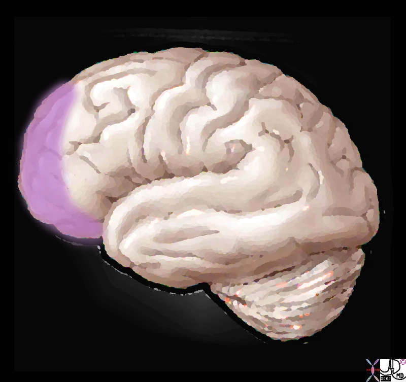
| In this artistic rendering of the brain, the prefrontal cortex is outlined in light purple. The prefrontal cortex of the brain is the anterior part of the frontal lobes and is positioned anterior to motor and premotor areas. It is divided into the lateral, orbitofrontal and medial prefrontal areas, Functionally it is said to have executive function in that it orchestrates thoughts and actions, discriminates between good and bad, positive and negative . It is responsible for planning, cognitive behaviors, personality expression and moderating correct social behavior. Courtesy Ashley Davidoff MD copyright 2008 copyright 2010 83029d.8s |
|

Prefrontal Cortex |
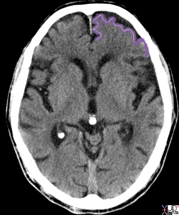
| The axial or transverse CTscan of the brain taken at the level of the third ventricle. The prefrontal cortex is outlined in light purple.The prefrontal cortex of the brain is the anterior part of the frontal lobes and is positioned anterior to motor and premotor areas. It is divided into the lateral, orbitofrontal and medial prefrontal areas.
Courtesy Ashley Davidoff MD copyright 2008 38568c02.8s |
|

The Prefrontal Cortex in Geographic Context |
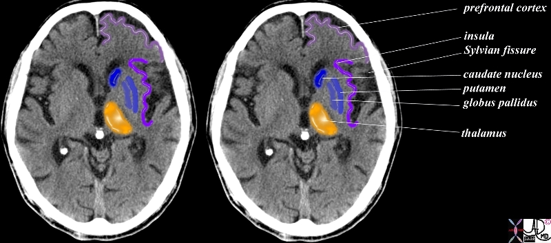
| This transverse image of the brain at the level of the third ventricle and thalamus, is presented to show some of the structures discussed above in context to the prefrontal cortex dark pink or light purple). In this image, the thalamus (orange), Sylvian fissure (black), insular cortex (dark purple) and components of the basal ganglia (blue) are shown.38568c12.8s Courtesy Ashley Davidoff MD copyright 2008 |
DOMElement Object
(
    [schemaTypeInfo] =>
    [tagName] => table
    [firstElementChild] => (object value omitted)
    [lastElementChild] => (object value omitted)
    [childElementCount] => 1
    [previousElementSibling] => (object value omitted)
    [nextElementSibling] =>
    [nodeName] => table
    [nodeValue] =>


The Prefrontal Cortex in Geographic Context


This transverse image of the brain at the level of the third ventricle and thalamus, is presented to show some of the structures discussed above in context to the prefrontal cortex dark pink or light purple).  In this image, the thalamus (orange), Sylvian fissure (black), insular cortex (dark purple) and components of the basal ganglia (blue) are shown.38568c12.8s Courtesy Ashley Davidoff MD copyright 2008


    [nodeType] => 1
    [parentNode] => (object value omitted)
    [childNodes] => (object value omitted)
    [firstChild] => (object value omitted)
    [lastChild] => (object value omitted)
    [previousSibling] => (object value omitted)
    [nextSibling] => (object value omitted)
    [attributes] => (object value omitted)
    [ownerDocument] => (object value omitted)
    [namespaceURI] =>
    [prefix] =>
    [localName] => table
    [baseURI] =>
    [textContent] =>


The Prefrontal Cortex in Geographic Context


This transverse image of the brain at the level of the third ventricle and thalamus, is presented to show some of the structures discussed above in context to the prefrontal cortex dark pink or light purple).  In this image, the thalamus (orange), Sylvian fissure (black), insular cortex (dark purple) and components of the basal ganglia (blue) are shown.38568c12.8s Courtesy Ashley Davidoff MD copyright 2008


)
DOMElement Object
(
    [schemaTypeInfo] =>
    [tagName] => td
    [firstElementChild] => (object value omitted)
    [lastElementChild] => (object value omitted)
    [childElementCount] => 2
    [previousElementSibling] =>
    [nextElementSibling] =>
    [nodeName] => td
    [nodeValue] => This transverse image of the brain at the level of the third ventricle and thalamus, is presented to show some of the structures discussed above in context to the prefrontal cortex dark pink or light purple).  In this image, the thalamus (orange), Sylvian fissure (black), insular cortex (dark purple) and components of the basal ganglia (blue) are shown.38568c12.8s Courtesy Ashley Davidoff MD copyright 2008
    [nodeType] => 1
    [parentNode] => (object value omitted)
    [childNodes] => (object value omitted)
    [firstChild] => (object value omitted)
    [lastChild] => (object value omitted)
    [previousSibling] => (object value omitted)
    [nextSibling] => (object value omitted)
    [attributes] => (object value omitted)
    [ownerDocument] => (object value omitted)
    [namespaceURI] =>
    [prefix] =>
    [localName] => td
    [baseURI] =>
    [textContent] => This transverse image of the brain at the level of the third ventricle and thalamus, is presented to show some of the structures discussed above in context to the prefrontal cortex dark pink or light purple).  In this image, the thalamus (orange), Sylvian fissure (black), insular cortex (dark purple) and components of the basal ganglia (blue) are shown.38568c12.8s Courtesy Ashley Davidoff MD copyright 2008
)
DOMElement Object
(
    [schemaTypeInfo] =>
    [tagName] => td
    [firstElementChild] => (object value omitted)
    [lastElementChild] => (object value omitted)
    [childElementCount] => 2
    [previousElementSibling] =>
    [nextElementSibling] =>
    [nodeName] => td
    [nodeValue] =>

The Prefrontal Cortex in Geographic Context
    [nodeType] => 1
    [parentNode] => (object value omitted)
    [childNodes] => (object value omitted)
    [firstChild] => (object value omitted)
    [lastChild] => (object value omitted)
    [previousSibling] => (object value omitted)
    [nextSibling] => (object value omitted)
    [attributes] => (object value omitted)
    [ownerDocument] => (object value omitted)
    [namespaceURI] =>
    [prefix] =>
    [localName] => td
    [baseURI] =>
    [textContent] =>

The Prefrontal Cortex in Geographic Context
)
DOMElement Object
(
    [schemaTypeInfo] =>
    [tagName] => table
    [firstElementChild] => (object value omitted)
    [lastElementChild] => (object value omitted)
    [childElementCount] => 1
    [previousElementSibling] => (object value omitted)
    [nextElementSibling] => (object value omitted)
    [nodeName] => table
    [nodeValue] =>


Prefrontal Cortex


The axial or transverse CTscan of the brain taken at the level of the third ventricle.  The prefrontal cortex is outlined in light purple.The prefrontal cortex of the brain is the anterior part of the frontal lobes and is positioned anterior to motor and premotor areas. It is divided into the lateral, orbitofrontal and medial prefrontal areas.
Courtesy Ashley Davidoff MD copyright 2008 38568c02.8s


    [nodeType] => 1
    [parentNode] => (object value omitted)
    [childNodes] => (object value omitted)
    [firstChild] => (object value omitted)
    [lastChild] => (object value omitted)
    [previousSibling] => (object value omitted)
    [nextSibling] => (object value omitted)
    [attributes] => (object value omitted)
    [ownerDocument] => (object value omitted)
    [namespaceURI] =>
    [prefix] =>
    [localName] => table
    [baseURI] =>
    [textContent] =>


Prefrontal Cortex


The axial or transverse CTscan of the brain taken at the level of the third ventricle.  The prefrontal cortex is outlined in light purple.The prefrontal cortex of the brain is the anterior part of the frontal lobes and is positioned anterior to motor and premotor areas. It is divided into the lateral, orbitofrontal and medial prefrontal areas.
Courtesy Ashley Davidoff MD copyright 2008 38568c02.8s


)
DOMElement Object
(
    [schemaTypeInfo] =>
    [tagName] => td
    [firstElementChild] => (object value omitted)
    [lastElementChild] => (object value omitted)
    [childElementCount] => 3
    [previousElementSibling] =>
    [nextElementSibling] =>
    [nodeName] => td
    [nodeValue] => The axial or transverse CTscan of the brain taken at the level of the third ventricle.  The prefrontal cortex is outlined in light purple.The prefrontal cortex of the brain is the anterior part of the frontal lobes and is positioned anterior to motor and premotor areas. It is divided into the lateral, orbitofrontal and medial prefrontal areas.
Courtesy Ashley Davidoff MD copyright 2008 38568c02.8s
    [nodeType] => 1
    [parentNode] => (object value omitted)
    [childNodes] => (object value omitted)
    [firstChild] => (object value omitted)
    [lastChild] => (object value omitted)
    [previousSibling] => (object value omitted)
    [nextSibling] => (object value omitted)
    [attributes] => (object value omitted)
    [ownerDocument] => (object value omitted)
    [namespaceURI] =>
    [prefix] =>
    [localName] => td
    [baseURI] =>
    [textContent] => The axial or transverse CTscan of the brain taken at the level of the third ventricle.  The prefrontal cortex is outlined in light purple.The prefrontal cortex of the brain is the anterior part of the frontal lobes and is positioned anterior to motor and premotor areas. It is divided into the lateral, orbitofrontal and medial prefrontal areas.
Courtesy Ashley Davidoff MD copyright 2008 38568c02.8s
)
DOMElement Object
(
    [schemaTypeInfo] =>
    [tagName] => td
    [firstElementChild] => (object value omitted)
    [lastElementChild] => (object value omitted)
    [childElementCount] => 2
    [previousElementSibling] =>
    [nextElementSibling] =>
    [nodeName] => td
    [nodeValue] =>

Prefrontal Cortex
    [nodeType] => 1
    [parentNode] => (object value omitted)
    [childNodes] => (object value omitted)
    [firstChild] => (object value omitted)
    [lastChild] => (object value omitted)
    [previousSibling] => (object value omitted)
    [nextSibling] => (object value omitted)
    [attributes] => (object value omitted)
    [ownerDocument] => (object value omitted)
    [namespaceURI] =>
    [prefix] =>
    [localName] => td
    [baseURI] =>
    [textContent] =>

Prefrontal Cortex
)
DOMElement Object
(
    [schemaTypeInfo] =>
    [tagName] => table
    [firstElementChild] => (object value omitted)
    [lastElementChild] => (object value omitted)
    [childElementCount] => 1
    [previousElementSibling] => (object value omitted)
    [nextElementSibling] => (object value omitted)
    [nodeName] => table
    [nodeValue] =>


The Prefrontal Cortex


In this artistic rendering of the brain, the prefrontal cortex is outlined in light purple.  The prefrontal cortex of the brain is the anterior part of the frontal lobes and is positioned anterior to motor and premotor areas. It is divided into the lateral, orbitofrontal and medial prefrontal areas, Functionally it is said to have executive function in that it orchestrates thoughts and actions, discriminates between good and bad, positive and negative . It is responsible for planning, cognitive behaviors, personality expression and moderating correct social behavior. Courtesy Ashley Davidoff MD copyright 2008 copyright 2010 83029d.8s


    [nodeType] => 1
    [parentNode] => (object value omitted)
    [childNodes] => (object value omitted)
    [firstChild] => (object value omitted)
    [lastChild] => (object value omitted)
    [previousSibling] => (object value omitted)
    [nextSibling] => (object value omitted)
    [attributes] => (object value omitted)
    [ownerDocument] => (object value omitted)
    [namespaceURI] =>
    [prefix] =>
    [localName] => table
    [baseURI] =>
    [textContent] =>


The Prefrontal Cortex


In this artistic rendering of the brain, the prefrontal cortex is outlined in light purple.  The prefrontal cortex of the brain is the anterior part of the frontal lobes and is positioned anterior to motor and premotor areas. It is divided into the lateral, orbitofrontal and medial prefrontal areas, Functionally it is said to have executive function in that it orchestrates thoughts and actions, discriminates between good and bad, positive and negative . It is responsible for planning, cognitive behaviors, personality expression and moderating correct social behavior. Courtesy Ashley Davidoff MD copyright 2008 copyright 2010 83029d.8s


)
DOMElement Object
(
    [schemaTypeInfo] =>
    [tagName] => td
    [firstElementChild] => (object value omitted)
    [lastElementChild] => (object value omitted)
    [childElementCount] => 2
    [previousElementSibling] =>
    [nextElementSibling] =>
    [nodeName] => td
    [nodeValue] => In this artistic rendering of the brain, the prefrontal cortex is outlined in light purple.  The prefrontal cortex of the brain is the anterior part of the frontal lobes and is positioned anterior to motor and premotor areas. It is divided into the lateral, orbitofrontal and medial prefrontal areas, Functionally it is said to have executive function in that it orchestrates thoughts and actions, discriminates between good and bad, positive and negative . It is responsible for planning, cognitive behaviors, personality expression and moderating correct social behavior. Courtesy Ashley Davidoff MD copyright 2008 copyright 2010 83029d.8s
    [nodeType] => 1
    [parentNode] => (object value omitted)
    [childNodes] => (object value omitted)
    [firstChild] => (object value omitted)
    [lastChild] => (object value omitted)
    [previousSibling] => (object value omitted)
    [nextSibling] => (object value omitted)
    [attributes] => (object value omitted)
    [ownerDocument] => (object value omitted)
    [namespaceURI] =>
    [prefix] =>
    [localName] => td
    [baseURI] =>
    [textContent] => In this artistic rendering of the brain, the prefrontal cortex is outlined in light purple.  The prefrontal cortex of the brain is the anterior part of the frontal lobes and is positioned anterior to motor and premotor areas. It is divided into the lateral, orbitofrontal and medial prefrontal areas, Functionally it is said to have executive function in that it orchestrates thoughts and actions, discriminates between good and bad, positive and negative . It is responsible for planning, cognitive behaviors, personality expression and moderating correct social behavior. Courtesy Ashley Davidoff MD copyright 2008 copyright 2010 83029d.8s
)
DOMElement Object
(
    [schemaTypeInfo] =>
    [tagName] => td
    [firstElementChild] => (object value omitted)
    [lastElementChild] => (object value omitted)
    [childElementCount] => 2
    [previousElementSibling] =>
    [nextElementSibling] =>
    [nodeName] => td
    [nodeValue] =>

The Prefrontal Cortex
    [nodeType] => 1
    [parentNode] => (object value omitted)
    [childNodes] => (object value omitted)
    [firstChild] => (object value omitted)
    [lastChild] => (object value omitted)
    [previousSibling] => (object value omitted)
    [nextSibling] => (object value omitted)
    [attributes] => (object value omitted)
    [ownerDocument] => (object value omitted)
    [namespaceURI] =>
    [prefix] =>
    [localName] => td
    [baseURI] =>
    [textContent] =>

The Prefrontal Cortex
)


