Ultrasound
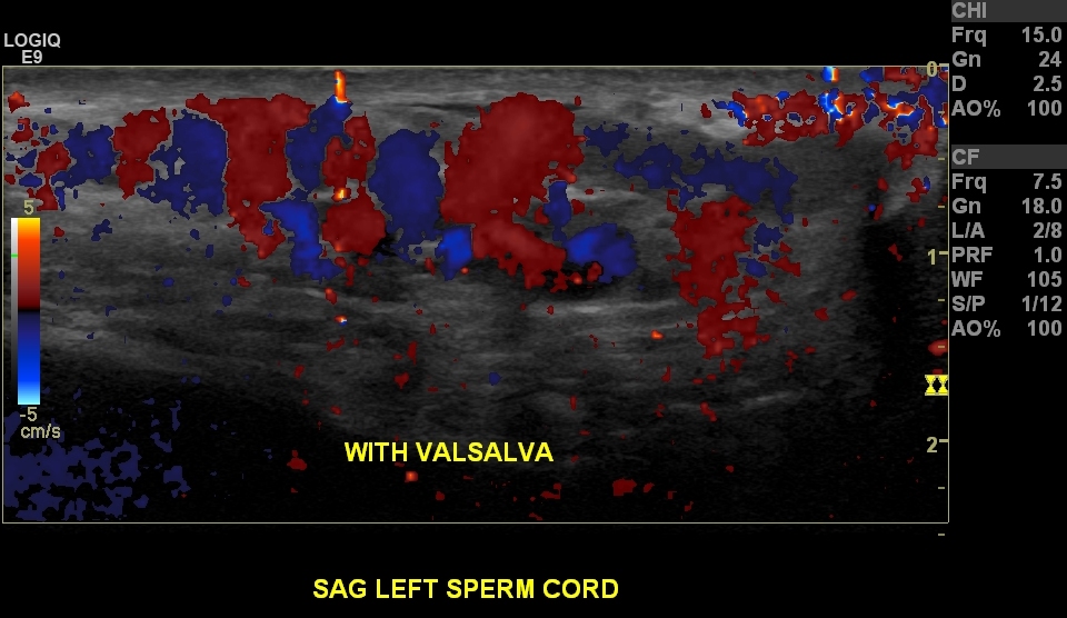
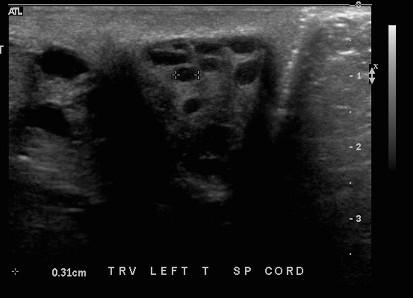
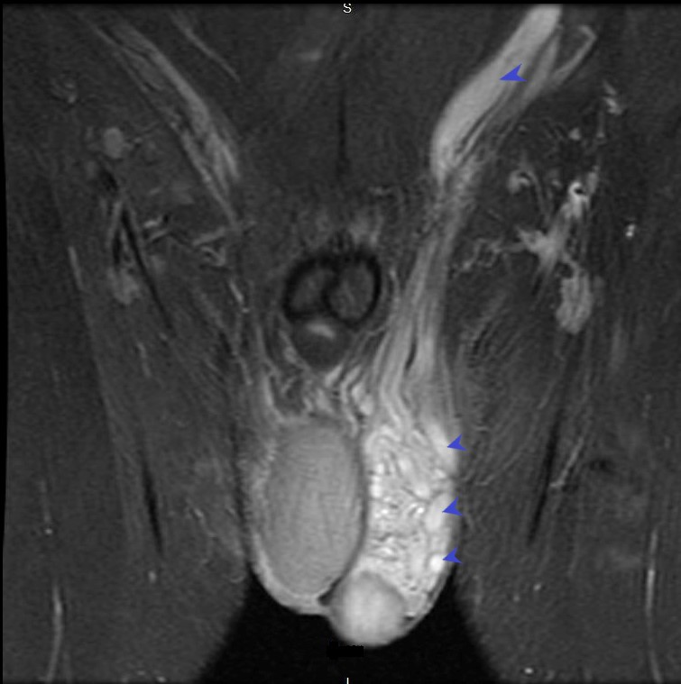
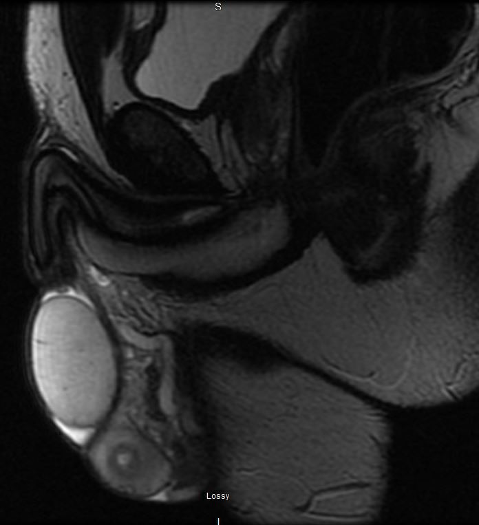
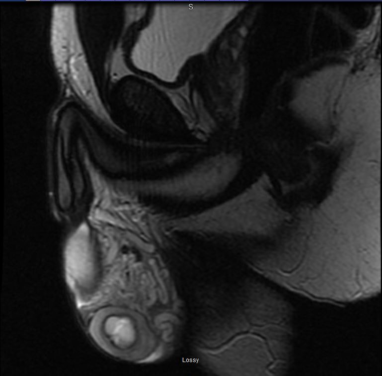
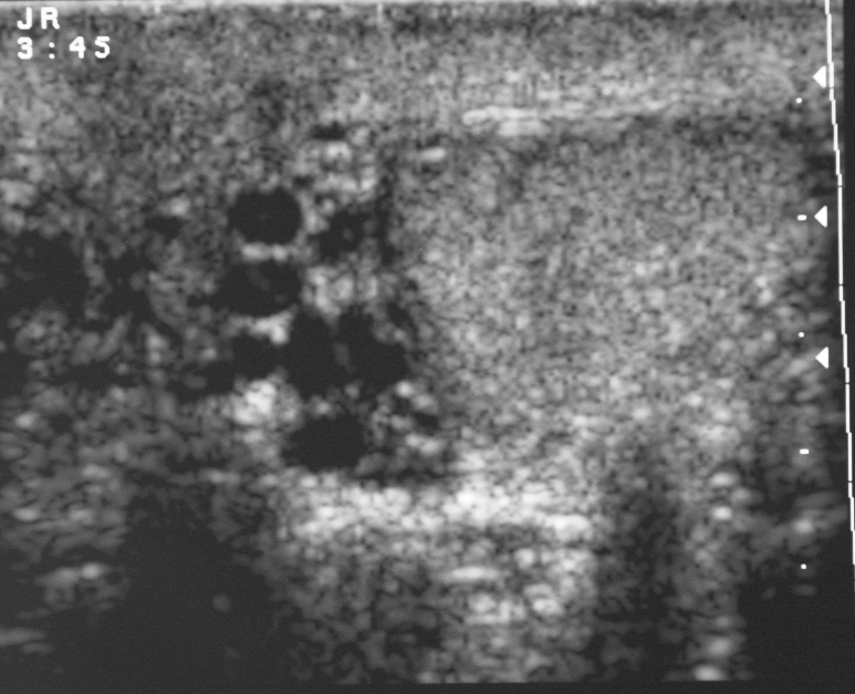
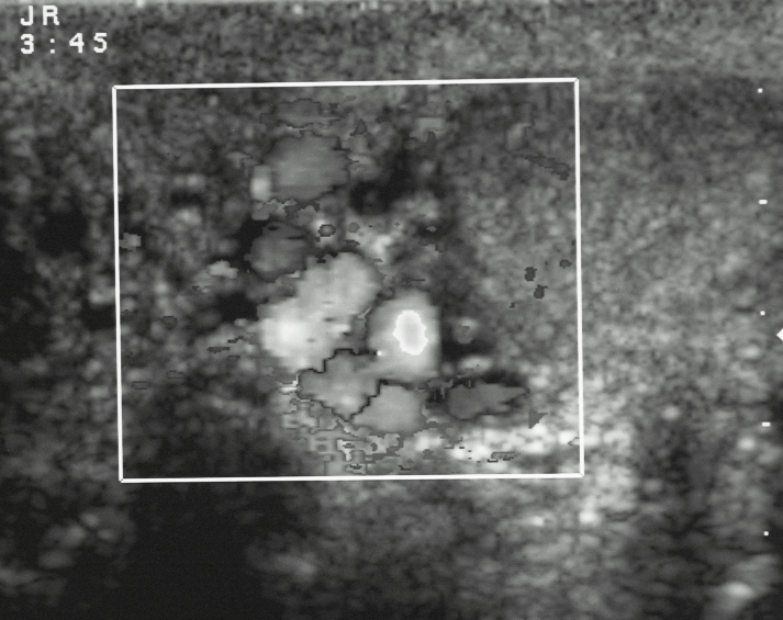
A varicocele, defined as a pathological enlargement of the pampiniform plexus with veins often running in a winding course,
The incidence of varicocele in males with primary (35%) and secondary (80%) infertility is significantly higher compared to the general male population (15%)
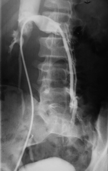
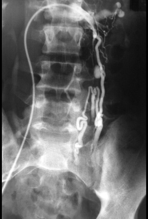
The etiology of varicocele is multifactorial. Differences in the course of testicular veins are considered to underlie the higher incidence of left-sided varicocele (>80% of cases). The left testicular vein runs vertically and enters the left renal vein at a right angle, which predisposes to turbulent blood flow and reverse pressure, while the right testicular vein opens directly into the inferior vena cava at a sharp angle(6, 7).
Compression of the left renal or testicular vein, either as a result of the so-called nutcracker mechanism or due to renal or retroperi-toneal tumors, is a relatively rare cause of varicocele(6, 11). A sudden onset of varicocele in a man over the age of 30 years requires the exclusion of renal tumors, particularly in elderly patients. In such cases it is necessary to extend diagnostic ultrasonography with abdominal examination.
- dilatation of pampiniform plexus veins >2-3 mm in diameter 3
- characteristically have a serpiginous appearance
- there can be flow reversal with the Valsalva maneuver 4
- Doppler ultrasound can be used to grade the degree of reflux
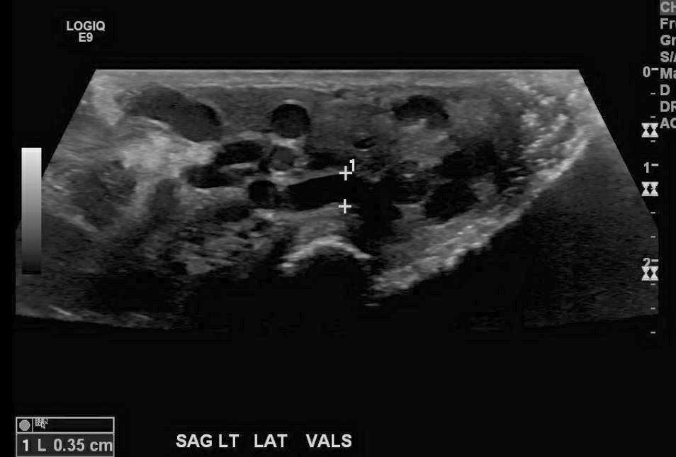
Ashley Davidoff MD 133546
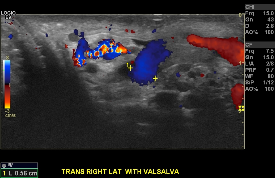
Ashley DAvidoff MD 133509
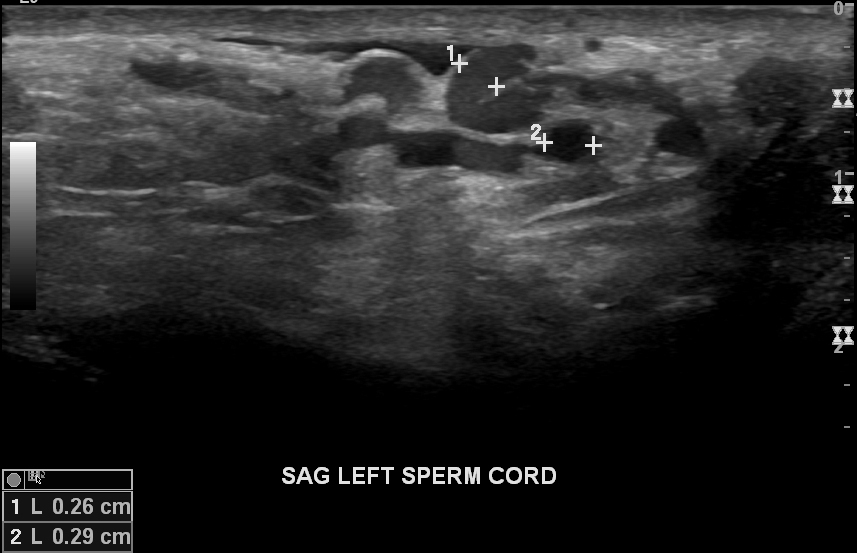
Ashley Davidoff MD 133354
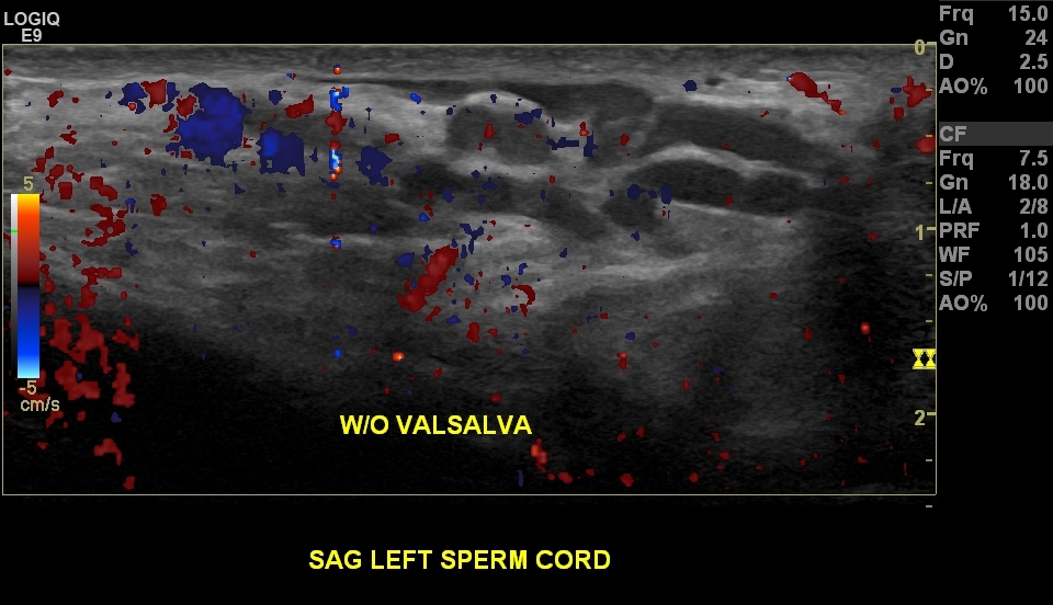
Without Valsalva
Ashley Davidoff MD 133356
Ashley Davidoff MD 133355
Ashley Davidoff MD 133640
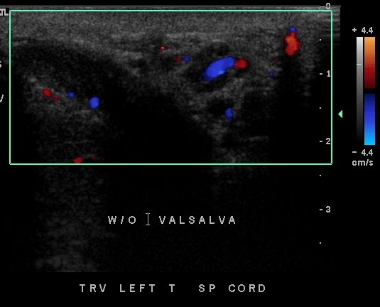
19 year old male with testicular discomfort. US in the supine position shows varicocele
Ashley Davidoff MD 133639
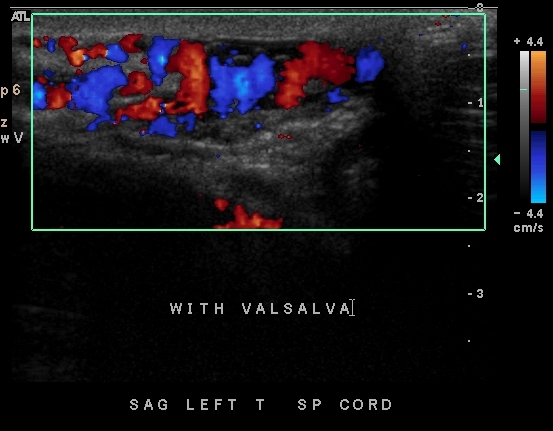
19 year old male with testicular discomfort. US in the supine position shows varicocele
Ashley Davidoff MD 133636
Ashley Davidoff MD 133580L
Ashley Davidoff MD 133630
Ashley Davidoff MD 133632
Ashley Davidoff MD
24905
Ashley Davidoff MD 24906
Ashley Davidoff MD 26573
Ashley Davidoff MD 26574
Links and References
Lorenc T et al Ultrasound and Varicocele
