Infarction Secondary to Infection
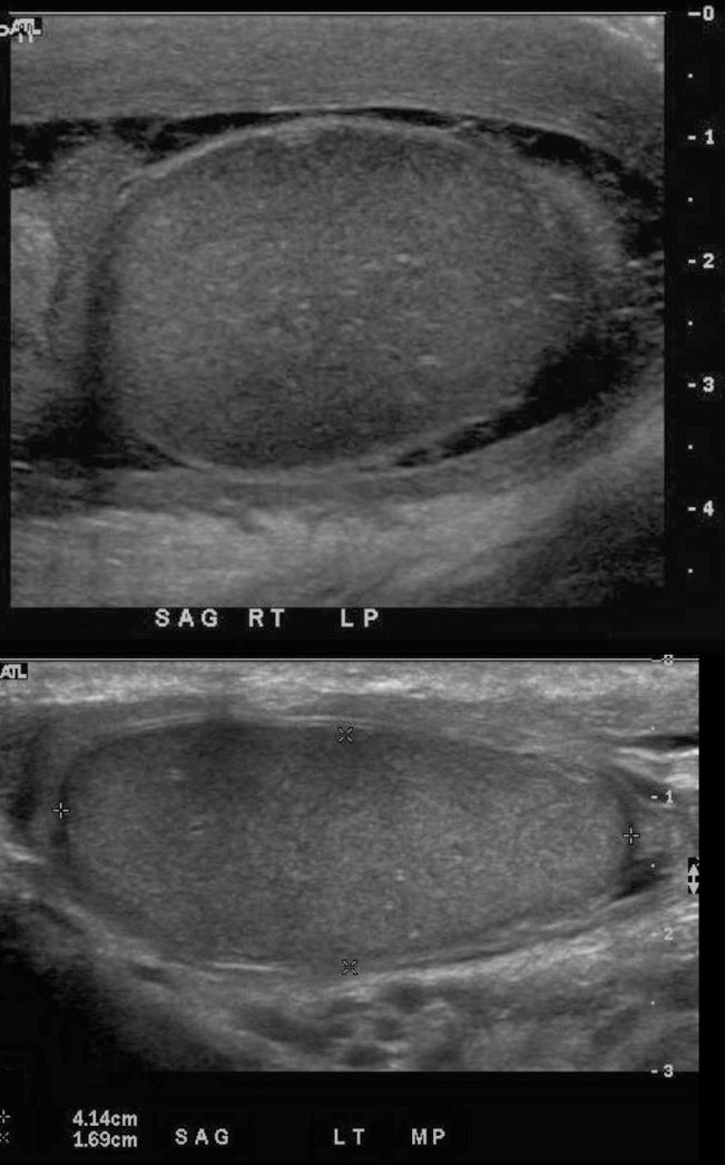
US in the sagittal plane of the right testis above and normal left testis below shows enlargement of the right epididymis, and testis, with thickening of the tunica albuginea, complex hydrocele with septations and thickening of the skin. The thickening of the tunica albuginea in this instance was a clue to the abnormality of the testis which was shown by Doppler to be infarcted
Ashley Davidoff MD 133227c
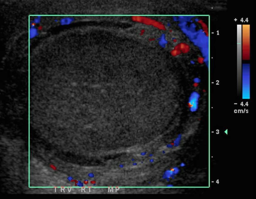
Doppler US in the transverse plane through the right testis shows absent vascularity in the testis with mild heterogeneity consistent with infarction of the right testis.
Ashley Davidoff MD 133221
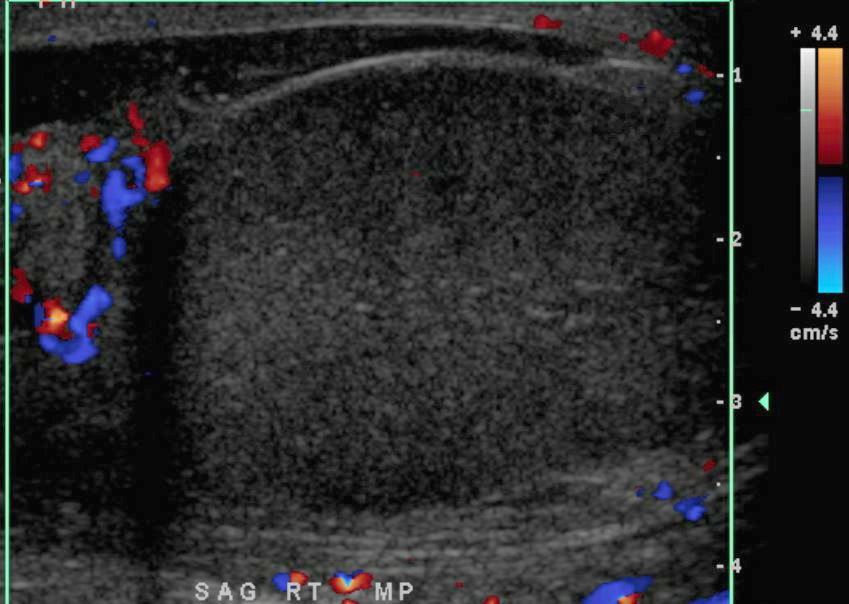
Doppler US in the transverse plane through the right testis shows absent vascularity in the testis with hypervascularity of the head of the epididymis consistent with acute epididymitis and infarction of the right testis.
Ashley Davidoff MD 133222

133237b01.jpg

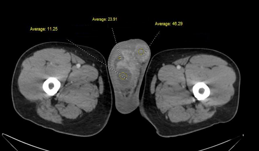
Axial CT scan through the scrotal sacs bilaterally show enlargement of the right epididymis (red arrow) and normal left epididymis (white arrow), infarcted right testis (black arrow) and normal left testis (yellow arrow).
Ashley Davidoff MD 133237b01
Coronal reconstruction of the CT scan of the pelvis shows hyperemic and enlarged spermatic cord, (orange arrow), enlargement of the right epididymis (red arrow) and normal left epididymis (white arrow)
Ashley Davidoff MD 133237b02
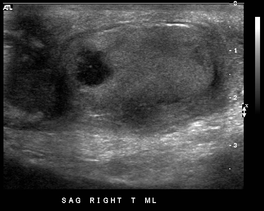
US performed 4 weeks later showed reduction in the size of the right testis and showing a 7mm focal area of necrosis in the parenchyma. Associated findings include persistent but improved enlargement of the epididymis and overlying skin thickening
Ashley Davidoff MD 133248