Seminoma
  • Testicular seminoma originates in the germinal epithelium ( epithelial layer of the seminiferous tubules of
  • 50% of germ cell tumors of the testicles are seminomas.

62-year-old male presents with a history of hypogonadism
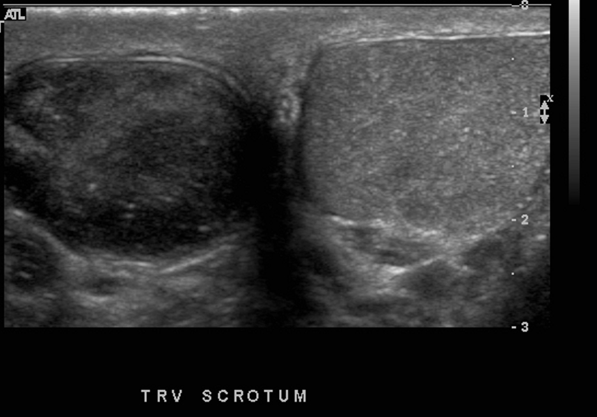
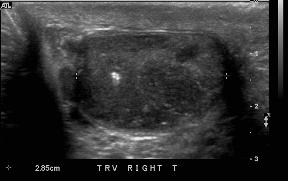
US in the transverse plane through both testes shows decrease echogenicity and diffuse heterogeneity of the smaller right testis, with foci of microlithiasis.
Ashley Davidoff MD 133711
US in the transverse plane through the right testis shows decrease echogenicity and diffuse heterogeneity of a small right testis, with foci of microlithiasis.
Ashley Davidoff MD 133714
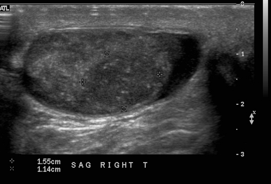
US in the sagittal plane through the right testis shows a dominant mass measuring 1.5 X 1.4 cms.
Ashley Davidoff MD 133729
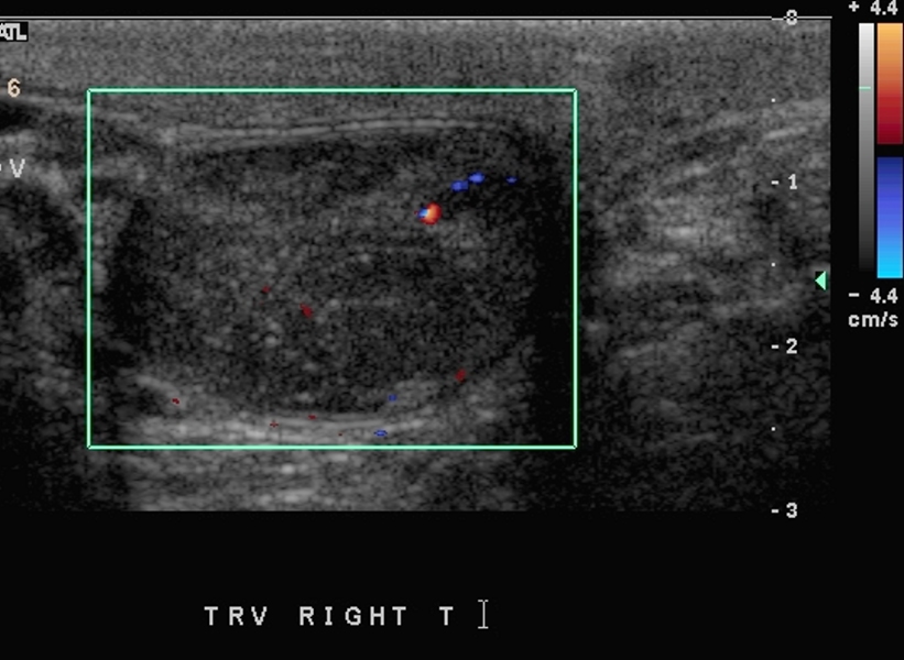
Doppler US in the sagittal plane through the right testis shows minimal vascularity in the dominant mass.
Ashley Davidoff MD 133727

Pathology revealed a stage 1 seminoma.

28-year-old male presents with a right testicular pain.

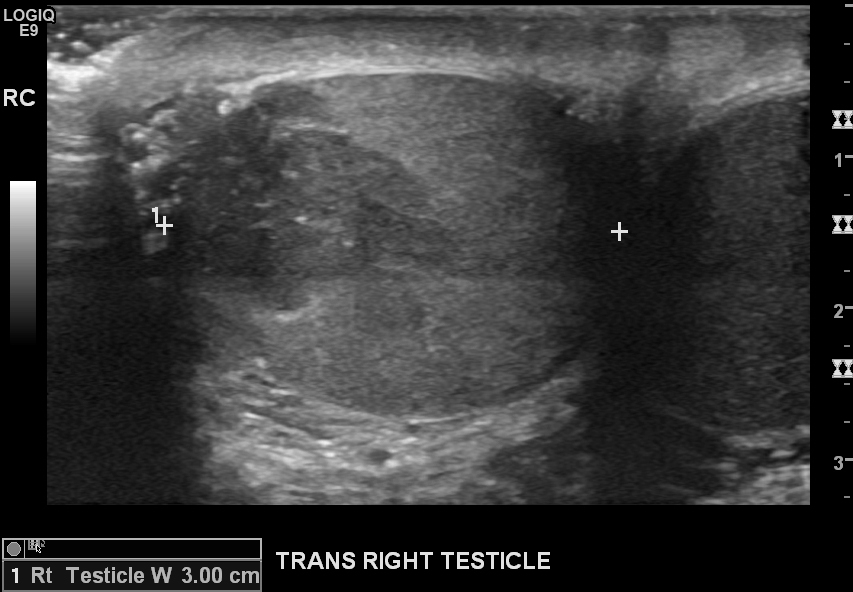
US in the transverse and sagittal planes through the right testicle shows multicentric hypoechoic nodules some of which are hyper vascular.  One of neoplastic nodules appears to extend into the region of the head of the epididymis and insertion of the cord.
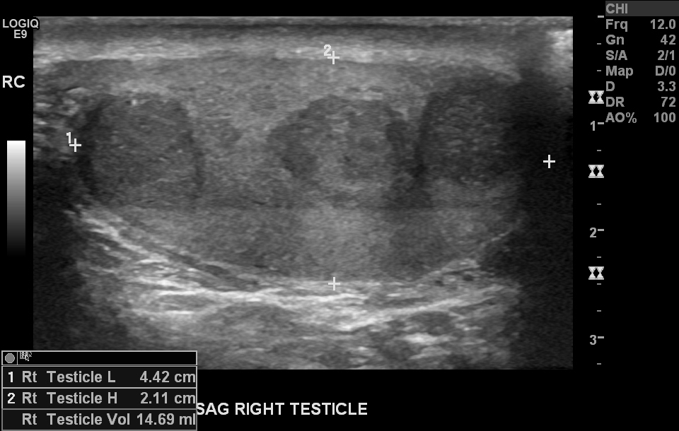
US in the sagittal plane through the normal sized right testicle shows multicentric hypoechoic nodules most likely reflecting malignant disease
Ashley Davidoff MD 133689
US in the transverse plane through the right testicle shows a hypoechoic nodule some of which appears to extend into the region of the head of the epididymis and insertion of the cord near the hilum.
Ashley Davidoff MD 133688
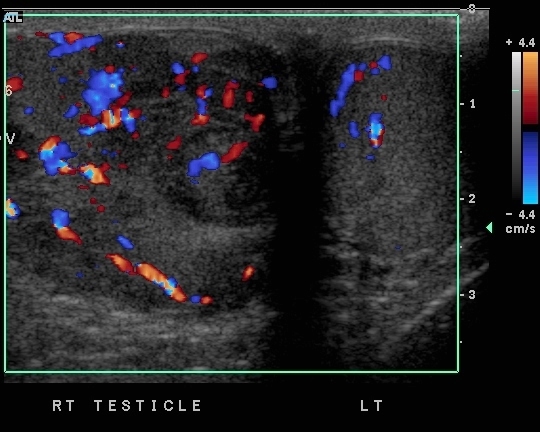
Doppler US in the transverse and sagittal planes through the right testicle shows multicentric hypoechoic nodules some of which are hyper vascular. One of neoplastic nodules appears to extend into the region of the head of the epididymis and insertion of the cord.
Ashley Davidoff MD 133699
Pathology:

Germ cell tumor with findings classical of seminoma.  There was involvement of the rete testis, and hilar stromal tissue.  The tumor penetrated the tunica albuginea. Multifocal angiolymphatic invasion TNM pT2 Nx Mx

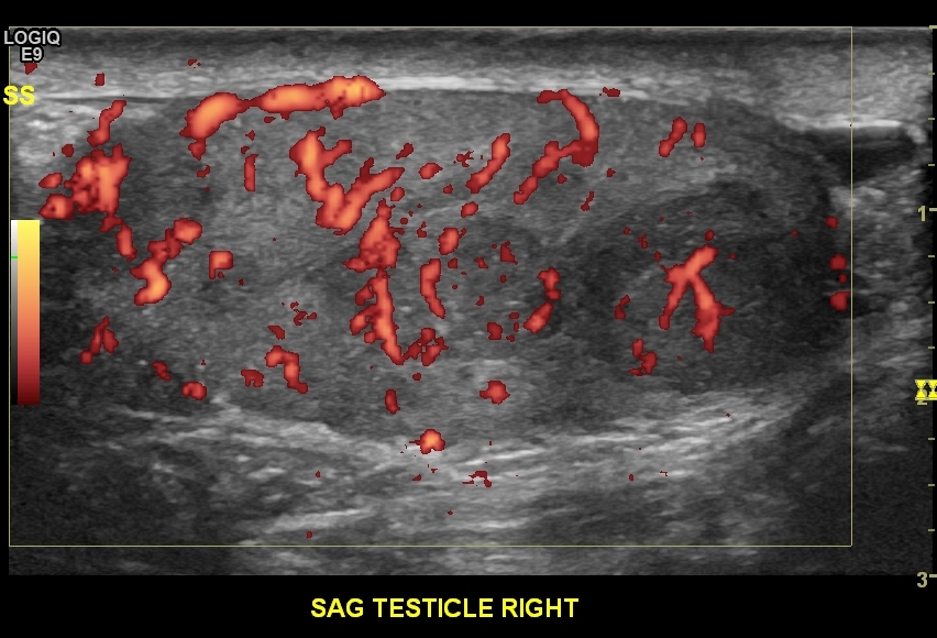
35-year-old male presents with a right testicular pain.
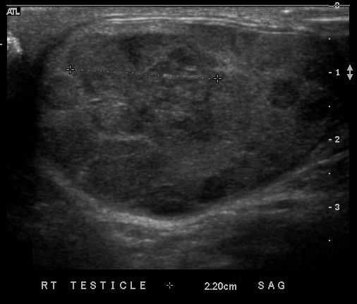
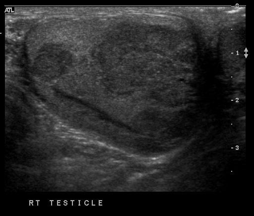
US in the sagittal plane through the right testicle shows multicentric hypoechoic nodules involving the entire testis. The neoplastic nodules appear to be confined to the testis
Ashley Davidoff MD
133703
US in the sagittal plane through the right testicle shows multicentric hypoechoic nodules involving the entire testis. The neoplastic nodules appear to be confined to the testis
Ashley Davidoff MD
133704
Doppler US through the right testicle shows nodular hypervascularity. The neoplastic nodules appear to be confined to the testis
Ashley Davidoff MD
133705
Pathology

Seminoma with syncitio-trophoblastic cells confined to the testis.  There was of the rete testis, and hilar stromal tissue.  The tumor penetrated the tunica albuginea. Multifocal angiolymphatic invasion TNM pT1 NX Mx

Case Studies

008Te Totally Infarcted Seminoma