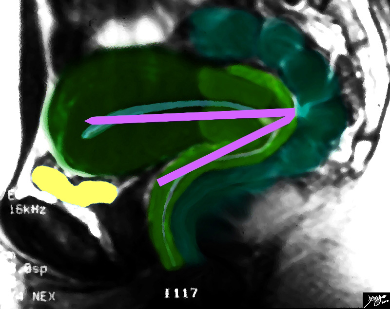
The normal position of uterus between the bladder and the rectum, and it projects superoanteriorly over the urinary bladder.
Normally and most commonly it is anteverted and anteflexed. The long axis of vagina forms a ninety degree angle with axis of cervix which is directed posteriorly and inferiorly, this is called as anteversion. The body of uterus is folded anteriorly making a 120 degree angle with cervix, this is called as anteflexion. The opposite position of anteversion is called a retroversion and cervix ix directed anteriorly and inferiorly; the opposite of anteflexion is retroflexion. This is sometimes the normal position of the uterus.
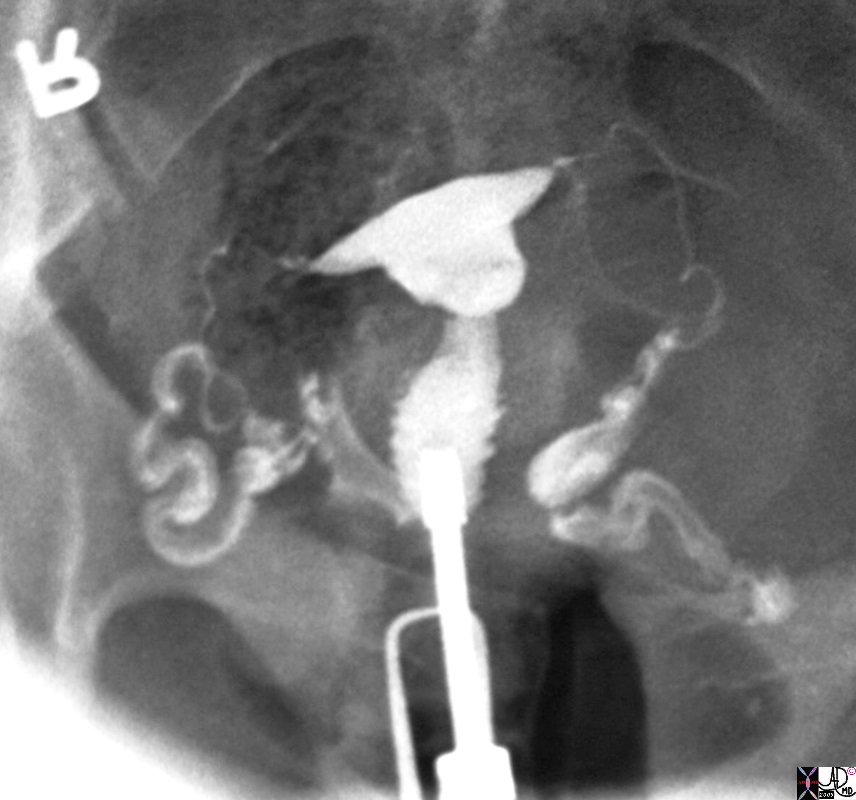
Normal HSG |
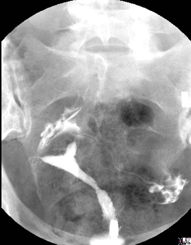
| 30594.8s Normal cervix. Note the serrated appearance of the cavity uterus cervix normal anatomy HSG hysterosalpingogram Courtes Ashley DAvidoff MD copyright 2009 all rights reserved |
|
Anteverted and “V Shaped Uterus with an Empty Bladder |
|
The diagram represents a sagittal view of the uterus reflecting an ?V? shaped structure of the uterus and vagina The uterus varies in position and in this case is anteverted, that converts the L shaped structure described to a “V” shaped structure Courtesy Ashley Davidoff MD copyright 2010 all rights reserved 14707.2kb04i01d03.81s |
|
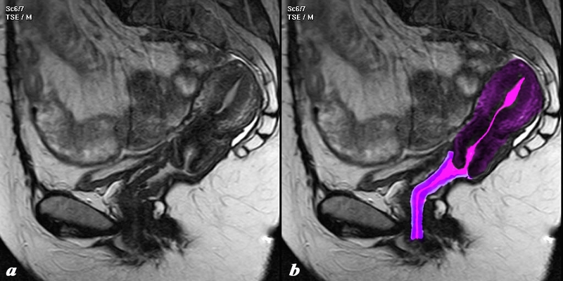
Retroverted Uterus |
|
The position and axis of the uterus is not always as outlined. The uterus is fairly mobile and in this sagittal MRI it is retrovertedand isnstead of being a “L” or a “V” configuration it is almost an “I” Courtesy Ashley Davidoff MD copyright 2010 93794c02.8s |
DOMElement Object
(
    [schemaTypeInfo] =>
    [tagName] => table
    [firstElementChild] => (object value omitted)
    [lastElementChild] => (object value omitted)
    [childElementCount] => 1
    [previousElementSibling] => (object value omitted)
    [nextElementSibling] => (object value omitted)
    [nodeName] => table
    [nodeValue] =>


Retroverted Uterus


The position and axis of the uterus is not always as outlined. The uterus is fairly mobile and in this sagittal MRI it is retrovertedand isnstead of being a “L” or a “V” configuration it is almost an “I”
Courtesy Ashley Davidoff MD copyright 2010 93794c02.8s


    [nodeType] => 1
    [parentNode] => (object value omitted)
    [childNodes] => (object value omitted)
    [firstChild] => (object value omitted)
    [lastChild] => (object value omitted)
    [previousSibling] => (object value omitted)
    [nextSibling] => (object value omitted)
    [attributes] => (object value omitted)
    [ownerDocument] => (object value omitted)
    [namespaceURI] =>
    [prefix] =>
    [localName] => table
    [baseURI] =>
    [textContent] =>


Retroverted Uterus


The position and axis of the uterus is not always as outlined. The uterus is fairly mobile and in this sagittal MRI it is retrovertedand isnstead of being a “L” or a “V” configuration it is almost an “I”
Courtesy Ashley Davidoff MD copyright 2010 93794c02.8s


)
DOMElement Object
(
    [schemaTypeInfo] =>
    [tagName] => td
    [firstElementChild] => (object value omitted)
    [lastElementChild] => (object value omitted)
    [childElementCount] => 2
    [previousElementSibling] =>
    [nextElementSibling] =>
    [nodeName] => td
    [nodeValue] =>
The position and axis of the uterus is not always as outlined. The uterus is fairly mobile and in this sagittal MRI it is retrovertedand isnstead of being a “L” or a “V” configuration it is almost an “I”
Courtesy Ashley Davidoff MD copyright 2010 93794c02.8s

    [nodeType] => 1
    [parentNode] => (object value omitted)
    [childNodes] => (object value omitted)
    [firstChild] => (object value omitted)
    [lastChild] => (object value omitted)
    [previousSibling] => (object value omitted)
    [nextSibling] => (object value omitted)
    [attributes] => (object value omitted)
    [ownerDocument] => (object value omitted)
    [namespaceURI] =>
    [prefix] =>
    [localName] => td
    [baseURI] =>
    [textContent] =>
The position and axis of the uterus is not always as outlined. The uterus is fairly mobile and in this sagittal MRI it is retrovertedand isnstead of being a “L” or a “V” configuration it is almost an “I”
Courtesy Ashley Davidoff MD copyright 2010 93794c02.8s

)
DOMElement Object
(
    [schemaTypeInfo] =>
    [tagName] => td
    [firstElementChild] => (object value omitted)
    [lastElementChild] => (object value omitted)
    [childElementCount] => 2
    [previousElementSibling] =>
    [nextElementSibling] =>
    [nodeName] => td
    [nodeValue] =>

Retroverted Uterus
    [nodeType] => 1
    [parentNode] => (object value omitted)
    [childNodes] => (object value omitted)
    [firstChild] => (object value omitted)
    [lastChild] => (object value omitted)
    [previousSibling] => (object value omitted)
    [nextSibling] => (object value omitted)
    [attributes] => (object value omitted)
    [ownerDocument] => (object value omitted)
    [namespaceURI] =>
    [prefix] =>
    [localName] => td
    [baseURI] =>
    [textContent] =>

Retroverted Uterus
)
DOMElement Object
(
    [schemaTypeInfo] =>
    [tagName] => table
    [firstElementChild] => (object value omitted)
    [lastElementChild] => (object value omitted)
    [childElementCount] => 1
    [previousElementSibling] => (object value omitted)
    [nextElementSibling] => (object value omitted)
    [nodeName] => table
    [nodeValue] =>


Anteverted and “V Shaped Uterus with an Empty Bladder


The diagram represents a sagittal view of the uterus reflecting an ?V? shaped structure of the uterus and vagina The uterus varies in position and in this case is anteverted, that converts the L shaped structure described to a “V” shaped structure
Courtesy Ashley Davidoff MD copyright 2010 all rights reserved 14707.2kb04i01d03.81s


    [nodeType] => 1
    [parentNode] => (object value omitted)
    [childNodes] => (object value omitted)
    [firstChild] => (object value omitted)
    [lastChild] => (object value omitted)
    [previousSibling] => (object value omitted)
    [nextSibling] => (object value omitted)
    [attributes] => (object value omitted)
    [ownerDocument] => (object value omitted)
    [namespaceURI] =>
    [prefix] =>
    [localName] => table
    [baseURI] =>
    [textContent] =>


Anteverted and “V Shaped Uterus with an Empty Bladder


The diagram represents a sagittal view of the uterus reflecting an ?V? shaped structure of the uterus and vagina The uterus varies in position and in this case is anteverted, that converts the L shaped structure described to a “V” shaped structure
Courtesy Ashley Davidoff MD copyright 2010 all rights reserved 14707.2kb04i01d03.81s


)
DOMElement Object
(
    [schemaTypeInfo] =>
    [tagName] => td
    [firstElementChild] => (object value omitted)
    [lastElementChild] => (object value omitted)
    [childElementCount] => 2
    [previousElementSibling] =>
    [nextElementSibling] =>
    [nodeName] => td
    [nodeValue] =>
The diagram represents a sagittal view of the uterus reflecting an ?V? shaped structure of the uterus and vagina The uterus varies in position and in this case is anteverted, that converts the L shaped structure described to a “V” shaped structure
Courtesy Ashley Davidoff MD copyright 2010 all rights reserved 14707.2kb04i01d03.81s

    [nodeType] => 1
    [parentNode] => (object value omitted)
    [childNodes] => (object value omitted)
    [firstChild] => (object value omitted)
    [lastChild] => (object value omitted)
    [previousSibling] => (object value omitted)
    [nextSibling] => (object value omitted)
    [attributes] => (object value omitted)
    [ownerDocument] => (object value omitted)
    [namespaceURI] =>
    [prefix] =>
    [localName] => td
    [baseURI] =>
    [textContent] =>
The diagram represents a sagittal view of the uterus reflecting an ?V? shaped structure of the uterus and vagina The uterus varies in position and in this case is anteverted, that converts the L shaped structure described to a “V” shaped structure
Courtesy Ashley Davidoff MD copyright 2010 all rights reserved 14707.2kb04i01d03.81s

)
DOMElement Object
(
    [schemaTypeInfo] =>
    [tagName] => td
    [firstElementChild] => (object value omitted)
    [lastElementChild] => (object value omitted)
    [childElementCount] => 2
    [previousElementSibling] =>
    [nextElementSibling] =>
    [nodeName] => td
    [nodeValue] =>

Anteverted and “V Shaped Uterus with an Empty Bladder
    [nodeType] => 1
    [parentNode] => (object value omitted)
    [childNodes] => (object value omitted)
    [firstChild] => (object value omitted)
    [lastChild] => (object value omitted)
    [previousSibling] => (object value omitted)
    [nextSibling] => (object value omitted)
    [attributes] => (object value omitted)
    [ownerDocument] => (object value omitted)
    [namespaceURI] =>
    [prefix] =>
    [localName] => td
    [baseURI] =>
    [textContent] =>

Anteverted and “V Shaped Uterus with an Empty Bladder
)
DOMElement Object
(
    [schemaTypeInfo] =>
    [tagName] => table
    [firstElementChild] => (object value omitted)
    [lastElementChild] => (object value omitted)
    [childElementCount] => 1
    [previousElementSibling] => (object value omitted)
    [nextElementSibling] => (object value omitted)
    [nodeName] => table
    [nodeValue] =>


The Normal Anteverted Uterus with A Filled Bladder


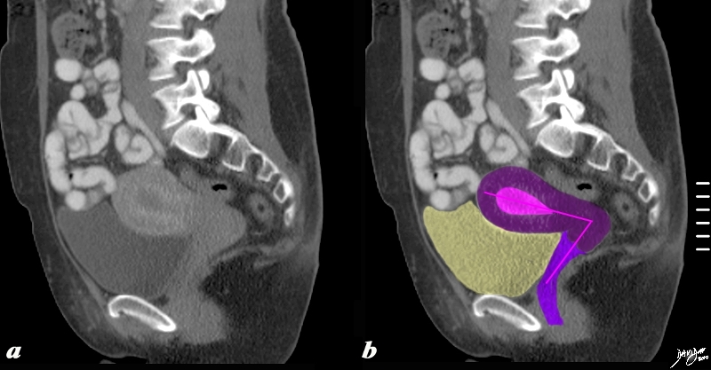
60385c04b.8s The sagittaly reconstructed CT shows an anteverted uterus buoyed and cushioned by partly filled bladder (yellow) In this sagittal view of the uterus an ?L? shaped structure of the uterus vagina, and the internal cavity is diagrammed in by the pink vectors of the tract. code uterus fundus body lower uterine segment cervix vagina anatomy structure normal tract concepts conceptual frameworks principles Davidoff art Courtesy Ashley Davidoff MD copyright 2010 all rights reserved


    [nodeType] => 1
    [parentNode] => (object value omitted)
    [childNodes] => (object value omitted)
    [firstChild] => (object value omitted)
    [lastChild] => (object value omitted)
    [previousSibling] => (object value omitted)
    [nextSibling] => (object value omitted)
    [attributes] => (object value omitted)
    [ownerDocument] => (object value omitted)
    [namespaceURI] =>
    [prefix] =>
    [localName] => table
    [baseURI] =>
    [textContent] =>


The Normal Anteverted Uterus with A Filled Bladder


60385c04b.8s The sagittaly reconstructed CT shows an anteverted uterus buoyed and cushioned by partly filled bladder (yellow) In this sagittal view of the uterus an ?L? shaped structure of the uterus vagina, and the internal cavity is diagrammed in by the pink vectors of the tract. code uterus fundus body lower uterine segment cervix vagina anatomy structure normal tract concepts conceptual frameworks principles Davidoff art Courtesy Ashley Davidoff MD copyright 2010 all rights reserved


)
DOMElement Object
(
    [schemaTypeInfo] =>
    [tagName] => td
    [firstElementChild] =>
    [lastElementChild] =>
    [childElementCount] => 0
    [previousElementSibling] =>
    [nextElementSibling] =>
    [nodeName] => td
    [nodeValue] => 60385c04b.8s The sagittaly reconstructed CT shows an anteverted uterus buoyed and cushioned by partly filled bladder (yellow) In this sagittal view of the uterus an ?L? shaped structure of the uterus vagina, and the internal cavity is diagrammed in by the pink vectors of the tract. code uterus fundus body lower uterine segment cervix vagina anatomy structure normal tract concepts conceptual frameworks principles Davidoff art Courtesy Ashley Davidoff MD copyright 2010 all rights reserved
    [nodeType] => 1
    [parentNode] => (object value omitted)
    [childNodes] => (object value omitted)
    [firstChild] => (object value omitted)
    [lastChild] => (object value omitted)
    [previousSibling] => (object value omitted)
    [nextSibling] => (object value omitted)
    [attributes] => (object value omitted)
    [ownerDocument] => (object value omitted)
    [namespaceURI] =>
    [prefix] =>
    [localName] => td
    [baseURI] =>
    [textContent] => 60385c04b.8s The sagittaly reconstructed CT shows an anteverted uterus buoyed and cushioned by partly filled bladder (yellow) In this sagittal view of the uterus an ?L? shaped structure of the uterus vagina, and the internal cavity is diagrammed in by the pink vectors of the tract. code uterus fundus body lower uterine segment cervix vagina anatomy structure normal tract concepts conceptual frameworks principles Davidoff art Courtesy Ashley Davidoff MD copyright 2010 all rights reserved
)
DOMElement Object
(
    [schemaTypeInfo] =>
    [tagName] => td
    [firstElementChild] => (object value omitted)
    [lastElementChild] => (object value omitted)
    [childElementCount] => 2
    [previousElementSibling] =>
    [nextElementSibling] =>
    [nodeName] => td
    [nodeValue] =>
The Normal Anteverted Uterus with A Filled Bladder
    [nodeType] => 1
    [parentNode] => (object value omitted)
    [childNodes] => (object value omitted)
    [firstChild] => (object value omitted)
    [lastChild] => (object value omitted)
    [previousSibling] => (object value omitted)
    [nextSibling] => (object value omitted)
    [attributes] => (object value omitted)
    [ownerDocument] => (object value omitted)
    [namespaceURI] =>
    [prefix] =>
    [localName] => td
    [baseURI] =>
    [textContent] =>
The Normal Anteverted Uterus with A Filled Bladder
)
DOMElement Object
(
    [schemaTypeInfo] =>
    [tagName] => table
    [firstElementChild] => (object value omitted)
    [lastElementChild] => (object value omitted)
    [childElementCount] => 1
    [previousElementSibling] => (object value omitted)
    [nextElementSibling] => (object value omitted)
    [nodeName] => table
    [nodeValue] =>


Position and Shape Relates to the Position of the Uterus


40 year old female with normal endometrial cavity and cervical canal and fallopian tubes normal anatomy cervix uterus HSG hysterosalpingogram
Courtesy Ashley Davidoff MD Copyright 2009 all rights reserved 83700.8s


    [nodeType] => 1
    [parentNode] => (object value omitted)
    [childNodes] => (object value omitted)
    [firstChild] => (object value omitted)
    [lastChild] => (object value omitted)
    [previousSibling] => (object value omitted)
    [nextSibling] => (object value omitted)
    [attributes] => (object value omitted)
    [ownerDocument] => (object value omitted)
    [namespaceURI] =>
    [prefix] =>
    [localName] => table
    [baseURI] =>
    [textContent] =>


Position and Shape Relates to the Position of the Uterus


40 year old female with normal endometrial cavity and cervical canal and fallopian tubes normal anatomy cervix uterus HSG hysterosalpingogram
Courtesy Ashley Davidoff MD Copyright 2009 all rights reserved 83700.8s


)
DOMElement Object
(
    [schemaTypeInfo] =>
    [tagName] => td
    [firstElementChild] => (object value omitted)
    [lastElementChild] => (object value omitted)
    [childElementCount] => 2
    [previousElementSibling] =>
    [nextElementSibling] =>
    [nodeName] => td
    [nodeValue] => 40 year old female with normal endometrial cavity and cervical canal and fallopian tubes normal anatomy cervix uterus HSG hysterosalpingogram
Courtesy Ashley Davidoff MD Copyright 2009 all rights reserved 83700.8s
    [nodeType] => 1
    [parentNode] => (object value omitted)
    [childNodes] => (object value omitted)
    [firstChild] => (object value omitted)
    [lastChild] => (object value omitted)
    [previousSibling] => (object value omitted)
    [nextSibling] => (object value omitted)
    [attributes] => (object value omitted)
    [ownerDocument] => (object value omitted)
    [namespaceURI] =>
    [prefix] =>
    [localName] => td
    [baseURI] =>
    [textContent] => 40 year old female with normal endometrial cavity and cervical canal and fallopian tubes normal anatomy cervix uterus HSG hysterosalpingogram
Courtesy Ashley Davidoff MD Copyright 2009 all rights reserved 83700.8s
)
DOMElement Object
(
    [schemaTypeInfo] =>
    [tagName] => td
    [firstElementChild] => (object value omitted)
    [lastElementChild] => (object value omitted)
    [childElementCount] => 2
    [previousElementSibling] =>
    [nextElementSibling] =>
    [nodeName] => td
    [nodeValue] =>
Position and Shape Relates to the Position of the Uterus

    [nodeType] => 1
    [parentNode] => (object value omitted)
    [childNodes] => (object value omitted)
    [firstChild] => (object value omitted)
    [lastChild] => (object value omitted)
    [previousSibling] => (object value omitted)
    [nextSibling] => (object value omitted)
    [attributes] => (object value omitted)
    [ownerDocument] => (object value omitted)
    [namespaceURI] =>
    [prefix] =>
    [localName] => td
    [baseURI] =>
    [textContent] =>
Position and Shape Relates to the Position of the Uterus

)
DOMElement Object
(
    [schemaTypeInfo] =>
    [tagName] => table
    [firstElementChild] => (object value omitted)
    [lastElementChild] => (object value omitted)
    [childElementCount] => 1
    [previousElementSibling] => (object value omitted)
    [nextElementSibling] => (object value omitted)
    [nodeName] => table
    [nodeValue] =>


Normal HSG


30594.8s Normal cervix. Note the serrated appearance of the cavity uterus cervix normal anatomy HSG hysterosalpingogram Courtes Ashley DAvidoff MD copyright 2009 all rights reserved


    [nodeType] => 1
    [parentNode] => (object value omitted)
    [childNodes] => (object value omitted)
    [firstChild] => (object value omitted)
    [lastChild] => (object value omitted)
    [previousSibling] => (object value omitted)
    [nextSibling] => (object value omitted)
    [attributes] => (object value omitted)
    [ownerDocument] => (object value omitted)
    [namespaceURI] =>
    [prefix] =>
    [localName] => table
    [baseURI] =>
    [textContent] =>


Normal HSG


30594.8s Normal cervix. Note the serrated appearance of the cavity uterus cervix normal anatomy HSG hysterosalpingogram Courtes Ashley DAvidoff MD copyright 2009 all rights reserved


)
DOMElement Object
(
    [schemaTypeInfo] =>
    [tagName] => td
    [firstElementChild] => (object value omitted)
    [lastElementChild] => (object value omitted)
    [childElementCount] => 1
    [previousElementSibling] =>
    [nextElementSibling] =>
    [nodeName] => td
    [nodeValue] => 30594.8s Normal cervix. Note the serrated appearance of the cavity uterus cervix normal anatomy HSG hysterosalpingogram Courtes Ashley DAvidoff MD copyright 2009 all rights reserved
    [nodeType] => 1
    [parentNode] => (object value omitted)
    [childNodes] => (object value omitted)
    [firstChild] => (object value omitted)
    [lastChild] => (object value omitted)
    [previousSibling] => (object value omitted)
    [nextSibling] => (object value omitted)
    [attributes] => (object value omitted)
    [ownerDocument] => (object value omitted)
    [namespaceURI] =>
    [prefix] =>
    [localName] => td
    [baseURI] =>
    [textContent] => 30594.8s Normal cervix. Note the serrated appearance of the cavity uterus cervix normal anatomy HSG hysterosalpingogram Courtes Ashley DAvidoff MD copyright 2009 all rights reserved
)
DOMElement Object
(
    [schemaTypeInfo] =>
    [tagName] => td
    [firstElementChild] => (object value omitted)
    [lastElementChild] => (object value omitted)
    [childElementCount] => 2
    [previousElementSibling] =>
    [nextElementSibling] =>
    [nodeName] => td
    [nodeValue] =>
Normal HSG

    [nodeType] => 1
    [parentNode] => (object value omitted)
    [childNodes] => (object value omitted)
    [firstChild] => (object value omitted)
    [lastChild] => (object value omitted)
    [previousSibling] => (object value omitted)
    [nextSibling] => (object value omitted)
    [attributes] => (object value omitted)
    [ownerDocument] => (object value omitted)
    [namespaceURI] =>
    [prefix] =>
    [localName] => td
    [baseURI] =>
    [textContent] =>
Normal HSG

)
DOMElement Object
(
    [schemaTypeInfo] =>
    [tagName] => table
    [firstElementChild] => (object value omitted)
    [lastElementChild] => (object value omitted)
    [childElementCount] => 1
    [previousElementSibling] => (object value omitted)
    [nextElementSibling] => (object value omitted)
    [nodeName] => table
    [nodeValue] =>


View from the Cul de Sac


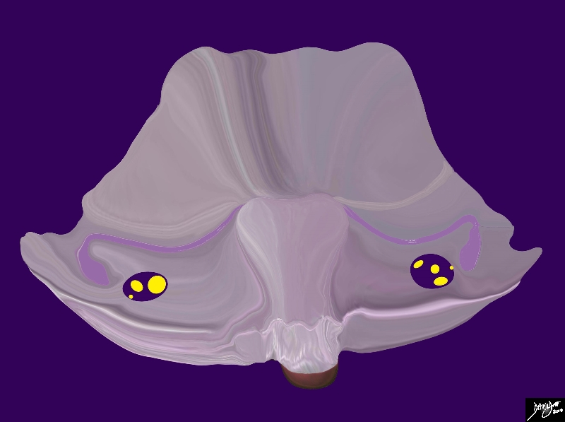
The diagram shows the peritoneal covering of the posterior aspect of the uterus and the intraperitoneal location of the ovaries The broad ligament contains the round ligament (superior band), the Fallopian tube, )mauve) and the ovarian ligament. Within the broad ligament the uterine/ovarian artery runs , uterine/ovarian vein lymphatics and nerves.
Courtesy Ashley Davidoff MD Copyright 2010 All rights reserved 04766b05b04.52kd01d15.8s


    [nodeType] => 1
    [parentNode] => (object value omitted)
    [childNodes] => (object value omitted)
    [firstChild] => (object value omitted)
    [lastChild] => (object value omitted)
    [previousSibling] => (object value omitted)
    [nextSibling] => (object value omitted)
    [attributes] => (object value omitted)
    [ownerDocument] => (object value omitted)
    [namespaceURI] =>
    [prefix] =>
    [localName] => table
    [baseURI] =>
    [textContent] =>


View from the Cul de Sac


The diagram shows the peritoneal covering of the posterior aspect of the uterus and the intraperitoneal location of the ovaries The broad ligament contains the round ligament (superior band), the Fallopian tube, )mauve) and the ovarian ligament. Within the broad ligament the uterine/ovarian artery runs , uterine/ovarian vein lymphatics and nerves.
Courtesy Ashley Davidoff MD Copyright 2010 All rights reserved 04766b05b04.52kd01d15.8s


)
DOMElement Object
(
    [schemaTypeInfo] =>
    [tagName] => td
    [firstElementChild] => (object value omitted)
    [lastElementChild] => (object value omitted)
    [childElementCount] => 2
    [previousElementSibling] =>
    [nextElementSibling] =>
    [nodeName] => td
    [nodeValue] =>
The diagram shows the peritoneal covering of the posterior aspect of the uterus and the intraperitoneal location of the ovaries The broad ligament contains the round ligament (superior band), the Fallopian tube, )mauve) and the ovarian ligament. Within the broad ligament the uterine/ovarian artery runs , uterine/ovarian vein lymphatics and nerves.
Courtesy Ashley Davidoff MD Copyright 2010 All rights reserved 04766b05b04.52kd01d15.8s

    [nodeType] => 1
    [parentNode] => (object value omitted)
    [childNodes] => (object value omitted)
    [firstChild] => (object value omitted)
    [lastChild] => (object value omitted)
    [previousSibling] => (object value omitted)
    [nextSibling] => (object value omitted)
    [attributes] => (object value omitted)
    [ownerDocument] => (object value omitted)
    [namespaceURI] =>
    [prefix] =>
    [localName] => td
    [baseURI] =>
    [textContent] =>
The diagram shows the peritoneal covering of the posterior aspect of the uterus and the intraperitoneal location of the ovaries The broad ligament contains the round ligament (superior band), the Fallopian tube, )mauve) and the ovarian ligament. Within the broad ligament the uterine/ovarian artery runs , uterine/ovarian vein lymphatics and nerves.
Courtesy Ashley Davidoff MD Copyright 2010 All rights reserved 04766b05b04.52kd01d15.8s

)
DOMElement Object
(
    [schemaTypeInfo] =>
    [tagName] => td
    [firstElementChild] => (object value omitted)
    [lastElementChild] => (object value omitted)
    [childElementCount] => 2
    [previousElementSibling] =>
    [nextElementSibling] =>
    [nodeName] => td
    [nodeValue] =>
View from the Cul de Sac
    [nodeType] => 1
    [parentNode] => (object value omitted)
    [childNodes] => (object value omitted)
    [firstChild] => (object value omitted)
    [lastChild] => (object value omitted)
    [previousSibling] => (object value omitted)
    [nextSibling] => (object value omitted)
    [attributes] => (object value omitted)
    [ownerDocument] => (object value omitted)
    [namespaceURI] =>
    [prefix] =>
    [localName] => td
    [baseURI] =>
    [textContent] =>
View from the Cul de Sac
)
DOMElement Object
(
    [schemaTypeInfo] =>
    [tagName] => table
    [firstElementChild] => (object value omitted)
    [lastElementChild] => (object value omitted)
    [childElementCount] => 1
    [previousElementSibling] => (object value omitted)
    [nextElementSibling] => (object value omitted)
    [nodeName] => table
    [nodeValue] =>


Peritoneal Relationships


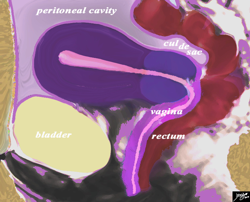
This diagram in the sagittal plane illustrates the peritoneal covering (light ourple lining) of the uterus reflected posteriorly off the rectum in the region of the fornix of the vagina creating a pouch called the recto-uterine pouch (aka cul de sac. pouch of Douglas recto-vaginal pouch Ehrhardt-Cole recess) This pouch is very important because it is the most posterior and inferior space of the peritoneal cavity and in the supine or upright position it will be the region where fluid will first accumulate. Anteriorly the peritoneal reflection is not as deep but a pouch is also formed called the uterovesical (aka vesico-uterine) pouch
Courtesy Ashley Davidoff MD Copyright 2010 All rights reserved 14707.2kb04i06.s.4ke06b02Lb.81s


    [nodeType] => 1
    [parentNode] => (object value omitted)
    [childNodes] => (object value omitted)
    [firstChild] => (object value omitted)
    [lastChild] => (object value omitted)
    [previousSibling] => (object value omitted)
    [nextSibling] => (object value omitted)
    [attributes] => (object value omitted)
    [ownerDocument] => (object value omitted)
    [namespaceURI] =>
    [prefix] =>
    [localName] => table
    [baseURI] =>
    [textContent] =>


Peritoneal Relationships


This diagram in the sagittal plane illustrates the peritoneal covering (light ourple lining) of the uterus reflected posteriorly off the rectum in the region of the fornix of the vagina creating a pouch called the recto-uterine pouch (aka cul de sac. pouch of Douglas recto-vaginal pouch Ehrhardt-Cole recess) This pouch is very important because it is the most posterior and inferior space of the peritoneal cavity and in the supine or upright position it will be the region where fluid will first accumulate. Anteriorly the peritoneal reflection is not as deep but a pouch is also formed called the uterovesical (aka vesico-uterine) pouch
Courtesy Ashley Davidoff MD Copyright 2010 All rights reserved 14707.2kb04i06.s.4ke06b02Lb.81s


)
DOMElement Object
(
    [schemaTypeInfo] =>
    [tagName] => td
    [firstElementChild] => (object value omitted)
    [lastElementChild] => (object value omitted)
    [childElementCount] => 2
    [previousElementSibling] =>
    [nextElementSibling] =>
    [nodeName] => td
    [nodeValue] =>
This diagram in the sagittal plane illustrates the peritoneal covering (light ourple lining) of the uterus reflected posteriorly off the rectum in the region of the fornix of the vagina creating a pouch called the recto-uterine pouch (aka cul de sac. pouch of Douglas recto-vaginal pouch Ehrhardt-Cole recess) This pouch is very important because it is the most posterior and inferior space of the peritoneal cavity and in the supine or upright position it will be the region where fluid will first accumulate. Anteriorly the peritoneal reflection is not as deep but a pouch is also formed called the uterovesical (aka vesico-uterine) pouch
Courtesy Ashley Davidoff MD Copyright 2010 All rights reserved 14707.2kb04i06.s.4ke06b02Lb.81s

    [nodeType] => 1
    [parentNode] => (object value omitted)
    [childNodes] => (object value omitted)
    [firstChild] => (object value omitted)
    [lastChild] => (object value omitted)
    [previousSibling] => (object value omitted)
    [nextSibling] => (object value omitted)
    [attributes] => (object value omitted)
    [ownerDocument] => (object value omitted)
    [namespaceURI] =>
    [prefix] =>
    [localName] => td
    [baseURI] =>
    [textContent] =>
This diagram in the sagittal plane illustrates the peritoneal covering (light ourple lining) of the uterus reflected posteriorly off the rectum in the region of the fornix of the vagina creating a pouch called the recto-uterine pouch (aka cul de sac. pouch of Douglas recto-vaginal pouch Ehrhardt-Cole recess) This pouch is very important because it is the most posterior and inferior space of the peritoneal cavity and in the supine or upright position it will be the region where fluid will first accumulate. Anteriorly the peritoneal reflection is not as deep but a pouch is also formed called the uterovesical (aka vesico-uterine) pouch
Courtesy Ashley Davidoff MD Copyright 2010 All rights reserved 14707.2kb04i06.s.4ke06b02Lb.81s

)
DOMElement Object
(
    [schemaTypeInfo] =>
    [tagName] => td
    [firstElementChild] => (object value omitted)
    [lastElementChild] => (object value omitted)
    [childElementCount] => 2
    [previousElementSibling] =>
    [nextElementSibling] =>
    [nodeName] => td
    [nodeValue] =>
Peritoneal Relationships
    [nodeType] => 1
    [parentNode] => (object value omitted)
    [childNodes] => (object value omitted)
    [firstChild] => (object value omitted)
    [lastChild] => (object value omitted)
    [previousSibling] => (object value omitted)
    [nextSibling] => (object value omitted)
    [attributes] => (object value omitted)
    [ownerDocument] => (object value omitted)
    [namespaceURI] =>
    [prefix] =>
    [localName] => td
    [baseURI] =>
    [textContent] =>
Peritoneal Relationships
)

