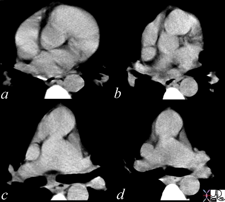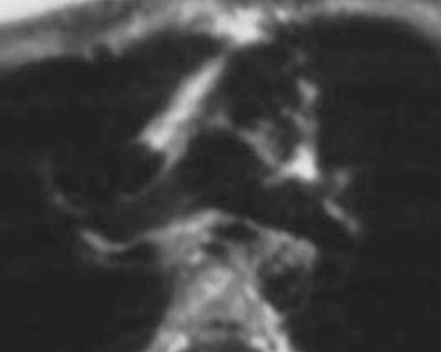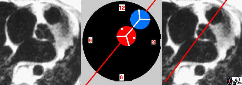
Position of the aorta relative to the subpulmonary conus |
| 07954eW.800 aorta aortic valve infundibulum RVOT right ventricular outflow tract position relation MRIscan Davidoff MD |
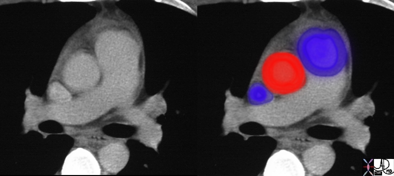
Aorta and Pulmonary Artery – Normal Size and Position |
| 27467c01 aorta pulmonary valve pulmonary artery SVC size position normal CTscan Davidoff MD |
|
Common Conotruncal Abnormalities |
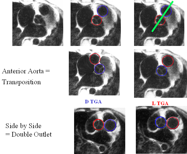
| 86778 01639 01639d02 01639d04 01639f03.jpg Anterior aorta = Transposition Copyright 2009 Courtesy Ashley Davidoff MD all rights reserved |
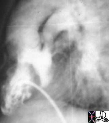
D Transposition of the Great Vessels
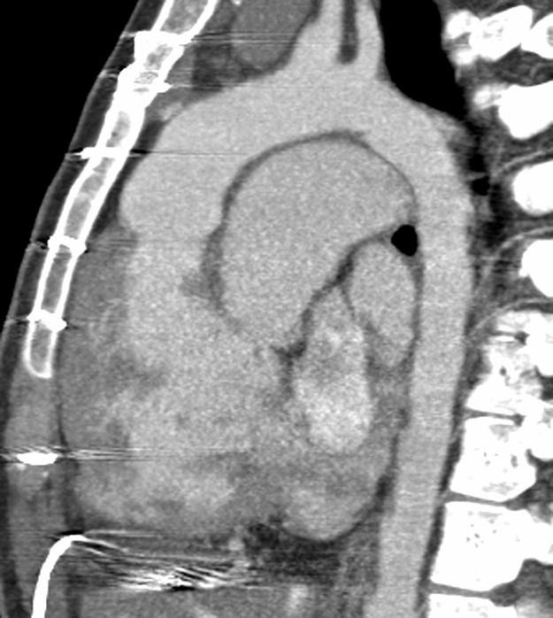
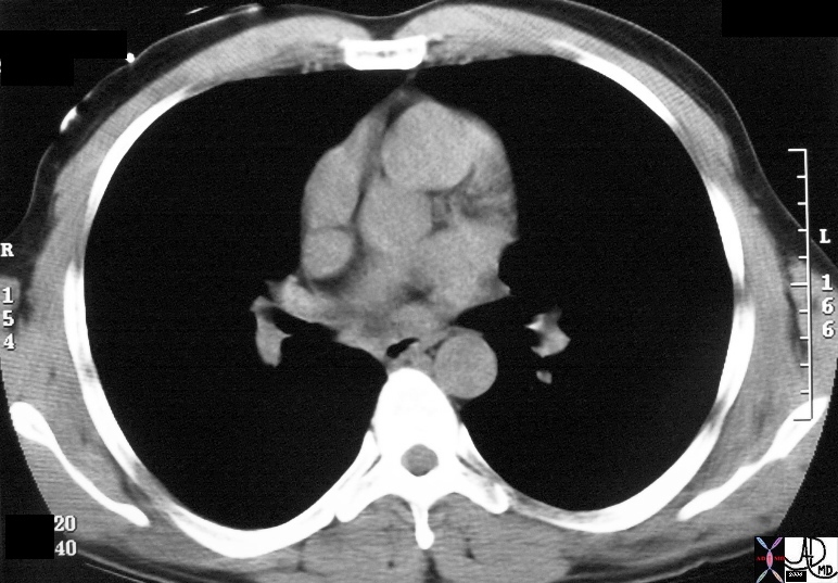
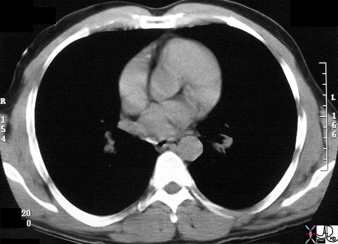
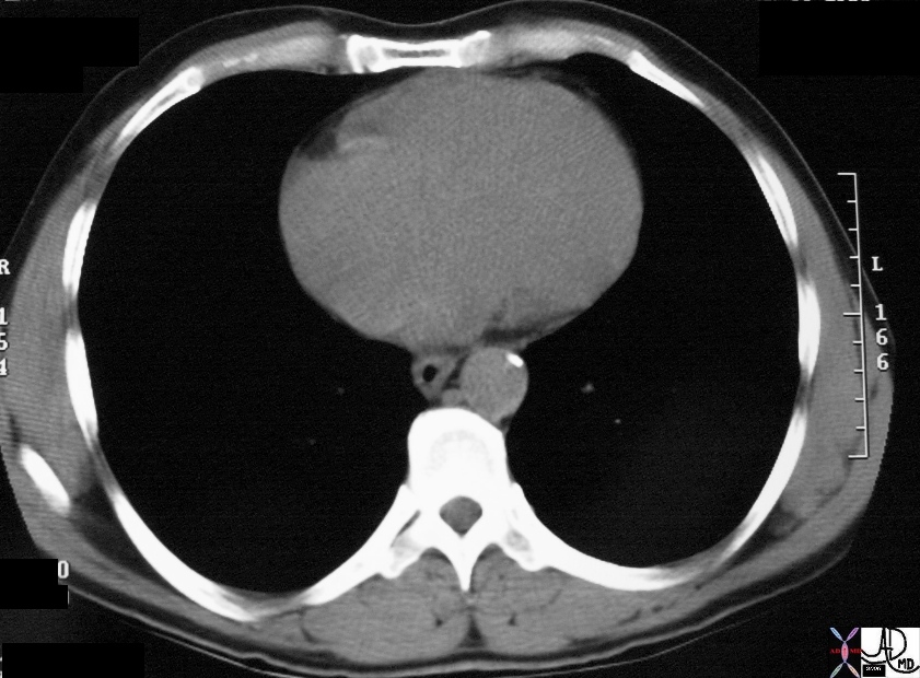
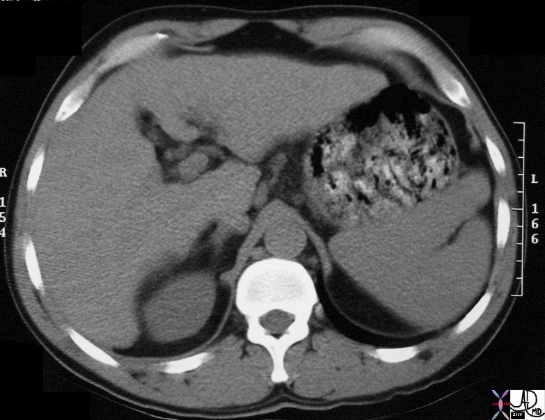
48320 |
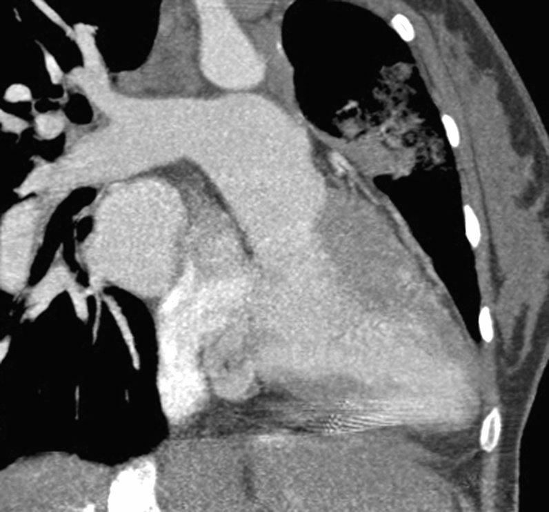
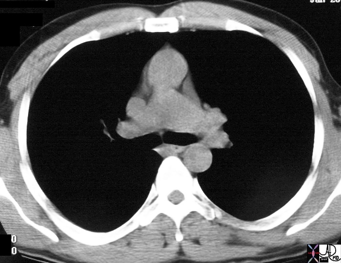
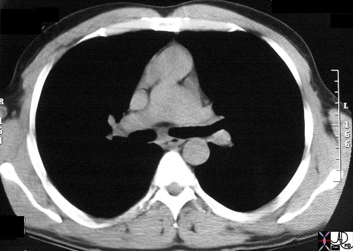
| 48320 CT of patient with an anterior aorta and a large posterior pulmonary artery TGA transposition 48320 48321 DTGA Courtesy Dr Hyun Woo Goo from Korea and Dr Laureen Sena |
|
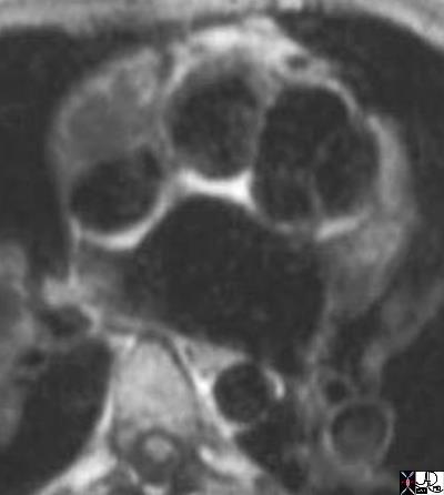
Anterior Aorta |
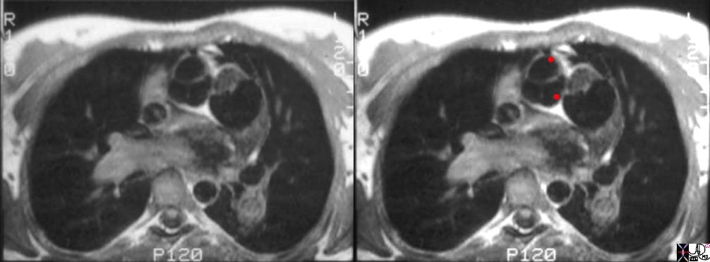
| 07249e01cs aorta pulmonary artery conus arteriosus displaced DTGA congenital heart disease heart cardiac MRI T1 weighted Courtesy Ashley Davidoff MD copyright 2009 |
Corrected Transposition
Transposition
|
Aorta Anterior and to the Left ? L TGA |
| 07304b01 anterior aorta posterior pulmonary artery position connection subaortic conus L TGA L TGV L transposition of the great vessels L transposition of the great arteries levo leftward MRI Davidoff MD |
|
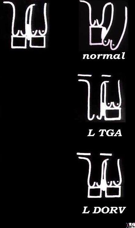
L ? Loop DORV |
| 06394c02Ls embryology bilateral conus DORV growth resorbtion normal mitral to aortic continuity transposition L transposition corrected transposition double outlet right ventricle Davidoff art copyright 2009 all rights reserved |
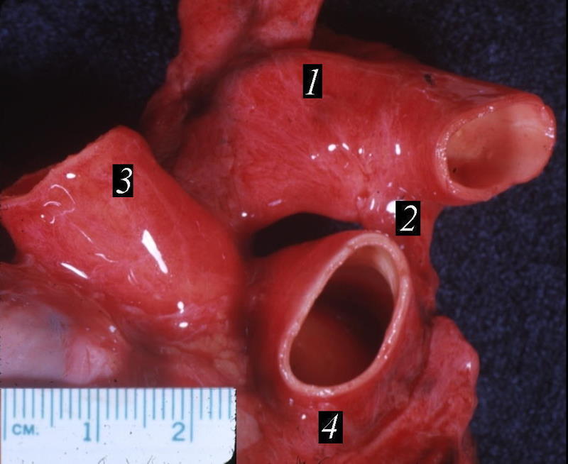
Branch Pulmonary Arteries |
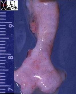
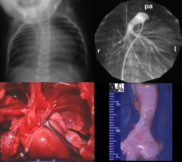
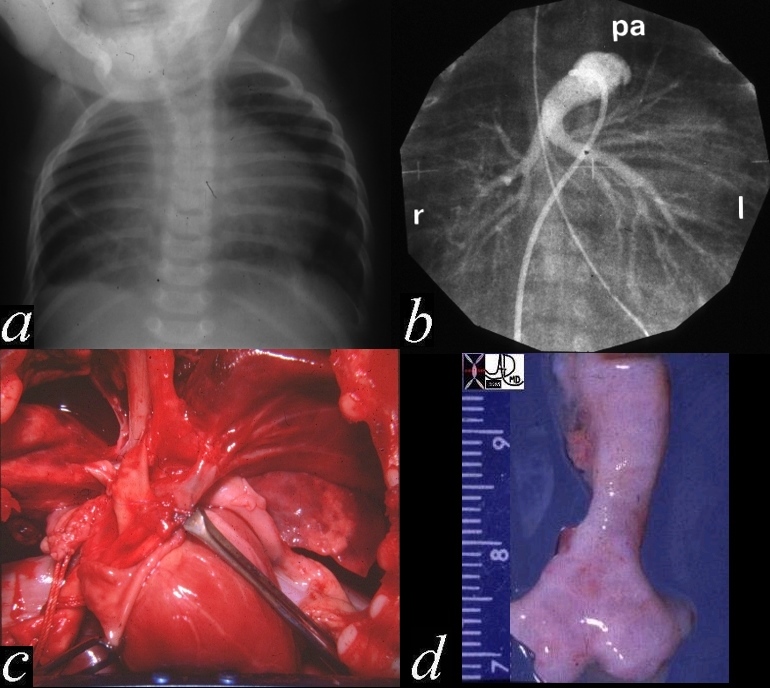
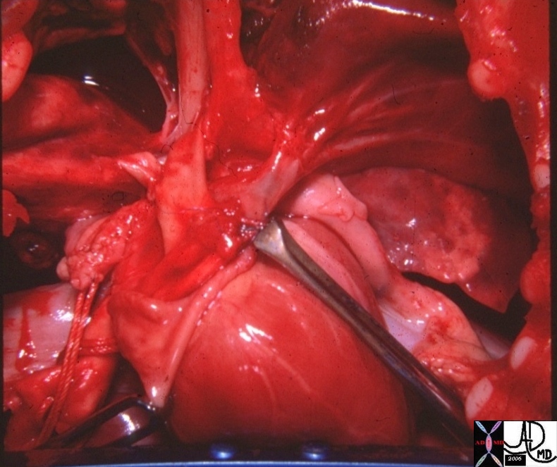
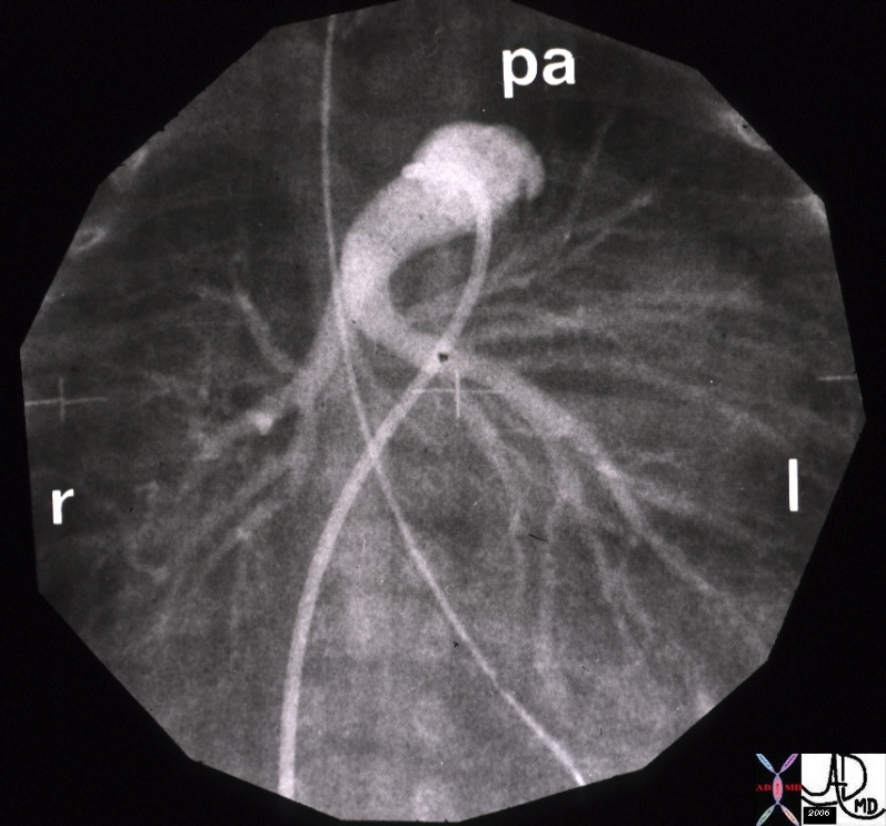
| 01559b03 aorta pulmonary artery lung ligamentum arteriosum aortic arch thickened walls relations grosspathology Davidoff MD |
Applied Biology
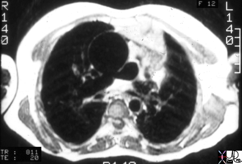
Truncus Arteriosus |
| 01651.800 aorta pulmonary arteries branch pulmonary artery fx origin of the branch PA fromthe posterior aspect of the aorta dx TA truncus arteriosus MRI T1 weighted CHD |
DOMElement Object
(
    [schemaTypeInfo] =>
    [tagName] => table
    [firstElementChild] => (object value omitted)
    [lastElementChild] => (object value omitted)
    [childElementCount] => 1
    [previousElementSibling] => (object value omitted)
    [nextElementSibling] =>
    [nodeName] => table
    [nodeValue] =>


Truncus Arteriosus


01651.800 aorta pulmonary arteries branch pulmonary artery fx origin of the branch PA fromthe posterior aspect of the aorta dx TA truncus arteriosus MRI T1 weighted CHD


    [nodeType] => 1
    [parentNode] => (object value omitted)
    [childNodes] => (object value omitted)
    [firstChild] => (object value omitted)
    [lastChild] => (object value omitted)
    [previousSibling] => (object value omitted)
    [nextSibling] => (object value omitted)
    [attributes] => (object value omitted)
    [ownerDocument] => (object value omitted)
    [namespaceURI] =>
    [prefix] =>
    [localName] => table
    [baseURI] =>
    [textContent] =>


Truncus Arteriosus


01651.800 aorta pulmonary arteries branch pulmonary artery fx origin of the branch PA fromthe posterior aspect of the aorta dx TA truncus arteriosus MRI T1 weighted CHD


)
DOMElement Object
(
    [schemaTypeInfo] =>
    [tagName] => td
    [firstElementChild] => (object value omitted)
    [lastElementChild] => (object value omitted)
    [childElementCount] => 1
    [previousElementSibling] =>
    [nextElementSibling] =>
    [nodeName] => td
    [nodeValue] => 01651.800 aorta pulmonary arteries branch pulmonary artery fx origin of the branch PA fromthe posterior aspect of the aorta dx TA truncus arteriosus MRI T1 weighted CHD
    [nodeType] => 1
    [parentNode] => (object value omitted)
    [childNodes] => (object value omitted)
    [firstChild] => (object value omitted)
    [lastChild] => (object value omitted)
    [previousSibling] => (object value omitted)
    [nextSibling] => (object value omitted)
    [attributes] => (object value omitted)
    [ownerDocument] => (object value omitted)
    [namespaceURI] =>
    [prefix] =>
    [localName] => td
    [baseURI] =>
    [textContent] => 01651.800 aorta pulmonary arteries branch pulmonary artery fx origin of the branch PA fromthe posterior aspect of the aorta dx TA truncus arteriosus MRI T1 weighted CHD
)
DOMElement Object
(
    [schemaTypeInfo] =>
    [tagName] => td
    [firstElementChild] => (object value omitted)
    [lastElementChild] => (object value omitted)
    [childElementCount] => 2
    [previousElementSibling] =>
    [nextElementSibling] =>
    [nodeName] => td
    [nodeValue] =>
Truncus Arteriosus
    [nodeType] => 1
    [parentNode] => (object value omitted)
    [childNodes] => (object value omitted)
    [firstChild] => (object value omitted)
    [lastChild] => (object value omitted)
    [previousSibling] => (object value omitted)
    [nextSibling] => (object value omitted)
    [attributes] => (object value omitted)
    [ownerDocument] => (object value omitted)
    [namespaceURI] =>
    [prefix] =>
    [localName] => td
    [baseURI] =>
    [textContent] =>
Truncus Arteriosus
)
DOMElement Object
(
    [schemaTypeInfo] =>
    [tagName] => table
    [firstElementChild] => (object value omitted)
    [lastElementChild] => (object value omitted)
    [childElementCount] => 1
    [previousElementSibling] => (object value omitted)
    [nextElementSibling] => (object value omitted)
    [nodeName] => table
    [nodeValue] =>


Anomalous Origin of the RPA


01533c02 lung trachea right pulmonary artery fx malposition left right pulmonary artery arising from right pulmonary artery fx tracheamalacia tracheomalacia fx hyperinflated right lung fx mediastinal shift dx pulmonary sling pulmonary angiogram pulmonary angiography Davidoff MD 01533c01 01531 01530.800 01532.800 01533.800 01533c02


    [nodeType] => 1
    [parentNode] => (object value omitted)
    [childNodes] => (object value omitted)
    [firstChild] => (object value omitted)
    [lastChild] => (object value omitted)
    [previousSibling] => (object value omitted)
    [nextSibling] => (object value omitted)
    [attributes] => (object value omitted)
    [ownerDocument] => (object value omitted)
    [namespaceURI] =>
    [prefix] =>
    [localName] => table
    [baseURI] =>
    [textContent] =>


Anomalous Origin of the RPA


01533c02 lung trachea right pulmonary artery fx malposition left right pulmonary artery arising from right pulmonary artery fx tracheamalacia tracheomalacia fx hyperinflated right lung fx mediastinal shift dx pulmonary sling pulmonary angiogram pulmonary angiography Davidoff MD 01533c01 01531 01530.800 01532.800 01533.800 01533c02


)
DOMElement Object
(
    [schemaTypeInfo] =>
    [tagName] => td
    [firstElementChild] => (object value omitted)
    [lastElementChild] => (object value omitted)
    [childElementCount] => 1
    [previousElementSibling] =>
    [nextElementSibling] =>
    [nodeName] => td
    [nodeValue] => 01533c02 lung trachea right pulmonary artery fx malposition left right pulmonary artery arising from right pulmonary artery fx tracheamalacia tracheomalacia fx hyperinflated right lung fx mediastinal shift dx pulmonary sling pulmonary angiogram pulmonary angiography Davidoff MD 01533c01 01531 01530.800 01532.800 01533.800 01533c02
    [nodeType] => 1
    [parentNode] => (object value omitted)
    [childNodes] => (object value omitted)
    [firstChild] => (object value omitted)
    [lastChild] => (object value omitted)
    [previousSibling] => (object value omitted)
    [nextSibling] => (object value omitted)
    [attributes] => (object value omitted)
    [ownerDocument] => (object value omitted)
    [namespaceURI] =>
    [prefix] =>
    [localName] => td
    [baseURI] =>
    [textContent] => 01533c02 lung trachea right pulmonary artery fx malposition left right pulmonary artery arising from right pulmonary artery fx tracheamalacia tracheomalacia fx hyperinflated right lung fx mediastinal shift dx pulmonary sling pulmonary angiogram pulmonary angiography Davidoff MD 01533c01 01531 01530.800 01532.800 01533.800 01533c02
)
DOMElement Object
(
    [schemaTypeInfo] =>
    [tagName] => td
    [firstElementChild] => (object value omitted)
    [lastElementChild] => (object value omitted)
    [childElementCount] => 2
    [previousElementSibling] =>
    [nextElementSibling] =>
    [nodeName] => td
    [nodeValue] =>
Anomalous Origin of the RPA
    [nodeType] => 1
    [parentNode] => (object value omitted)
    [childNodes] => (object value omitted)
    [firstChild] => (object value omitted)
    [lastChild] => (object value omitted)
    [previousSibling] => (object value omitted)
    [nextSibling] => (object value omitted)
    [attributes] => (object value omitted)
    [ownerDocument] => (object value omitted)
    [namespaceURI] =>
    [prefix] =>
    [localName] => td
    [baseURI] =>
    [textContent] =>
Anomalous Origin of the RPA
)
https://beta.thecommonvein.net/wp-content/uploads/2023/04/01533.800.jpg https://beta.thecommonvein.net/wp-content/uploads/2023/04/01532.800.jpg https://beta.thecommonvein.net/wp-content/uploads/2023/04/01530.800.jpg https://beta.thecommonvein.net/wp-content/uploads/2023/09/01533c02.jpg https://beta.thecommonvein.net/wp-content/uploads/2023/04/01533c01.jpg https://beta.thecommonvein.net/wp-content/uploads/2023/04/01531.jpg
http://thecommonvein.net/media/01533c02.jpg
DOMElement Object
(
    [schemaTypeInfo] =>
    [tagName] => table
    [firstElementChild] => (object value omitted)
    [lastElementChild] => (object value omitted)
    [childElementCount] => 1
    [previousElementSibling] => (object value omitted)
    [nextElementSibling] => (object value omitted)
    [nodeName] => table
    [nodeValue] =>


Anomalous Origin of the RPA


01533.800 lung trachea right pulmonary artery fx malposition left right pulmonary artery arising from right pulmonary artery fx tracheamalacia tracheomalacia fx hyperinflated right lung fx mediastinal shift dx pulmonary sling pulmonary angiogram pulmonary angiography Davidoff MD 01533c01 01531 01530.800 01532.800 01533.800 01533c02


    [nodeType] => 1
    [parentNode] => (object value omitted)
    [childNodes] => (object value omitted)
    [firstChild] => (object value omitted)
    [lastChild] => (object value omitted)
    [previousSibling] => (object value omitted)
    [nextSibling] => (object value omitted)
    [attributes] => (object value omitted)
    [ownerDocument] => (object value omitted)
    [namespaceURI] =>
    [prefix] =>
    [localName] => table
    [baseURI] =>
    [textContent] =>


Anomalous Origin of the RPA


01533.800 lung trachea right pulmonary artery fx malposition left right pulmonary artery arising from right pulmonary artery fx tracheamalacia tracheomalacia fx hyperinflated right lung fx mediastinal shift dx pulmonary sling pulmonary angiogram pulmonary angiography Davidoff MD 01533c01 01531 01530.800 01532.800 01533.800 01533c02


)
DOMElement Object
(
    [schemaTypeInfo] =>
    [tagName] => td
    [firstElementChild] => (object value omitted)
    [lastElementChild] => (object value omitted)
    [childElementCount] => 1
    [previousElementSibling] =>
    [nextElementSibling] =>
    [nodeName] => td
    [nodeValue] => 01533.800 lung trachea right pulmonary artery fx malposition left right pulmonary artery arising from right pulmonary artery fx tracheamalacia tracheomalacia fx hyperinflated right lung fx mediastinal shift dx pulmonary sling pulmonary angiogram pulmonary angiography Davidoff MD 01533c01 01531 01530.800 01532.800 01533.800 01533c02
    [nodeType] => 1
    [parentNode] => (object value omitted)
    [childNodes] => (object value omitted)
    [firstChild] => (object value omitted)
    [lastChild] => (object value omitted)
    [previousSibling] => (object value omitted)
    [nextSibling] => (object value omitted)
    [attributes] => (object value omitted)
    [ownerDocument] => (object value omitted)
    [namespaceURI] =>
    [prefix] =>
    [localName] => td
    [baseURI] =>
    [textContent] => 01533.800 lung trachea right pulmonary artery fx malposition left right pulmonary artery arising from right pulmonary artery fx tracheamalacia tracheomalacia fx hyperinflated right lung fx mediastinal shift dx pulmonary sling pulmonary angiogram pulmonary angiography Davidoff MD 01533c01 01531 01530.800 01532.800 01533.800 01533c02
)
DOMElement Object
(
    [schemaTypeInfo] =>
    [tagName] => td
    [firstElementChild] => (object value omitted)
    [lastElementChild] => (object value omitted)
    [childElementCount] => 2
    [previousElementSibling] =>
    [nextElementSibling] =>
    [nodeName] => td
    [nodeValue] =>
Anomalous Origin of the RPA
    [nodeType] => 1
    [parentNode] => (object value omitted)
    [childNodes] => (object value omitted)
    [firstChild] => (object value omitted)
    [lastChild] => (object value omitted)
    [previousSibling] => (object value omitted)
    [nextSibling] => (object value omitted)
    [attributes] => (object value omitted)
    [ownerDocument] => (object value omitted)
    [namespaceURI] =>
    [prefix] =>
    [localName] => td
    [baseURI] =>
    [textContent] =>
Anomalous Origin of the RPA
)
https://beta.thecommonvein.net/wp-content/uploads/2023/04/01533.800.jpg https://beta.thecommonvein.net/wp-content/uploads/2023/04/01532.800.jpg https://beta.thecommonvein.net/wp-content/uploads/2023/04/01530.800.jpg https://beta.thecommonvein.net/wp-content/uploads/2023/09/01533c02.jpg https://beta.thecommonvein.net/wp-content/uploads/2023/04/01533c01.jpg https://beta.thecommonvein.net/wp-content/uploads/2023/04/01531.jpg
http://thecommonvein.net/media/01533.800.jpg
DOMElement Object
(
    [schemaTypeInfo] =>
    [tagName] => table
    [firstElementChild] => (object value omitted)
    [lastElementChild] => (object value omitted)
    [childElementCount] => 1
    [previousElementSibling] => (object value omitted)
    [nextElementSibling] => (object value omitted)
    [nodeName] => table
    [nodeValue] =>


Normal relationship and Size – Arterioles and Bronchioles


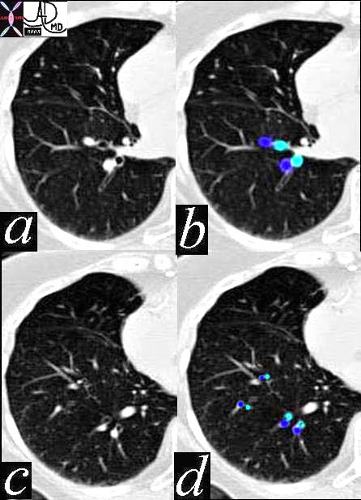
In this CT of a normal patient we see two levels of the RLL with the segmental bronchioles and arterioles branching dichotomously and simultaneously. Note again the similarity in size and shape through these levels of division, a form that is maintained in the normal person until they reach the terminal bronchiole.
42455Courtesy Ashley Davidoff MD 42455 lung artery bronchus normal anatomy size applied anatomy applied biology


    [nodeType] => 1
    [parentNode] => (object value omitted)
    [childNodes] => (object value omitted)
    [firstChild] => (object value omitted)
    [lastChild] => (object value omitted)
    [previousSibling] => (object value omitted)
    [nextSibling] => (object value omitted)
    [attributes] => (object value omitted)
    [ownerDocument] => (object value omitted)
    [namespaceURI] =>
    [prefix] =>
    [localName] => table
    [baseURI] =>
    [textContent] =>


Normal relationship and Size – Arterioles and Bronchioles


In this CT of a normal patient we see two levels of the RLL with the segmental bronchioles and arterioles branching dichotomously and simultaneously. Note again the similarity in size and shape through these levels of division, a form that is maintained in the normal person until they reach the terminal bronchiole.
42455Courtesy Ashley Davidoff MD 42455 lung artery bronchus normal anatomy size applied anatomy applied biology


)
DOMElement Object
(
    [schemaTypeInfo] =>
    [tagName] => td
    [firstElementChild] => (object value omitted)
    [lastElementChild] => (object value omitted)
    [childElementCount] => 2
    [previousElementSibling] =>
    [nextElementSibling] =>
    [nodeName] => td
    [nodeValue] => In this CT of a normal patient we see two levels of the RLL with the segmental bronchioles and arterioles branching dichotomously and simultaneously. Note again the similarity in size and shape through these levels of division, a form that is maintained in the normal person until they reach the terminal bronchiole.
42455Courtesy Ashley Davidoff MD 42455 lung artery bronchus normal anatomy size applied anatomy applied biology
    [nodeType] => 1
    [parentNode] => (object value omitted)
    [childNodes] => (object value omitted)
    [firstChild] => (object value omitted)
    [lastChild] => (object value omitted)
    [previousSibling] => (object value omitted)
    [nextSibling] => (object value omitted)
    [attributes] => (object value omitted)
    [ownerDocument] => (object value omitted)
    [namespaceURI] =>
    [prefix] =>
    [localName] => td
    [baseURI] =>
    [textContent] => In this CT of a normal patient we see two levels of the RLL with the segmental bronchioles and arterioles branching dichotomously and simultaneously. Note again the similarity in size and shape through these levels of division, a form that is maintained in the normal person until they reach the terminal bronchiole.
42455Courtesy Ashley Davidoff MD 42455 lung artery bronchus normal anatomy size applied anatomy applied biology
)
DOMElement Object
(
    [schemaTypeInfo] =>
    [tagName] => td
    [firstElementChild] => (object value omitted)
    [lastElementChild] => (object value omitted)
    [childElementCount] => 2
    [previousElementSibling] =>
    [nextElementSibling] =>
    [nodeName] => td
    [nodeValue] =>
Normal relationship and Size – Arterioles and Bronchioles

    [nodeType] => 1
    [parentNode] => (object value omitted)
    [childNodes] => (object value omitted)
    [firstChild] => (object value omitted)
    [lastChild] => (object value omitted)
    [previousSibling] => (object value omitted)
    [nextSibling] => (object value omitted)
    [attributes] => (object value omitted)
    [ownerDocument] => (object value omitted)
    [namespaceURI] =>
    [prefix] =>
    [localName] => td
    [baseURI] =>
    [textContent] =>
Normal relationship and Size – Arterioles and Bronchioles

)
DOMElement Object
(
    [schemaTypeInfo] =>
    [tagName] => table
    [firstElementChild] => (object value omitted)
    [lastElementChild] => (object value omitted)
    [childElementCount] => 1
    [previousElementSibling] => (object value omitted)
    [nextElementSibling] => (object value omitted)
    [nodeName] => table
    [nodeValue] =>


Branch Pulmonary Arteries


01559b03 aorta pulmonary artery lung ligamentum arteriosum aortic arch thickened walls relations grosspathology Davidoff MD


    [nodeType] => 1
    [parentNode] => (object value omitted)
    [childNodes] => (object value omitted)
    [firstChild] => (object value omitted)
    [lastChild] => (object value omitted)
    [previousSibling] => (object value omitted)
    [nextSibling] => (object value omitted)
    [attributes] => (object value omitted)
    [ownerDocument] => (object value omitted)
    [namespaceURI] =>
    [prefix] =>
    [localName] => table
    [baseURI] =>
    [textContent] =>


Branch Pulmonary Arteries


01559b03 aorta pulmonary artery lung ligamentum arteriosum aortic arch thickened walls relations grosspathology Davidoff MD


)
DOMElement Object
(
    [schemaTypeInfo] =>
    [tagName] => td
    [firstElementChild] => (object value omitted)
    [lastElementChild] => (object value omitted)
    [childElementCount] => 1
    [previousElementSibling] =>
    [nextElementSibling] =>
    [nodeName] => td
    [nodeValue] => 01559b03 aorta pulmonary artery lung ligamentum arteriosum aortic arch thickened walls relations grosspathology Davidoff MD
    [nodeType] => 1
    [parentNode] => (object value omitted)
    [childNodes] => (object value omitted)
    [firstChild] => (object value omitted)
    [lastChild] => (object value omitted)
    [previousSibling] => (object value omitted)
    [nextSibling] => (object value omitted)
    [attributes] => (object value omitted)
    [ownerDocument] => (object value omitted)
    [namespaceURI] =>
    [prefix] =>
    [localName] => td
    [baseURI] =>
    [textContent] => 01559b03 aorta pulmonary artery lung ligamentum arteriosum aortic arch thickened walls relations grosspathology Davidoff MD
)
DOMElement Object
(
    [schemaTypeInfo] =>
    [tagName] => td
    [firstElementChild] => (object value omitted)
    [lastElementChild] => (object value omitted)
    [childElementCount] => 2
    [previousElementSibling] =>
    [nextElementSibling] =>
    [nodeName] => td
    [nodeValue] =>
Branch Pulmonary Arteries
    [nodeType] => 1
    [parentNode] => (object value omitted)
    [childNodes] => (object value omitted)
    [firstChild] => (object value omitted)
    [lastChild] => (object value omitted)
    [previousSibling] => (object value omitted)
    [nextSibling] => (object value omitted)
    [attributes] => (object value omitted)
    [ownerDocument] => (object value omitted)
    [namespaceURI] =>
    [prefix] =>
    [localName] => td
    [baseURI] =>
    [textContent] =>
Branch Pulmonary Arteries
)
DOMElement Object
(
    [schemaTypeInfo] =>
    [tagName] => table
    [firstElementChild] => (object value omitted)
    [lastElementChild] => (object value omitted)
    [childElementCount] => 1
    [previousElementSibling] => (object value omitted)
    [nextElementSibling] => (object value omitted)
    [nodeName] => table
    [nodeValue] =>


L ? Loop DORV


06394c02Ls embryology bilateral conus DORV growth resorbtion normal mitral to aortic continuity transposition L transposition corrected transposition double outlet right ventricle Davidoff art copyright 2009 all rights reserved


    [nodeType] => 1
    [parentNode] => (object value omitted)
    [childNodes] => (object value omitted)
    [firstChild] => (object value omitted)
    [lastChild] => (object value omitted)
    [previousSibling] => (object value omitted)
    [nextSibling] => (object value omitted)
    [attributes] => (object value omitted)
    [ownerDocument] => (object value omitted)
    [namespaceURI] =>
    [prefix] =>
    [localName] => table
    [baseURI] =>
    [textContent] =>


L ? Loop DORV


06394c02Ls embryology bilateral conus DORV growth resorbtion normal mitral to aortic continuity transposition L transposition corrected transposition double outlet right ventricle Davidoff art copyright 2009 all rights reserved


)
DOMElement Object
(
    [schemaTypeInfo] =>
    [tagName] => td
    [firstElementChild] => (object value omitted)
    [lastElementChild] => (object value omitted)
    [childElementCount] => 1
    [previousElementSibling] =>
    [nextElementSibling] =>
    [nodeName] => td
    [nodeValue] => 06394c02Ls embryology bilateral conus DORV growth resorbtion normal mitral to aortic continuity transposition L transposition corrected transposition double outlet right ventricle Davidoff art copyright 2009 all rights reserved
    [nodeType] => 1
    [parentNode] => (object value omitted)
    [childNodes] => (object value omitted)
    [firstChild] => (object value omitted)
    [lastChild] => (object value omitted)
    [previousSibling] => (object value omitted)
    [nextSibling] => (object value omitted)
    [attributes] => (object value omitted)
    [ownerDocument] => (object value omitted)
    [namespaceURI] =>
    [prefix] =>
    [localName] => td
    [baseURI] =>
    [textContent] => 06394c02Ls embryology bilateral conus DORV growth resorbtion normal mitral to aortic continuity transposition L transposition corrected transposition double outlet right ventricle Davidoff art copyright 2009 all rights reserved
)
DOMElement Object
(
    [schemaTypeInfo] =>
    [tagName] => td
    [firstElementChild] => (object value omitted)
    [lastElementChild] => (object value omitted)
    [childElementCount] => 2
    [previousElementSibling] =>
    [nextElementSibling] =>
    [nodeName] => td
    [nodeValue] =>

L ? Loop DORV

    [nodeType] => 1
    [parentNode] => (object value omitted)
    [childNodes] => (object value omitted)
    [firstChild] => (object value omitted)
    [lastChild] => (object value omitted)
    [previousSibling] => (object value omitted)
    [nextSibling] => (object value omitted)
    [attributes] => (object value omitted)
    [ownerDocument] => (object value omitted)
    [namespaceURI] =>
    [prefix] =>
    [localName] => td
    [baseURI] =>
    [textContent] =>

L ? Loop DORV

)
DOMElement Object
(
    [schemaTypeInfo] =>
    [tagName] => table
    [firstElementChild] => (object value omitted)
    [lastElementChild] => (object value omitted)
    [childElementCount] => 1
    [previousElementSibling] => (object value omitted)
    [nextElementSibling] => (object value omitted)
    [nodeName] => table
    [nodeValue] =>


Aorta Anterior and to the Left ? L TGA


07304b01 anterior aorta posterior pulmonary artery position connection subaortic conus L TGA L TGV L transposition of the great vessels L transposition of the great arteries levo leftward MRI Davidoff MD


    [nodeType] => 1
    [parentNode] => (object value omitted)
    [childNodes] => (object value omitted)
    [firstChild] => (object value omitted)
    [lastChild] => (object value omitted)
    [previousSibling] => (object value omitted)
    [nextSibling] => (object value omitted)
    [attributes] => (object value omitted)
    [ownerDocument] => (object value omitted)
    [namespaceURI] =>
    [prefix] =>
    [localName] => table
    [baseURI] =>
    [textContent] =>


Aorta Anterior and to the Left ? L TGA


07304b01 anterior aorta posterior pulmonary artery position connection subaortic conus L TGA L TGV L transposition of the great vessels L transposition of the great arteries levo leftward MRI Davidoff MD


)
DOMElement Object
(
    [schemaTypeInfo] =>
    [tagName] => td
    [firstElementChild] => (object value omitted)
    [lastElementChild] => (object value omitted)
    [childElementCount] => 1
    [previousElementSibling] =>
    [nextElementSibling] =>
    [nodeName] => td
    [nodeValue] => 07304b01 anterior aorta posterior pulmonary artery position connection subaortic conus L TGA L TGV L transposition of the great vessels L transposition of the great arteries levo leftward MRI Davidoff MD
    [nodeType] => 1
    [parentNode] => (object value omitted)
    [childNodes] => (object value omitted)
    [firstChild] => (object value omitted)
    [lastChild] => (object value omitted)
    [previousSibling] => (object value omitted)
    [nextSibling] => (object value omitted)
    [attributes] => (object value omitted)
    [ownerDocument] => (object value omitted)
    [namespaceURI] =>
    [prefix] =>
    [localName] => td
    [baseURI] =>
    [textContent] => 07304b01 anterior aorta posterior pulmonary artery position connection subaortic conus L TGA L TGV L transposition of the great vessels L transposition of the great arteries levo leftward MRI Davidoff MD
)
DOMElement Object
(
    [schemaTypeInfo] =>
    [tagName] => td
    [firstElementChild] => (object value omitted)
    [lastElementChild] => (object value omitted)
    [childElementCount] => 2
    [previousElementSibling] =>
    [nextElementSibling] =>
    [nodeName] => td
    [nodeValue] =>

Aorta Anterior and to the Left ? L TGA

    [nodeType] => 1
    [parentNode] => (object value omitted)
    [childNodes] => (object value omitted)
    [firstChild] => (object value omitted)
    [lastChild] => (object value omitted)
    [previousSibling] => (object value omitted)
    [nextSibling] => (object value omitted)
    [attributes] => (object value omitted)
    [ownerDocument] => (object value omitted)
    [namespaceURI] =>
    [prefix] =>
    [localName] => td
    [baseURI] =>
    [textContent] =>

Aorta Anterior and to the Left ? L TGA

)
DOMElement Object
(
    [schemaTypeInfo] =>
    [tagName] => table
    [firstElementChild] => (object value omitted)
    [lastElementChild] => (object value omitted)
    [childElementCount] => 1
    [previousElementSibling] => (object value omitted)
    [nextElementSibling] => (object value omitted)
    [nodeName] => table
    [nodeValue] =>


LTGA


28999b01 heart cardiac aorta pulmonary artery RVOT conotruncal malformation LTGA L TGV transposition of the great vessels transposition of the geat arteries corrected transposition position connection relation embryology CTscan Davidoff MD 28994 28995 28996 28997 28998 28999


    [nodeType] => 1
    [parentNode] => (object value omitted)
    [childNodes] => (object value omitted)
    [firstChild] => (object value omitted)
    [lastChild] => (object value omitted)
    [previousSibling] => (object value omitted)
    [nextSibling] => (object value omitted)
    [attributes] => (object value omitted)
    [ownerDocument] => (object value omitted)
    [namespaceURI] =>
    [prefix] =>
    [localName] => table
    [baseURI] =>
    [textContent] =>


LTGA


28999b01 heart cardiac aorta pulmonary artery RVOT conotruncal malformation LTGA L TGV transposition of the great vessels transposition of the geat arteries corrected transposition position connection relation embryology CTscan Davidoff MD 28994 28995 28996 28997 28998 28999


)
DOMElement Object
(
    [schemaTypeInfo] =>
    [tagName] => td
    [firstElementChild] => (object value omitted)
    [lastElementChild] => (object value omitted)
    [childElementCount] => 1
    [previousElementSibling] =>
    [nextElementSibling] =>
    [nodeName] => td
    [nodeValue] => 28999b01 heart cardiac aorta pulmonary artery RVOT conotruncal malformation LTGA L TGV transposition of the great vessels transposition of the geat arteries corrected transposition position connection relation embryology CTscan Davidoff MD 28994 28995 28996 28997 28998 28999
    [nodeType] => 1
    [parentNode] => (object value omitted)
    [childNodes] => (object value omitted)
    [firstChild] => (object value omitted)
    [lastChild] => (object value omitted)
    [previousSibling] => (object value omitted)
    [nextSibling] => (object value omitted)
    [attributes] => (object value omitted)
    [ownerDocument] => (object value omitted)
    [namespaceURI] =>
    [prefix] =>
    [localName] => td
    [baseURI] =>
    [textContent] => 28999b01 heart cardiac aorta pulmonary artery RVOT conotruncal malformation LTGA L TGV transposition of the great vessels transposition of the geat arteries corrected transposition position connection relation embryology CTscan Davidoff MD 28994 28995 28996 28997 28998 28999
)
DOMElement Object
(
    [schemaTypeInfo] =>
    [tagName] => td
    [firstElementChild] => (object value omitted)
    [lastElementChild] => (object value omitted)
    [childElementCount] => 2
    [previousElementSibling] =>
    [nextElementSibling] =>
    [nodeName] => td
    [nodeValue] => LTGA
    [nodeType] => 1
    [parentNode] => (object value omitted)
    [childNodes] => (object value omitted)
    [firstChild] => (object value omitted)
    [lastChild] => (object value omitted)
    [previousSibling] => (object value omitted)
    [nextSibling] => (object value omitted)
    [attributes] => (object value omitted)
    [ownerDocument] => (object value omitted)
    [namespaceURI] =>
    [prefix] =>
    [localName] => td
    [baseURI] =>
    [textContent] => LTGA
)
https://beta.thecommonvein.net/wp-content/uploads/2023/09/28999b01.jpg https://beta.thecommonvein.net/wp-content/uploads/2023/05/28999.jpg https://beta.thecommonvein.net/wp-content/uploads/2023/05/28998.jpg https://beta.thecommonvein.net/wp-content/uploads/2023/05/28997.jpg https://beta.thecommonvein.net/wp-content/uploads/2023/05/28996.jpg https://beta.thecommonvein.net/wp-content/uploads/2023/05/28995.jpg https://beta.thecommonvein.net/wp-content/uploads/2023/05/28994.jpg
http://thecommonvein.net/media/28999b01.jpg
DOMElement Object
(
    [schemaTypeInfo] =>
    [tagName] => table
    [firstElementChild] => (object value omitted)
    [lastElementChild] => (object value omitted)
    [childElementCount] => 1
    [previousElementSibling] => (object value omitted)
    [nextElementSibling] => (object value omitted)
    [nodeName] => table
    [nodeValue] =>


DTGA


07258b02 anterior aorta posterior pulmonary artery position connection subaortic conus D TGA TGV D transposition of the great vessels D transposition of the great arteries dextro rightward MRI Davidoff MD


    [nodeType] => 1
    [parentNode] => (object value omitted)
    [childNodes] => (object value omitted)
    [firstChild] => (object value omitted)
    [lastChild] => (object value omitted)
    [previousSibling] => (object value omitted)
    [nextSibling] => (object value omitted)
    [attributes] => (object value omitted)
    [ownerDocument] => (object value omitted)
    [namespaceURI] =>
    [prefix] =>
    [localName] => table
    [baseURI] =>
    [textContent] =>


DTGA


07258b02 anterior aorta posterior pulmonary artery position connection subaortic conus D TGA TGV D transposition of the great vessels D transposition of the great arteries dextro rightward MRI Davidoff MD


)
DOMElement Object
(
    [schemaTypeInfo] =>
    [tagName] => td
    [firstElementChild] => (object value omitted)
    [lastElementChild] => (object value omitted)
    [childElementCount] => 1
    [previousElementSibling] =>
    [nextElementSibling] =>
    [nodeName] => td
    [nodeValue] => 07258b02 anterior aorta posterior pulmonary artery position connection subaortic conus D TGA TGV D transposition of the great vessels D transposition of the great arteries dextro rightward MRI Davidoff MD
    [nodeType] => 1
    [parentNode] => (object value omitted)
    [childNodes] => (object value omitted)
    [firstChild] => (object value omitted)
    [lastChild] => (object value omitted)
    [previousSibling] => (object value omitted)
    [nextSibling] => (object value omitted)
    [attributes] => (object value omitted)
    [ownerDocument] => (object value omitted)
    [namespaceURI] =>
    [prefix] =>
    [localName] => td
    [baseURI] =>
    [textContent] => 07258b02 anterior aorta posterior pulmonary artery position connection subaortic conus D TGA TGV D transposition of the great vessels D transposition of the great arteries dextro rightward MRI Davidoff MD
)
DOMElement Object
(
    [schemaTypeInfo] =>
    [tagName] => td
    [firstElementChild] => (object value omitted)
    [lastElementChild] => (object value omitted)
    [childElementCount] => 2
    [previousElementSibling] =>
    [nextElementSibling] =>
    [nodeName] => td
    [nodeValue] =>
DTGA

    [nodeType] => 1
    [parentNode] => (object value omitted)
    [childNodes] => (object value omitted)
    [firstChild] => (object value omitted)
    [lastChild] => (object value omitted)
    [previousSibling] => (object value omitted)
    [nextSibling] => (object value omitted)
    [attributes] => (object value omitted)
    [ownerDocument] => (object value omitted)
    [namespaceURI] =>
    [prefix] =>
    [localName] => td
    [baseURI] =>
    [textContent] =>
DTGA

)
DOMElement Object
(
    [schemaTypeInfo] =>
    [tagName] => table
    [firstElementChild] => (object value omitted)
    [lastElementChild] => (object value omitted)
    [childElementCount] => 1
    [previousElementSibling] => (object value omitted)
    [nextElementSibling] => (object value omitted)
    [nodeName] => table
    [nodeValue] =>


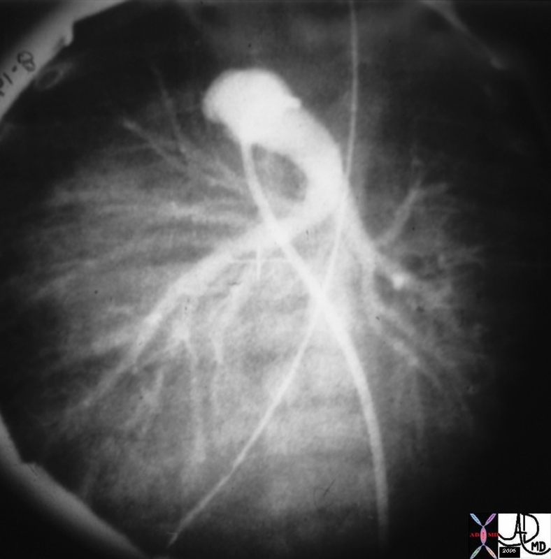
Transposition of the Great Vessels


This is an angiogram of the RV, showing an anteriorly placed aorta, a VSD filling the LV, and a posteriorly positioned smaller MPA. The catheter courses via the IVC into the RV. The findings are consistent with TGA and in this case a D-TGA, though it is impossible to identify the position of the aortic valve in relation to the PA in this lateral projection. An associated VSD and subpulmonary stenosis and or PS is implied by the small sized PA. Courtesy Ashley Davidoff MD 01487 code CVS heart cardiac transpoistion of the great arteries DTGA VSD subpulmonary stenosis imaging radiology angiography


    [nodeType] => 1
    [parentNode] => (object value omitted)
    [childNodes] => (object value omitted)
    [firstChild] => (object value omitted)
    [lastChild] => (object value omitted)
    [previousSibling] => (object value omitted)
    [nextSibling] => (object value omitted)
    [attributes] => (object value omitted)
    [ownerDocument] => (object value omitted)
    [namespaceURI] =>
    [prefix] =>
    [localName] => table
    [baseURI] =>
    [textContent] =>


Transposition of the Great Vessels


This is an angiogram of the RV, showing an anteriorly placed aorta, a VSD filling the LV, and a posteriorly positioned smaller MPA. The catheter courses via the IVC into the RV. The findings are consistent with TGA and in this case a D-TGA, though it is impossible to identify the position of the aortic valve in relation to the PA in this lateral projection. An associated VSD and subpulmonary stenosis and or PS is implied by the small sized PA. Courtesy Ashley Davidoff MD 01487 code CVS heart cardiac transpoistion of the great arteries DTGA VSD subpulmonary stenosis imaging radiology angiography


)
DOMElement Object
(
    [schemaTypeInfo] =>
    [tagName] => td
    [firstElementChild] => (object value omitted)
    [lastElementChild] => (object value omitted)
    [childElementCount] => 1
    [previousElementSibling] =>
    [nextElementSibling] =>
    [nodeName] => td
    [nodeValue] => This is an angiogram of the RV, showing an anteriorly placed aorta, a VSD filling the LV, and a posteriorly positioned smaller MPA. The catheter courses via the IVC into the RV. The findings are consistent with TGA and in this case a D-TGA, though it is impossible to identify the position of the aortic valve in relation to the PA in this lateral projection. An associated VSD and subpulmonary stenosis and or PS is implied by the small sized PA. Courtesy Ashley Davidoff MD 01487 code CVS heart cardiac transpoistion of the great arteries DTGA VSD subpulmonary stenosis imaging radiology angiography
    [nodeType] => 1
    [parentNode] => (object value omitted)
    [childNodes] => (object value omitted)
    [firstChild] => (object value omitted)
    [lastChild] => (object value omitted)
    [previousSibling] => (object value omitted)
    [nextSibling] => (object value omitted)
    [attributes] => (object value omitted)
    [ownerDocument] => (object value omitted)
    [namespaceURI] =>
    [prefix] =>
    [localName] => td
    [baseURI] =>
    [textContent] => This is an angiogram of the RV, showing an anteriorly placed aorta, a VSD filling the LV, and a posteriorly positioned smaller MPA. The catheter courses via the IVC into the RV. The findings are consistent with TGA and in this case a D-TGA, though it is impossible to identify the position of the aortic valve in relation to the PA in this lateral projection. An associated VSD and subpulmonary stenosis and or PS is implied by the small sized PA. Courtesy Ashley Davidoff MD 01487 code CVS heart cardiac transpoistion of the great arteries DTGA VSD subpulmonary stenosis imaging radiology angiography
)
DOMElement Object
(
    [schemaTypeInfo] =>
    [tagName] => td
    [firstElementChild] => (object value omitted)
    [lastElementChild] => (object value omitted)
    [childElementCount] => 2
    [previousElementSibling] =>
    [nextElementSibling] =>
    [nodeName] => td
    [nodeValue] =>
Transposition of the Great Vessels

    [nodeType] => 1
    [parentNode] => (object value omitted)
    [childNodes] => (object value omitted)
    [firstChild] => (object value omitted)
    [lastChild] => (object value omitted)
    [previousSibling] => (object value omitted)
    [nextSibling] => (object value omitted)
    [attributes] => (object value omitted)
    [ownerDocument] => (object value omitted)
    [namespaceURI] =>
    [prefix] =>
    [localName] => td
    [baseURI] =>
    [textContent] =>
Transposition of the Great Vessels

)
DOMElement Object
(
    [schemaTypeInfo] =>
    [tagName] => table
    [firstElementChild] => (object value omitted)
    [lastElementChild] => (object value omitted)
    [childElementCount] => 1
    [previousElementSibling] => (object value omitted)
    [nextElementSibling] => (object value omitted)
    [nodeName] => table
    [nodeValue] =>


Anterior Aorta


07249e01cs aorta pulmonary artery conus arteriosus displaced DTGA congenital heart disease heart cardiac MRI T1 weighted Courtesy Ashley Davidoff MD copyright 2009


    [nodeType] => 1
    [parentNode] => (object value omitted)
    [childNodes] => (object value omitted)
    [firstChild] => (object value omitted)
    [lastChild] => (object value omitted)
    [previousSibling] => (object value omitted)
    [nextSibling] => (object value omitted)
    [attributes] => (object value omitted)
    [ownerDocument] => (object value omitted)
    [namespaceURI] =>
    [prefix] =>
    [localName] => table
    [baseURI] =>
    [textContent] =>


Anterior Aorta


07249e01cs aorta pulmonary artery conus arteriosus displaced DTGA congenital heart disease heart cardiac MRI T1 weighted Courtesy Ashley Davidoff MD copyright 2009


)
DOMElement Object
(
    [schemaTypeInfo] =>
    [tagName] => td
    [firstElementChild] => (object value omitted)
    [lastElementChild] => (object value omitted)
    [childElementCount] => 1
    [previousElementSibling] =>
    [nextElementSibling] =>
    [nodeName] => td
    [nodeValue] => 07249e01cs aorta pulmonary artery conus arteriosus displaced DTGA congenital heart disease heart cardiac MRI T1 weighted Courtesy Ashley Davidoff MD copyright 2009
    [nodeType] => 1
    [parentNode] => (object value omitted)
    [childNodes] => (object value omitted)
    [firstChild] => (object value omitted)
    [lastChild] => (object value omitted)
    [previousSibling] => (object value omitted)
    [nextSibling] => (object value omitted)
    [attributes] => (object value omitted)
    [ownerDocument] => (object value omitted)
    [namespaceURI] =>
    [prefix] =>
    [localName] => td
    [baseURI] =>
    [textContent] => 07249e01cs aorta pulmonary artery conus arteriosus displaced DTGA congenital heart disease heart cardiac MRI T1 weighted Courtesy Ashley Davidoff MD copyright 2009
)
DOMElement Object
(
    [schemaTypeInfo] =>
    [tagName] => td
    [firstElementChild] => (object value omitted)
    [lastElementChild] => (object value omitted)
    [childElementCount] => 2
    [previousElementSibling] =>
    [nextElementSibling] =>
    [nodeName] => td
    [nodeValue] =>

Anterior Aorta

    [nodeType] => 1
    [parentNode] => (object value omitted)
    [childNodes] => (object value omitted)
    [firstChild] => (object value omitted)
    [lastChild] => (object value omitted)
    [previousSibling] => (object value omitted)
    [nextSibling] => (object value omitted)
    [attributes] => (object value omitted)
    [ownerDocument] => (object value omitted)
    [namespaceURI] =>
    [prefix] =>
    [localName] => td
    [baseURI] =>
    [textContent] =>

Anterior Aorta

)
DOMElement Object
(
    [schemaTypeInfo] =>
    [tagName] => table
    [firstElementChild] => (object value omitted)
    [lastElementChild] => (object value omitted)
    [childElementCount] => 1
    [previousElementSibling] => (object value omitted)
    [nextElementSibling] => (object value omitted)
    [nodeName] => table
    [nodeValue] =>


48320


48320 CT of patient with an anterior aorta and a large posterior pulmonary artery TGA transposition 48320 48321 DTGA Courtesy Dr Hyun Woo Goo from Korea and Dr Laureen Sena


    [nodeType] => 1
    [parentNode] => (object value omitted)
    [childNodes] => (object value omitted)
    [firstChild] => (object value omitted)
    [lastChild] => (object value omitted)
    [previousSibling] => (object value omitted)
    [nextSibling] => (object value omitted)
    [attributes] => (object value omitted)
    [ownerDocument] => (object value omitted)
    [namespaceURI] =>
    [prefix] =>
    [localName] => table
    [baseURI] =>
    [textContent] =>


48320


48320 CT of patient with an anterior aorta and a large posterior pulmonary artery TGA transposition 48320 48321 DTGA Courtesy Dr Hyun Woo Goo from Korea and Dr Laureen Sena


)
DOMElement Object
(
    [schemaTypeInfo] =>
    [tagName] => td
    [firstElementChild] => (object value omitted)
    [lastElementChild] => (object value omitted)
    [childElementCount] => 1
    [previousElementSibling] =>
    [nextElementSibling] =>
    [nodeName] => td
    [nodeValue] => 48320 CT of patient with an anterior aorta and a large posterior pulmonary artery TGA transposition 48320 48321 DTGA Courtesy Dr Hyun Woo Goo from Korea and Dr Laureen Sena
    [nodeType] => 1
    [parentNode] => (object value omitted)
    [childNodes] => (object value omitted)
    [firstChild] => (object value omitted)
    [lastChild] => (object value omitted)
    [previousSibling] => (object value omitted)
    [nextSibling] => (object value omitted)
    [attributes] => (object value omitted)
    [ownerDocument] => (object value omitted)
    [namespaceURI] =>
    [prefix] =>
    [localName] => td
    [baseURI] =>
    [textContent] => 48320 CT of patient with an anterior aorta and a large posterior pulmonary artery TGA transposition 48320 48321 DTGA Courtesy Dr Hyun Woo Goo from Korea and Dr Laureen Sena
)
DOMElement Object
(
    [schemaTypeInfo] =>
    [tagName] => td
    [firstElementChild] => (object value omitted)
    [lastElementChild] => (object value omitted)
    [childElementCount] => 2
    [previousElementSibling] =>
    [nextElementSibling] =>
    [nodeName] => td
    [nodeValue] =>

48320

    [nodeType] => 1
    [parentNode] => (object value omitted)
    [childNodes] => (object value omitted)
    [firstChild] => (object value omitted)
    [lastChild] => (object value omitted)
    [previousSibling] => (object value omitted)
    [nextSibling] => (object value omitted)
    [attributes] => (object value omitted)
    [ownerDocument] => (object value omitted)
    [namespaceURI] =>
    [prefix] =>
    [localName] => td
    [baseURI] =>
    [textContent] =>

48320

)
https://beta.thecommonvein.net/wp-content/uploads/2023/09/48321.jpg https://beta.thecommonvein.net/wp-content/uploads/2023/09/48320.jpg
http://www.thecommonvein.net/search/assets/48320.jpg
DOMElement Object
(
    [schemaTypeInfo] =>
    [tagName] => table
    [firstElementChild] => (object value omitted)
    [lastElementChild] => (object value omitted)
    [childElementCount] => 1
    [previousElementSibling] => (object value omitted)
    [nextElementSibling] => (object value omitted)
    [nodeName] => table
    [nodeValue] =>


Anterior Aorta and Posterior Pulmonary Artery


48321 CT of patient with an anterior aorta and a large posterior pulmonary artery TGA transposition heart aorta position 48320 48321 DTGA Courtesy Dr Hyun Woo Goo from Korea and Dr Laureen Sena


    [nodeType] => 1
    [parentNode] => (object value omitted)
    [childNodes] => (object value omitted)
    [firstChild] => (object value omitted)
    [lastChild] => (object value omitted)
    [previousSibling] => (object value omitted)
    [nextSibling] => (object value omitted)
    [attributes] => (object value omitted)
    [ownerDocument] => (object value omitted)
    [namespaceURI] =>
    [prefix] =>
    [localName] => table
    [baseURI] =>
    [textContent] =>


Anterior Aorta and Posterior Pulmonary Artery


48321 CT of patient with an anterior aorta and a large posterior pulmonary artery TGA transposition heart aorta position 48320 48321 DTGA Courtesy Dr Hyun Woo Goo from Korea and Dr Laureen Sena


)
DOMElement Object
(
    [schemaTypeInfo] =>
    [tagName] => td
    [firstElementChild] => (object value omitted)
    [lastElementChild] => (object value omitted)
    [childElementCount] => 1
    [previousElementSibling] =>
    [nextElementSibling] =>
    [nodeName] => td
    [nodeValue] => 48321 CT of patient with an anterior aorta and a large posterior pulmonary artery TGA transposition heart aorta position 48320 48321 DTGA Courtesy Dr Hyun Woo Goo from Korea and Dr Laureen Sena
    [nodeType] => 1
    [parentNode] => (object value omitted)
    [childNodes] => (object value omitted)
    [firstChild] => (object value omitted)
    [lastChild] => (object value omitted)
    [previousSibling] => (object value omitted)
    [nextSibling] => (object value omitted)
    [attributes] => (object value omitted)
    [ownerDocument] => (object value omitted)
    [namespaceURI] =>
    [prefix] =>
    [localName] => td
    [baseURI] =>
    [textContent] => 48321 CT of patient with an anterior aorta and a large posterior pulmonary artery TGA transposition heart aorta position 48320 48321 DTGA Courtesy Dr Hyun Woo Goo from Korea and Dr Laureen Sena
)
DOMElement Object
(
    [schemaTypeInfo] =>
    [tagName] => td
    [firstElementChild] => (object value omitted)
    [lastElementChild] => (object value omitted)
    [childElementCount] => 2
    [previousElementSibling] =>
    [nextElementSibling] =>
    [nodeName] => td
    [nodeValue] =>

Anterior Aorta and Posterior Pulmonary Artery

    [nodeType] => 1
    [parentNode] => (object value omitted)
    [childNodes] => (object value omitted)
    [firstChild] => (object value omitted)
    [lastChild] => (object value omitted)
    [previousSibling] => (object value omitted)
    [nextSibling] => (object value omitted)
    [attributes] => (object value omitted)
    [ownerDocument] => (object value omitted)
    [namespaceURI] =>
    [prefix] =>
    [localName] => td
    [baseURI] =>
    [textContent] =>

Anterior Aorta and Posterior Pulmonary Artery

)
https://beta.thecommonvein.net/wp-content/uploads/2023/09/48321.jpg https://beta.thecommonvein.net/wp-content/uploads/2023/09/48320.jpg
http://www.thecommonvein.net/search/assets/48321.jpg
DOMElement Object
(
    [schemaTypeInfo] =>
    [tagName] => table
    [firstElementChild] => (object value omitted)
    [lastElementChild] => (object value omitted)
    [childElementCount] => 1
    [previousElementSibling] => (object value omitted)
    [nextElementSibling] => (object value omitted)
    [nodeName] => table
    [nodeValue] =>


Common Conotruncal Abnormalities


86778 01639 01639d02 01639d04 01639f03.jpg Anterior aorta = Transposition Copyright 2009 Courtesy Ashley Davidoff MD all rights reserved


    [nodeType] => 1
    [parentNode] => (object value omitted)
    [childNodes] => (object value omitted)
    [firstChild] => (object value omitted)
    [lastChild] => (object value omitted)
    [previousSibling] => (object value omitted)
    [nextSibling] => (object value omitted)
    [attributes] => (object value omitted)
    [ownerDocument] => (object value omitted)
    [namespaceURI] =>
    [prefix] =>
    [localName] => table
    [baseURI] =>
    [textContent] =>


Common Conotruncal Abnormalities


86778 01639 01639d02 01639d04 01639f03.jpg Anterior aorta = Transposition Copyright 2009 Courtesy Ashley Davidoff MD all rights reserved


)
DOMElement Object
(
    [schemaTypeInfo] =>
    [tagName] => td
    [firstElementChild] => (object value omitted)
    [lastElementChild] => (object value omitted)
    [childElementCount] => 1
    [previousElementSibling] =>
    [nextElementSibling] =>
    [nodeName] => td
    [nodeValue] => 86778 01639 01639d02 01639d04 01639f03.jpg Anterior aorta = Transposition Copyright 2009 Courtesy Ashley Davidoff MD all rights reserved
    [nodeType] => 1
    [parentNode] => (object value omitted)
    [childNodes] => (object value omitted)
    [firstChild] => (object value omitted)
    [lastChild] => (object value omitted)
    [previousSibling] => (object value omitted)
    [nextSibling] => (object value omitted)
    [attributes] => (object value omitted)
    [ownerDocument] => (object value omitted)
    [namespaceURI] =>
    [prefix] =>
    [localName] => td
    [baseURI] =>
    [textContent] => 86778 01639 01639d02 01639d04 01639f03.jpg Anterior aorta = Transposition Copyright 2009 Courtesy Ashley Davidoff MD all rights reserved
)
DOMElement Object
(
    [schemaTypeInfo] =>
    [tagName] => td
    [firstElementChild] => (object value omitted)
    [lastElementChild] => (object value omitted)
    [childElementCount] => 2
    [previousElementSibling] =>
    [nextElementSibling] =>
    [nodeName] => td
    [nodeValue] =>

Common Conotruncal Abnormalities

    [nodeType] => 1
    [parentNode] => (object value omitted)
    [childNodes] => (object value omitted)
    [firstChild] => (object value omitted)
    [lastChild] => (object value omitted)
    [previousSibling] => (object value omitted)
    [nextSibling] => (object value omitted)
    [attributes] => (object value omitted)
    [ownerDocument] => (object value omitted)
    [namespaceURI] =>
    [prefix] =>
    [localName] => td
    [baseURI] =>
    [textContent] =>

Common Conotruncal Abnormalities

)
DOMElement Object
(
    [schemaTypeInfo] =>
    [tagName] => table
    [firstElementChild] => (object value omitted)
    [lastElementChild] => (object value omitted)
    [childElementCount] => 1
    [previousElementSibling] => (object value omitted)
    [nextElementSibling] => (object value omitted)
    [nodeName] => table
    [nodeValue] =>


Aorta and Pulmonary Artery –  Normal Size and Position


27467c01 aorta pulmonary valve pulmonary artery SVC size position normal CTscan Davidoff MD


    [nodeType] => 1
    [parentNode] => (object value omitted)
    [childNodes] => (object value omitted)
    [firstChild] => (object value omitted)
    [lastChild] => (object value omitted)
    [previousSibling] => (object value omitted)
    [nextSibling] => (object value omitted)
    [attributes] => (object value omitted)
    [ownerDocument] => (object value omitted)
    [namespaceURI] =>
    [prefix] =>
    [localName] => table
    [baseURI] =>
    [textContent] =>


Aorta and Pulmonary Artery –  Normal Size and Position


27467c01 aorta pulmonary valve pulmonary artery SVC size position normal CTscan Davidoff MD


)
DOMElement Object
(
    [schemaTypeInfo] =>
    [tagName] => td
    [firstElementChild] => (object value omitted)
    [lastElementChild] => (object value omitted)
    [childElementCount] => 1
    [previousElementSibling] =>
    [nextElementSibling] =>
    [nodeName] => td
    [nodeValue] => 27467c01 aorta pulmonary valve pulmonary artery SVC size position normal CTscan Davidoff MD
    [nodeType] => 1
    [parentNode] => (object value omitted)
    [childNodes] => (object value omitted)
    [firstChild] => (object value omitted)
    [lastChild] => (object value omitted)
    [previousSibling] => (object value omitted)
    [nextSibling] => (object value omitted)
    [attributes] => (object value omitted)
    [ownerDocument] => (object value omitted)
    [namespaceURI] =>
    [prefix] =>
    [localName] => td
    [baseURI] =>
    [textContent] => 27467c01 aorta pulmonary valve pulmonary artery SVC size position normal CTscan Davidoff MD
)
DOMElement Object
(
    [schemaTypeInfo] =>
    [tagName] => td
    [firstElementChild] => (object value omitted)
    [lastElementChild] => (object value omitted)
    [childElementCount] => 2
    [previousElementSibling] =>
    [nextElementSibling] =>
    [nodeName] => td
    [nodeValue] =>
Aorta and Pulmonary Artery –  Normal Size and Position
    [nodeType] => 1
    [parentNode] => (object value omitted)
    [childNodes] => (object value omitted)
    [firstChild] => (object value omitted)
    [lastChild] => (object value omitted)
    [previousSibling] => (object value omitted)
    [nextSibling] => (object value omitted)
    [attributes] => (object value omitted)
    [ownerDocument] => (object value omitted)
    [namespaceURI] =>
    [prefix] =>
    [localName] => td
    [baseURI] =>
    [textContent] =>
Aorta and Pulmonary Artery –  Normal Size and Position
)
DOMElement Object
(
    [schemaTypeInfo] =>
    [tagName] => table
    [firstElementChild] => (object value omitted)
    [lastElementChild] => (object value omitted)
    [childElementCount] => 1
    [previousElementSibling] => (object value omitted)
    [nextElementSibling] => (object value omitted)
    [nodeName] => table
    [nodeValue] =>


Position of the aorta relative to the subpulmonary conus


07954eW.800 aorta aortic valve infundibulum RVOT right ventricular outflow tract position relation MRIscan Davidoff MD


    [nodeType] => 1
    [parentNode] => (object value omitted)
    [childNodes] => (object value omitted)
    [firstChild] => (object value omitted)
    [lastChild] => (object value omitted)
    [previousSibling] => (object value omitted)
    [nextSibling] => (object value omitted)
    [attributes] => (object value omitted)
    [ownerDocument] => (object value omitted)
    [namespaceURI] =>
    [prefix] =>
    [localName] => table
    [baseURI] =>
    [textContent] =>


Position of the aorta relative to the subpulmonary conus


07954eW.800 aorta aortic valve infundibulum RVOT right ventricular outflow tract position relation MRIscan Davidoff MD


)
DOMElement Object
(
    [schemaTypeInfo] =>
    [tagName] => td
    [firstElementChild] => (object value omitted)
    [lastElementChild] => (object value omitted)
    [childElementCount] => 1
    [previousElementSibling] =>
    [nextElementSibling] =>
    [nodeName] => td
    [nodeValue] => 07954eW.800 aorta aortic valve infundibulum RVOT right ventricular outflow tract position relation MRIscan Davidoff MD
    [nodeType] => 1
    [parentNode] => (object value omitted)
    [childNodes] => (object value omitted)
    [firstChild] => (object value omitted)
    [lastChild] => (object value omitted)
    [previousSibling] => (object value omitted)
    [nextSibling] => (object value omitted)
    [attributes] => (object value omitted)
    [ownerDocument] => (object value omitted)
    [namespaceURI] =>
    [prefix] =>
    [localName] => td
    [baseURI] =>
    [textContent] => 07954eW.800 aorta aortic valve infundibulum RVOT right ventricular outflow tract position relation MRIscan Davidoff MD
)
DOMElement Object
(
    [schemaTypeInfo] =>
    [tagName] => td
    [firstElementChild] => (object value omitted)
    [lastElementChild] => (object value omitted)
    [childElementCount] => 2
    [previousElementSibling] =>
    [nextElementSibling] =>
    [nodeName] => td
    [nodeValue] =>
Position of the aorta relative to the subpulmonary conus
    [nodeType] => 1
    [parentNode] => (object value omitted)
    [childNodes] => (object value omitted)
    [firstChild] => (object value omitted)
    [lastChild] => (object value omitted)
    [previousSibling] => (object value omitted)
    [nextSibling] => (object value omitted)
    [attributes] => (object value omitted)
    [ownerDocument] => (object value omitted)
    [namespaceURI] =>
    [prefix] =>
    [localName] => td
    [baseURI] =>
    [textContent] =>
Position of the aorta relative to the subpulmonary conus
)
DOMElement Object
(
    [schemaTypeInfo] =>
    [tagName] => table
    [firstElementChild] => (object value omitted)
    [lastElementChild] => (object value omitted)
    [childElementCount] => 1
    [previousElementSibling] =>
    [nextElementSibling] => (object value omitted)
    [nodeName] => table
    [nodeValue] =>


Aortic Valve amd Pulmonary Valve – Oblique Section through the Valves


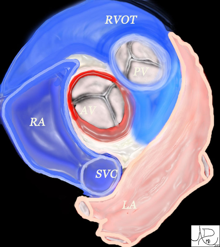
The aortic valve (AV) lies central to many structures including the right ventricular outflow tract (RVOT), pulmonary valve(PV), left atrium (LA), right atrium (RA), and superior vena cava (SVC). aorta from above
47681 heart cardiac aortic valve aorta right atrium right atrial appendage left atrial appendage LAA RAA pulmonary valve right ventricular outflow tract left atrium LA RA RVOT anatomy normal position relations Davidoff art Davidoff MD


    [nodeType] => 1
    [parentNode] => (object value omitted)
    [childNodes] => (object value omitted)
    [firstChild] => (object value omitted)
    [lastChild] => (object value omitted)
    [previousSibling] =>
    [nextSibling] => (object value omitted)
    [attributes] => (object value omitted)
    [ownerDocument] => (object value omitted)
    [namespaceURI] =>
    [prefix] =>
    [localName] => table
    [baseURI] =>
    [textContent] =>


Aortic Valve amd Pulmonary Valve – Oblique Section through the Valves


The aortic valve (AV) lies central to many structures including the right ventricular outflow tract (RVOT), pulmonary valve(PV), left atrium (LA), right atrium (RA), and superior vena cava (SVC). aorta from above
47681 heart cardiac aortic valve aorta right atrium right atrial appendage left atrial appendage LAA RAA pulmonary valve right ventricular outflow tract left atrium LA RA RVOT anatomy normal position relations Davidoff art Davidoff MD


)
DOMElement Object
(
    [schemaTypeInfo] =>
    [tagName] => td
    [firstElementChild] => (object value omitted)
    [lastElementChild] => (object value omitted)
    [childElementCount] => 2
    [previousElementSibling] =>
    [nextElementSibling] =>
    [nodeName] => td
    [nodeValue] => The aortic valve (AV) lies central to many structures including the right ventricular outflow tract (RVOT), pulmonary valve(PV), left atrium (LA), right atrium (RA), and superior vena cava (SVC). aorta from above
47681 heart cardiac aortic valve aorta right atrium right atrial appendage left atrial appendage LAA RAA pulmonary valve right ventricular outflow tract left atrium LA RA RVOT anatomy normal position relations Davidoff art Davidoff MD

    [nodeType] => 1
    [parentNode] => (object value omitted)
    [childNodes] => (object value omitted)
    [firstChild] => (object value omitted)
    [lastChild] => (object value omitted)
    [previousSibling] => (object value omitted)
    [nextSibling] => (object value omitted)
    [attributes] => (object value omitted)
    [ownerDocument] => (object value omitted)
    [namespaceURI] =>
    [prefix] =>
    [localName] => td
    [baseURI] =>
    [textContent] => The aortic valve (AV) lies central to many structures including the right ventricular outflow tract (RVOT), pulmonary valve(PV), left atrium (LA), right atrium (RA), and superior vena cava (SVC). aorta from above
47681 heart cardiac aortic valve aorta right atrium right atrial appendage left atrial appendage LAA RAA pulmonary valve right ventricular outflow tract left atrium LA RA RVOT anatomy normal position relations Davidoff art Davidoff MD

)
DOMElement Object
(
    [schemaTypeInfo] =>
    [tagName] => td
    [firstElementChild] => (object value omitted)
    [lastElementChild] => (object value omitted)
    [childElementCount] => 2
    [previousElementSibling] =>
    [nextElementSibling] =>
    [nodeName] => td
    [nodeValue] =>
Aortic Valve amd Pulmonary Valve – Oblique Section through the Valves
    [nodeType] => 1
    [parentNode] => (object value omitted)
    [childNodes] => (object value omitted)
    [firstChild] => (object value omitted)
    [lastChild] => (object value omitted)
    [previousSibling] => (object value omitted)
    [nextSibling] => (object value omitted)
    [attributes] => (object value omitted)
    [ownerDocument] => (object value omitted)
    [namespaceURI] =>
    [prefix] =>
    [localName] => td
    [baseURI] =>
    [textContent] =>
Aortic Valve amd Pulmonary Valve – Oblique Section through the Valves
)

