The gallbladder is nestled in the gallbladder fossa located on the undersurface of the liver between the right and left lobes.
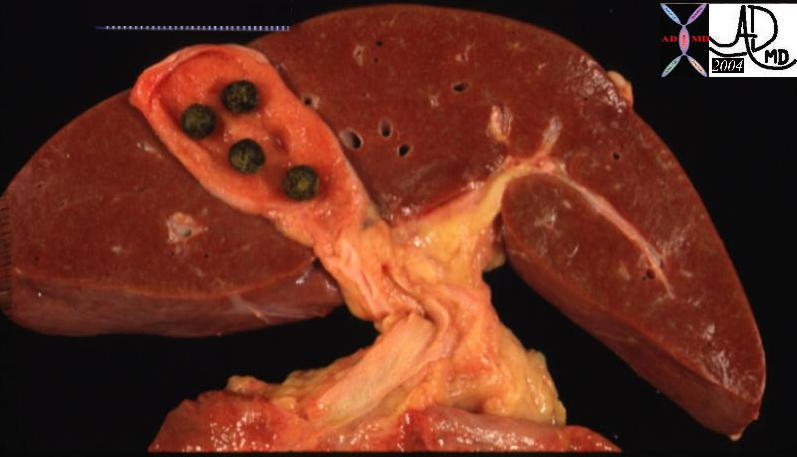
Position of the Gallbladder (with Stones) – between Right and Left Lobes of the Liver
|
| 13456 liver normal anatomy gallbladder stones cholelithiasis grosspathology |
If we remove the gallbladder we expose the gallbladder fossa.
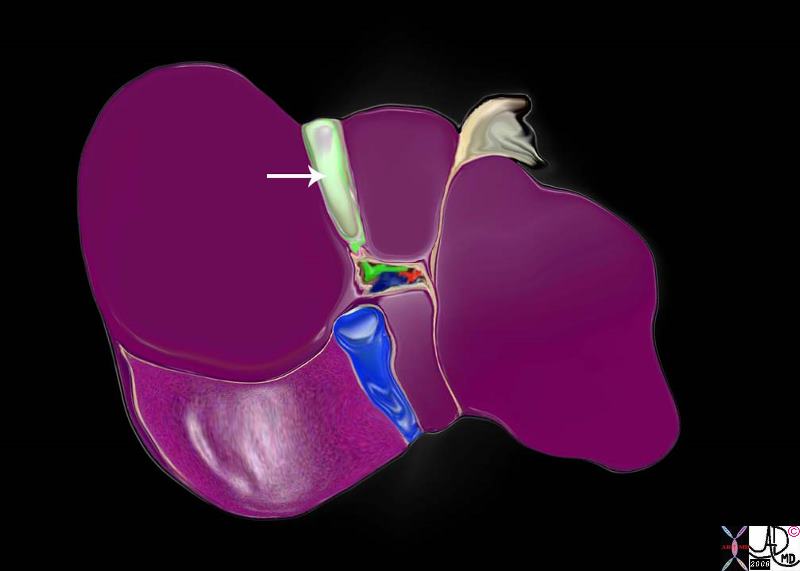
The Gallbladder Fossa on the Undersurface of the Gallbladder
|
|
The gallbladder fossa is a long and relatively narrow bed within which the gallbladder lies. It consists of loose connective tissue and vessels that anchor and connect the gallbladder to the liver.
82220b05.8s liver gallbladder porta hepatis hepatic artery portal vein IVC inferior vena cava falciform ligament ligamentum teres bare area of the liver left lobe segment IV segment I caudate lobe quadrate lobe gastrohepatic ligament right lobe hepatic Davidoff art copyright 2008
|
The liver is divided into 5 major segments based on the hepatic venous anatomy .(Couinard) The gallbladder lies between segment V of the right lobe and segment IVb (aka quadrate lobe or the medial segment of the left lobe ).
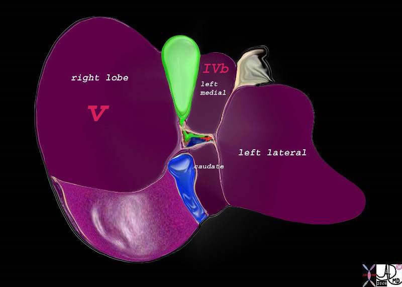
Undersurface of the Liver
|
|
Using surgical terminology of Couinard the gallbladder more speifically is situated between the the right lobe (segment V) and the medial segment odf the left lobe (segment IVb)
82220b01.2k05.82s liver gallbladder porta hepatis hepatic artery portal vein IVC inferior vena cava falciform ligament ligamentum teres bare area of the liver left lobe segment IV segment I caudate lobe quadrate lobe medial segment left ;obe lateral segment left lobe gastrohepatic ligament gallbladder right lobe hepatic Davidoff art copyright 2008 |
Its position and axis can also be described as lying within Cantlie’s line which is an oblique plane drawn from the middle of the gallbladder to left border of the inferior vena cava – generally and roughly considered a line of division between the right and left lobes.
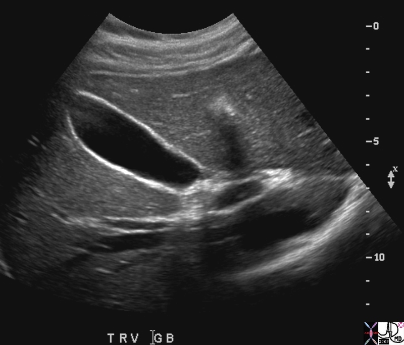 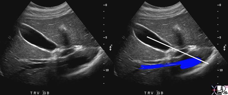
Cantlie’s Line
Transverse Section of the Gallbladder Dividing Liver into Left and Right Lobes
|
|
The ultrasound image taken in the transverse plane shows the gallbladder positioned between the right and left lobes. Cantlie’s line is an oblique plane drawn from the middle of the gallbladder to left border of the inferior vena cava – generally considered a line of division between the right and left lobes.
71327s gallbladder liver left lobe right lobe position interlobar fissure middle hepatic vein normal anatomy USscan copyright 2008 Courtesy Ashley Davidoff MD |
The plane of the gallbladder also lies in the same plane as the middle hepatic vein which is the true and more accurate division of the right and left lobes.
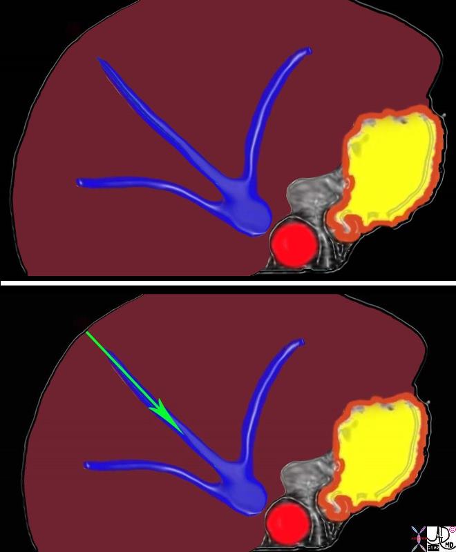
Middle Hepatic Vein, Plane of the Gallbladder |
| 82221c.8s liver middle hepatic vein normal anatomy right lobe left lobe parts anatmical classification overlay |
The fundus of the gallbladder is the most anterior, lateral, and inferior part of the gallbladder.
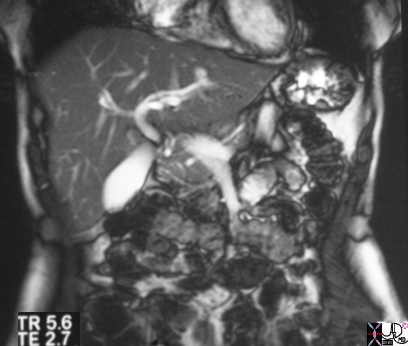
Position of the Gallbladder
|
|
This T2 weighted MRI is acquired in the coronal plane showing the gallbladder as an oval white structure. The fundus is lateral and inferior relative to the body and neck which lie medial and posterior
28839.8s gallbladder liver normal anatomy MRI T2 weighted Courtesy Ashley Davidoff MD copyright 2008 |
The fundus lies just under the anterior abdominal wall and is the only portion of the gallbladder that lies unprotected. The exposed position of the fundus is well known to examining clinical hand as the being ” below the liver edge, in the midclavicular line, just below the 9the rib.”
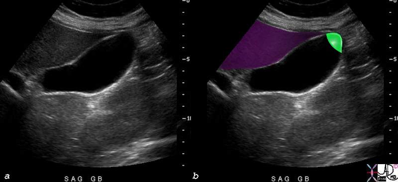
Normal Position of the Fasting Gallbladder on Ultrasound
|
|
The ultrasound reveals a longitudinal view of the gallbladder in the fasting state. The fundus is anterior to the body which in turn is anterior to the neck of the gallbladder. Only a small portion of the fundus (green in b) is exposed. The rest of the gallbladder lies posterior to the liver and are relatively well protected by it.
82021c01.8s gallbladder normal anatomy size shape position character width 3.5cms length = 10cms pear shape pyriform shape fundus body neck fundus anterior neck posterior USscan ultrasound Courtesy Ashley Davidoff MD copyright 2008 pear shape |
The body and neck lie medial, posterior, and superior to the fundus. In this position they are relatively protected by the liver. This is an important consideration since leakage of bile from traumatic injury into the peritoneal cavity can result in devastating and sometimes fatal peritonitis.
The position of the parts of the gallbladder in relation to each other is also very important in the functioning of the gallbladder. The body and and fundus lie inferior to the neck and they accumulate, concentrate and store bile making its SG a slightly higher value than the “new” incoming bile. As bile is concentrated it sinks to the dependant portion of the gallbladder, which in the upright position is in the fundus. Stratification thus occurs dividing the bile into “old”, “new”, and “fresh”, each relatively positioned in fundus, body and neck.

Inferior Positioning of the Gallbladder Allows Old Bile to be Stored and Concentrated
|
|
This ultrasound of a normal gallbladder of a 20 year old male has been turned to simulate a standing and or seated position which is the dominant position during the day. The inferiorly positioned fundus and body enable a stratification of new and old bile. With the person in the standing or sitting position, the shape of this gallbladder allows new bile to come into the neck and then quickly spill over into the body where it will become layered on the top of the old bile since it has a SG that is lower than the concentrated bile. It will be the “new” within the body and fundus, and will then be subjected to concentration. Thus the position of the gallbladder is designed to optimize the function.
82257c06b04.8s 20 male gallbladder normal position function storage concentration fundus inferior shape anatomy USscan ultrasound Courtesy Ashley Davidoff MD copyright 2008
|
Position of the Gallbladder in Relation to Other Structures
The gallbladder’s immediate neighbours besides the the liver lobes, include the porta hepatis which contains the portal triad (hepatic artery, portal vein, bile duct), lyphatics and nerves, surrounded by Glisson’s capsule (extension of the gastrohepatic ligament). More posteriorly the intrahepatic portion of the IVC is present.
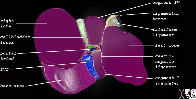
Surrounding Structures
|
| 82220b06.8s liver gallbladder porta hepatis hepatic artery portal vein IVC inferior vena cava falciform ligament ligamentum teres bare area of the liver left lobe segment IV segment I caudate lobe quadrate lobe gastrohepatic ligament gallbladder right lobe hepatic IVC Davidoff art copyright 2008 |
Outside of the liver, an inferior neighour of the fundus is the hepatic flexure, while the neck and body lie in close association to the descending portion of the duodenum.
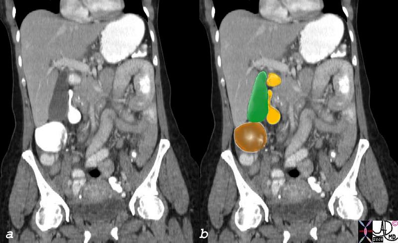
Relation to the Colon and Duodenum
|
| The gallbladder (green) is seen in close relation to the hepatic flexure (dark orange) and the descending duodenum (light orange)
37957c01.8s gallbladder position relations colon duodenum abdomen normal anatomy CTscan Courtesy Ashley Davidoff MD copyright 2008 |
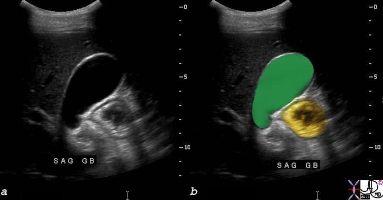
Relation to the Antrum and Duodenum
|
| The ultrasound in this instance demonstrates a transverse view of the gallbladder (green) with antrum or duodenum (orange) noted medial to the body and neck of the gallbladder.
71326c01.8s gallbladder antrum duodenum anatomy normal relations position Courtesy Ashley Davidoff MD copyright 200871326c01.8s |
Applied Biology
The gallbladder is not usually papable but when enlarged it is felt just below the liver edge in the midclavicular line, below the 9th rib.
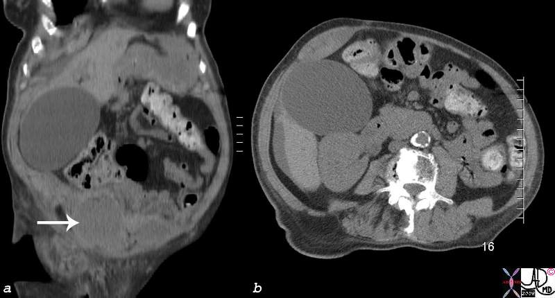
Significantly Enlarged Gallbladder that was Clinically Palpable
|
| The sagittal image (a) from a CTscan, and the transverse image reveal an enormous ballon like rotund gallbladder that was clinically palpable. This 80 year old female had recently undergone cardiac catherization and had a bleed (arrow) into the right rectus sheath.
82304c01.8s 80f s/p cardiac cath spontaneous anterior wall muscle bleed (arrow) distended gallbladder huge large dilated distended cholestasis CTscan copyright 2008 Courtesy Ashley Davidoff MD |
Courvoisier’s sign is a palpable gallbladder in the presence of obstructive jaundice most commonly caused by pancreatic cancer, but sometimes caused by a cancer at the ampulla..
The close relationship of the gallbladder to the liver, creates a potential pathway for the spread of disease. Thus exension of the inflammatory and infectious process of acute cholecystids will be identified in the liver.
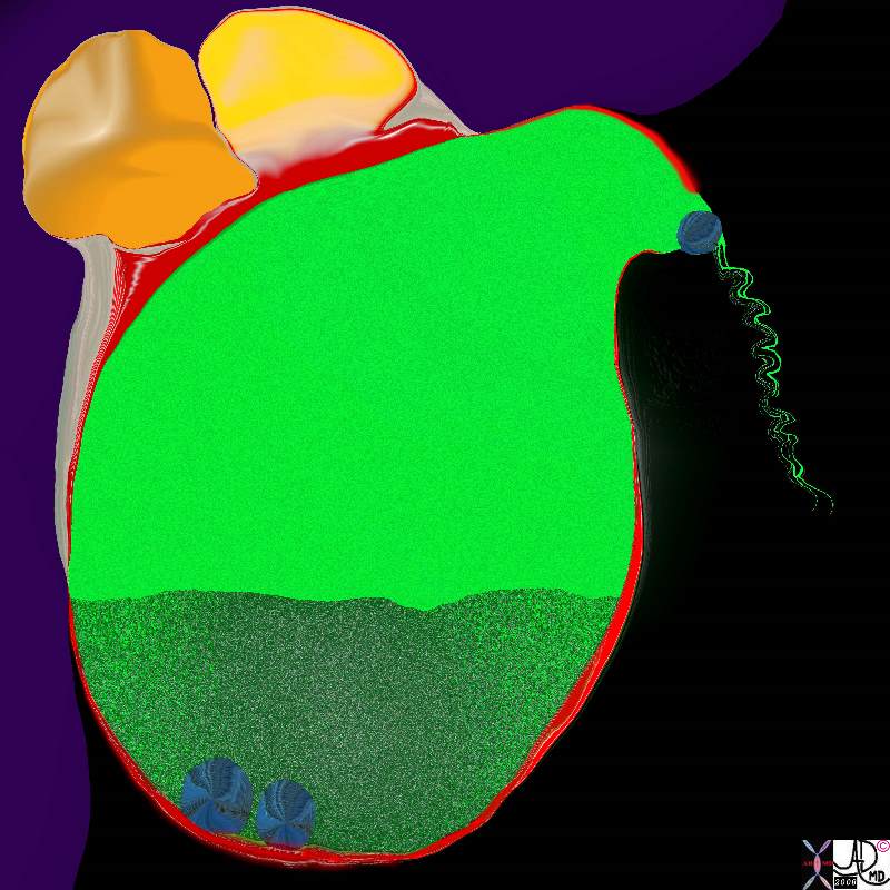
Cholecystitis Complicated by Abscess Formation in the Gallbladder Fossa
|
|
The artistic rendition shows 2 stones lying free in the lumen and one impacted in the neck of the gallbladder with secondary inflammatory changes in the wall(red) and abscess formation in the gallbladder fossa and extension into the neighbouring liver.
11921.8b05b037.8s gallbladder cystic duct gallstones cholelithiasis stone impacted in the cystic duct distended enlarged hyperemic wall complex fluid in the gallbladder fossa tumefactive bile cholestasis sludge acute cholecytitis abscess formation Davidoff Art copyright 2008 |
Similalrly when gallbladder cancer spreads, its closest neighbour, the liver is invvolved, though sometimes, and usually at a later stage the colon may be involved as well. The reverse is also true when colon carcinoma of the hepatic flexure can spread to the gallbladder.
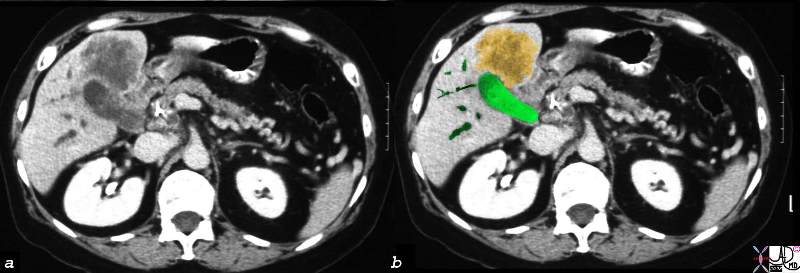
Direct Invasion into the Liver and Bile Duct Obstruction
|
|
A gallbladder cancer (orange) has extended from the gallbladder (green) directly into the liver. Associated ductal obstruction is present.
16254c02b.8s gallbladder anterior wall liver invasion space occupatopn obstruction bile ducts aggressive gallbladder carcinoma complicated by direct invasion metastasis liver windows narroe windws tumor settings gallbladder fossa GBF CTscan Courtesy Ashley Davidoff copyright 2008
|
Sometimes patients are born with an abnormal position of the gallbladder. This occurs with situs inversus but also occurs in rare instances in situs ambiguus states such as asplenia and polysplenia syndromes.
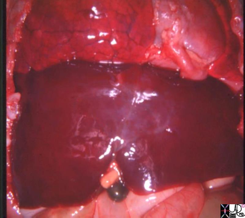
Central Gallbladder
Situs Ambiguus
|
| In this post mortem specimen of the unfortnate child with asplenia syndrome the gallbladder is noted in the middle of the body. (dark green) This ambiguus position of the gallbladder is coupled in this image with an ambiguus liver that appears to have two large right lobes. The whole entity is combined with other ambiguus anomalies such as bilateral right atria, or bilateral IVC’s for example. Many of these anomalies are incompatible with life.
82222.8s liver gallbladder bilateral right lobe asplenia syndrome Ivemark syndrome central gallbladder situs ambiguus congenital position gross pathology Courtesy Ashley DAvidoff MD copyright 2008 |
Cavity and Spaces:
The gallbladder lies within the abdominal cavity and in the peritoneal cavity on the undersurface of the right lobe of the liver in the gallbladder fossa.
Rightward and Leftward Positioning:
It is a right sided structure.
The fundus of the gallbladder usually projects below the inferior margin of the liver and lies in midclavicular line just below the right 9th rib.
Anteroposterior Positioning:
As a relatively anterior structure, it lies in close contact with the anterior abdominal wall.
Craniocaudal Positioning:
The infundibulum is medial and superior while the fundus is inferior and lateral.
Long Axis:
Its axis follows the axis of the middle hepatic vein and is obliquely and mostly vertically oriented. divides the liver into right and left lobes
The body of the gallbladder is directed upwards backward and to the left.
The neck becomes continuous with the cystic duct which turns into the lesser omentum to join the right side of the common hepatic duct to form the common bile duct.
The average position is at the level of the lower third of the second lumbar vertebra.
Touches and indents the duodenum
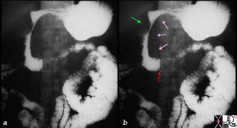
Relation to the Duodenum (Green Arrow)
|
| 01979c01.8s gallbladder duodenum extrinsic impression pancreas SMA superior mesenteric artery mass enlarged SMA syndrome X-ray upper GI barium Courtesy Ashley Davidoff copyright 2008 |
RELATIONS
Overall, the most important and relevant relations of the gallbladder are to the anterior abdominal wall, the liver surface, the portal triad, the porta heptis, amd the duodenum
Anterior Relations:
The anterior abdominal wall and the inferior surface of the liver.
Posterior Relations:
The transverse colon and the first and second parts of the duodenum.
The fundus is in contact with the anterior abdominal wall at the level of the tip of the ninth right costal cartilage.
The body lies in contact with the visceral surface of the liver.
Anatomically, the gallbladder can touch the liver, duodenum, transverse colon, and the posterior surface of the anterior abdominal wall. Because of its close proximity to all these structures and due to the fact that it does not have a natural capsule, gallbladder disease is often reflected by abnormalities in these structures. For example, in cholecystitis, the fundus can irritate the peritoneum of the anterior abdominal wall leading to a positive Murphy’s sign on deep palpation of the right upper quadrant of the abdomin as the patient inhales. Liver function tests are often abnormal in gallbladder disease. An ileus can also manifest in the setting of gallbladder problems.
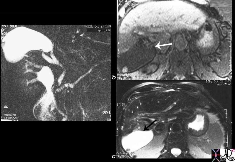
Abnormal Axis Due to Atrophy of Right Lobe – Sclerosing Cholangitis
|
| 27094c01.8s 77 male liver gallbladder bile ducts narrowed dilated atrophy right lobe abnormal position abnormal axis of the gallbladder sclerosing cholangitis MRI FSE T1 weighted T2 weighted gallstone cholelithiaisis filling defect Courtesy Ashley Davidoff MD copyright 2008 |
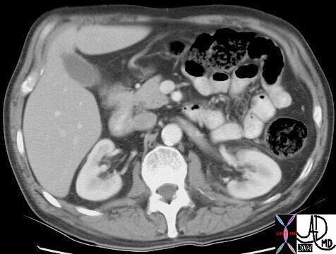 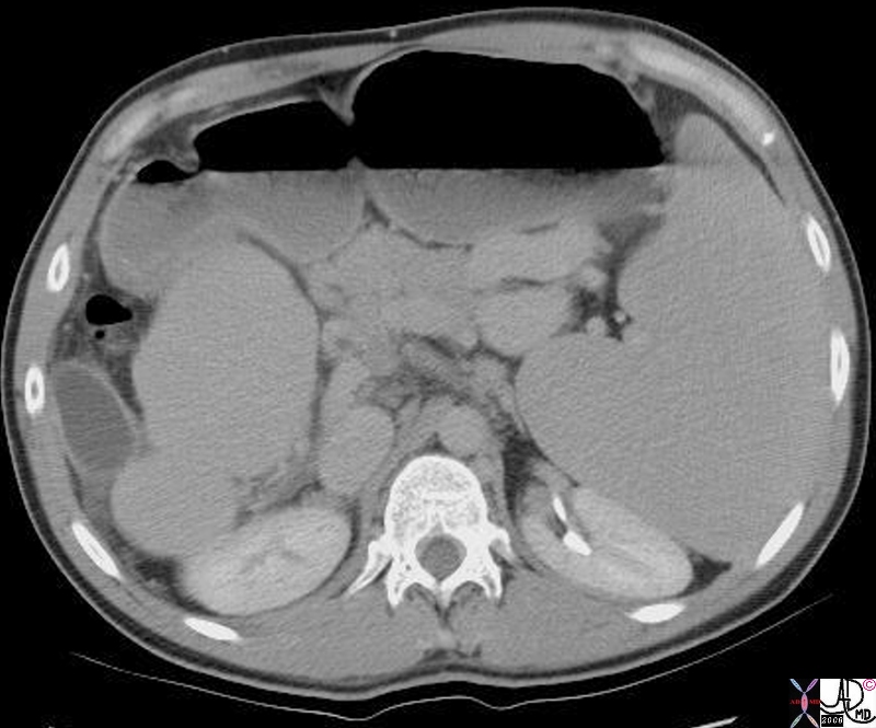
Normal and Malposition
|
|
16854 Courtesy Ashley Davidoff MD code pancreas + code liver right lobe left lobe interlobar plane fx normal + dx normal + code gallbladder imaging radiology CTscan
16792.8s gallbladder fx malposition naked dx cirrhosis sclerosing cholangitis liver imaging radiology CTscan spleen splenomegaly Courtesy Ashley DAvidoff MD copyright 2008
|
DOMElement Object
(
    [schemaTypeInfo] =>
    [tagName] => table
    [firstElementChild] => (object value omitted)
    [lastElementChild] => (object value omitted)
    [childElementCount] => 1
    [previousElementSibling] => (object value omitted)
    [nextElementSibling] => (object value omitted)
    [nodeName] => table
    [nodeValue] =>


Normal and Malposition


16854 Courtesy Ashley Davidoff MD code pancreas + code liver right lobe left lobe interlobar plane fx normal + dx normal + code gallbladder imaging radiology CTscan
16792.8s gallbladder fx malposition naked dx cirrhosis sclerosing cholangitis liver imaging radiology CTscan spleen splenomegaly Courtesy Ashley DAvidoff MD copyright 2008


    [nodeType] => 1
    [parentNode] => (object value omitted)
    [childNodes] => (object value omitted)
    [firstChild] => (object value omitted)
    [lastChild] => (object value omitted)
    [previousSibling] => (object value omitted)
    [nextSibling] => (object value omitted)
    [attributes] => (object value omitted)
    [ownerDocument] => (object value omitted)
    [namespaceURI] =>
    [prefix] =>
    [localName] => table
    [baseURI] =>
    [textContent] =>


Normal and Malposition


16854 Courtesy Ashley Davidoff MD code pancreas + code liver right lobe left lobe interlobar plane fx normal + dx normal + code gallbladder imaging radiology CTscan
16792.8s gallbladder fx malposition naked dx cirrhosis sclerosing cholangitis liver imaging radiology CTscan spleen splenomegaly Courtesy Ashley DAvidoff MD copyright 2008


)
DOMElement Object
(
    [schemaTypeInfo] =>
    [tagName] => td
    [firstElementChild] => (object value omitted)
    [lastElementChild] => (object value omitted)
    [childElementCount] => 2
    [previousElementSibling] =>
    [nextElementSibling] =>
    [nodeName] => td
    [nodeValue] =>
16854 Courtesy Ashley Davidoff MD code pancreas + code liver right lobe left lobe interlobar plane fx normal + dx normal + code gallbladder imaging radiology CTscan
16792.8s gallbladder fx malposition naked dx cirrhosis sclerosing cholangitis liver imaging radiology CTscan spleen splenomegaly Courtesy Ashley DAvidoff MD copyright 2008

    [nodeType] => 1
    [parentNode] => (object value omitted)
    [childNodes] => (object value omitted)
    [firstChild] => (object value omitted)
    [lastChild] => (object value omitted)
    [previousSibling] => (object value omitted)
    [nextSibling] => (object value omitted)
    [attributes] => (object value omitted)
    [ownerDocument] => (object value omitted)
    [namespaceURI] =>
    [prefix] =>
    [localName] => td
    [baseURI] =>
    [textContent] =>
16854 Courtesy Ashley Davidoff MD code pancreas + code liver right lobe left lobe interlobar plane fx normal + dx normal + code gallbladder imaging radiology CTscan
16792.8s gallbladder fx malposition naked dx cirrhosis sclerosing cholangitis liver imaging radiology CTscan spleen splenomegaly Courtesy Ashley DAvidoff MD copyright 2008

)
DOMElement Object
(
    [schemaTypeInfo] =>
    [tagName] => td
    [firstElementChild] => (object value omitted)
    [lastElementChild] => (object value omitted)
    [childElementCount] => 3
    [previousElementSibling] =>
    [nextElementSibling] =>
    [nodeName] => td
    [nodeValue] =>
Normal and Malposition

    [nodeType] => 1
    [parentNode] => (object value omitted)
    [childNodes] => (object value omitted)
    [firstChild] => (object value omitted)
    [lastChild] => (object value omitted)
    [previousSibling] => (object value omitted)
    [nextSibling] => (object value omitted)
    [attributes] => (object value omitted)
    [ownerDocument] => (object value omitted)
    [namespaceURI] =>
    [prefix] =>
    [localName] => td
    [baseURI] =>
    [textContent] =>
Normal and Malposition

)
DOMElement Object
(
    [schemaTypeInfo] =>
    [tagName] => table
    [firstElementChild] => (object value omitted)
    [lastElementChild] => (object value omitted)
    [childElementCount] => 1
    [previousElementSibling] => (object value omitted)
    [nextElementSibling] => (object value omitted)
    [nodeName] => table
    [nodeValue] =>


Abnormal Axis Due to Atrophy of Right Lobe – Sclerosing Cholangitis


27094c01.8s 77 male liver gallbladder bile ducts narrowed dilated atrophy right lobe abnormal position abnormal axis of the gallbladder sclerosing cholangitis MRI FSE T1 weighted T2 weighted gallstone cholelithiaisis filling defect Courtesy Ashley Davidoff MD copyright 2008


    [nodeType] => 1
    [parentNode] => (object value omitted)
    [childNodes] => (object value omitted)
    [firstChild] => (object value omitted)
    [lastChild] => (object value omitted)
    [previousSibling] => (object value omitted)
    [nextSibling] => (object value omitted)
    [attributes] => (object value omitted)
    [ownerDocument] => (object value omitted)
    [namespaceURI] =>
    [prefix] =>
    [localName] => table
    [baseURI] =>
    [textContent] =>


Abnormal Axis Due to Atrophy of Right Lobe – Sclerosing Cholangitis


27094c01.8s 77 male liver gallbladder bile ducts narrowed dilated atrophy right lobe abnormal position abnormal axis of the gallbladder sclerosing cholangitis MRI FSE T1 weighted T2 weighted gallstone cholelithiaisis filling defect Courtesy Ashley Davidoff MD copyright 2008


)
DOMElement Object
(
    [schemaTypeInfo] =>
    [tagName] => td
    [firstElementChild] => (object value omitted)
    [lastElementChild] => (object value omitted)
    [childElementCount] => 1
    [previousElementSibling] =>
    [nextElementSibling] =>
    [nodeName] => td
    [nodeValue] => 27094c01.8s 77 male liver gallbladder bile ducts narrowed dilated atrophy right lobe abnormal position abnormal axis of the gallbladder sclerosing cholangitis MRI FSE T1 weighted T2 weighted gallstone cholelithiaisis filling defect Courtesy Ashley Davidoff MD copyright 2008
    [nodeType] => 1
    [parentNode] => (object value omitted)
    [childNodes] => (object value omitted)
    [firstChild] => (object value omitted)
    [lastChild] => (object value omitted)
    [previousSibling] => (object value omitted)
    [nextSibling] => (object value omitted)
    [attributes] => (object value omitted)
    [ownerDocument] => (object value omitted)
    [namespaceURI] =>
    [prefix] =>
    [localName] => td
    [baseURI] =>
    [textContent] => 27094c01.8s 77 male liver gallbladder bile ducts narrowed dilated atrophy right lobe abnormal position abnormal axis of the gallbladder sclerosing cholangitis MRI FSE T1 weighted T2 weighted gallstone cholelithiaisis filling defect Courtesy Ashley Davidoff MD copyright 2008
)
DOMElement Object
(
    [schemaTypeInfo] =>
    [tagName] => td
    [firstElementChild] => (object value omitted)
    [lastElementChild] => (object value omitted)
    [childElementCount] => 2
    [previousElementSibling] =>
    [nextElementSibling] =>
    [nodeName] => td
    [nodeValue] =>
Abnormal Axis Due to Atrophy of Right Lobe – Sclerosing Cholangitis

    [nodeType] => 1
    [parentNode] => (object value omitted)
    [childNodes] => (object value omitted)
    [firstChild] => (object value omitted)
    [lastChild] => (object value omitted)
    [previousSibling] => (object value omitted)
    [nextSibling] => (object value omitted)
    [attributes] => (object value omitted)
    [ownerDocument] => (object value omitted)
    [namespaceURI] =>
    [prefix] =>
    [localName] => td
    [baseURI] =>
    [textContent] =>
Abnormal Axis Due to Atrophy of Right Lobe – Sclerosing Cholangitis

)
DOMElement Object
(
    [schemaTypeInfo] =>
    [tagName] => table
    [firstElementChild] => (object value omitted)
    [lastElementChild] => (object value omitted)
    [childElementCount] => 1
    [previousElementSibling] => (object value omitted)
    [nextElementSibling] => (object value omitted)
    [nodeName] => table
    [nodeValue] =>


Relation to the Duodenum (Green Arrow)


01979c01.8s gallbladder duodenum extrinsic impression pancreas SMA superior mesenteric artery mass enlarged SMA syndrome X-ray upper GI barium Courtesy Ashley Davidoff copyright 2008


    [nodeType] => 1
    [parentNode] => (object value omitted)
    [childNodes] => (object value omitted)
    [firstChild] => (object value omitted)
    [lastChild] => (object value omitted)
    [previousSibling] => (object value omitted)
    [nextSibling] => (object value omitted)
    [attributes] => (object value omitted)
    [ownerDocument] => (object value omitted)
    [namespaceURI] =>
    [prefix] =>
    [localName] => table
    [baseURI] =>
    [textContent] =>


Relation to the Duodenum (Green Arrow)


01979c01.8s gallbladder duodenum extrinsic impression pancreas SMA superior mesenteric artery mass enlarged SMA syndrome X-ray upper GI barium Courtesy Ashley Davidoff copyright 2008


)
DOMElement Object
(
    [schemaTypeInfo] =>
    [tagName] => td
    [firstElementChild] =>
    [lastElementChild] =>
    [childElementCount] => 0
    [previousElementSibling] =>
    [nextElementSibling] =>
    [nodeName] => td
    [nodeValue] => 01979c01.8s gallbladder duodenum extrinsic impression pancreas SMA superior mesenteric artery mass enlarged SMA syndrome X-ray upper GI barium Courtesy Ashley Davidoff copyright 2008
    [nodeType] => 1
    [parentNode] => (object value omitted)
    [childNodes] => (object value omitted)
    [firstChild] => (object value omitted)
    [lastChild] => (object value omitted)
    [previousSibling] => (object value omitted)
    [nextSibling] => (object value omitted)
    [attributes] => (object value omitted)
    [ownerDocument] => (object value omitted)
    [namespaceURI] =>
    [prefix] =>
    [localName] => td
    [baseURI] =>
    [textContent] => 01979c01.8s gallbladder duodenum extrinsic impression pancreas SMA superior mesenteric artery mass enlarged SMA syndrome X-ray upper GI barium Courtesy Ashley Davidoff copyright 2008
)
DOMElement Object
(
    [schemaTypeInfo] =>
    [tagName] => td
    [firstElementChild] => (object value omitted)
    [lastElementChild] => (object value omitted)
    [childElementCount] => 2
    [previousElementSibling] =>
    [nextElementSibling] =>
    [nodeName] => td
    [nodeValue] =>
Relation to the Duodenum (Green Arrow)

    [nodeType] => 1
    [parentNode] => (object value omitted)
    [childNodes] => (object value omitted)
    [firstChild] => (object value omitted)
    [lastChild] => (object value omitted)
    [previousSibling] => (object value omitted)
    [nextSibling] => (object value omitted)
    [attributes] => (object value omitted)
    [ownerDocument] => (object value omitted)
    [namespaceURI] =>
    [prefix] =>
    [localName] => td
    [baseURI] =>
    [textContent] =>
Relation to the Duodenum (Green Arrow)

)
DOMElement Object
(
    [schemaTypeInfo] =>
    [tagName] => table
    [firstElementChild] => (object value omitted)
    [lastElementChild] => (object value omitted)
    [childElementCount] => 1
    [previousElementSibling] => (object value omitted)
    [nextElementSibling] => (object value omitted)
    [nodeName] => table
    [nodeValue] =>


Central Gallbladder
Situs Ambiguus


In this post mortem specimen of the unfortnate child with asplenia syndrome the gallbladder is noted in the middle of the body.  (dark green) This ambiguus position of the gallbladder is coupled in this image with an ambiguus liver that appears to have two large right lobes.  The whole entity is combined with other ambiguus anomalies such as bilateral right atria, or bilateral IVC’s for example.  Many of these anomalies are incompatible with life.
82222.8s liver gallbladder bilateral right lobe asplenia syndrome Ivemark syndrome central gallbladder situs ambiguus congenital position gross pathology Courtesy Ashley DAvidoff MD copyright 2008


    [nodeType] => 1
    [parentNode] => (object value omitted)
    [childNodes] => (object value omitted)
    [firstChild] => (object value omitted)
    [lastChild] => (object value omitted)
    [previousSibling] => (object value omitted)
    [nextSibling] => (object value omitted)
    [attributes] => (object value omitted)
    [ownerDocument] => (object value omitted)
    [namespaceURI] =>
    [prefix] =>
    [localName] => table
    [baseURI] =>
    [textContent] =>


Central Gallbladder
Situs Ambiguus


In this post mortem specimen of the unfortnate child with asplenia syndrome the gallbladder is noted in the middle of the body.  (dark green) This ambiguus position of the gallbladder is coupled in this image with an ambiguus liver that appears to have two large right lobes.  The whole entity is combined with other ambiguus anomalies such as bilateral right atria, or bilateral IVC’s for example.  Many of these anomalies are incompatible with life.
82222.8s liver gallbladder bilateral right lobe asplenia syndrome Ivemark syndrome central gallbladder situs ambiguus congenital position gross pathology Courtesy Ashley DAvidoff MD copyright 2008


)
DOMElement Object
(
    [schemaTypeInfo] =>
    [tagName] => td
    [firstElementChild] => (object value omitted)
    [lastElementChild] => (object value omitted)
    [childElementCount] => 2
    [previousElementSibling] =>
    [nextElementSibling] =>
    [nodeName] => td
    [nodeValue] => In this post mortem specimen of the unfortnate child with asplenia syndrome the gallbladder is noted in the middle of the body.  (dark green) This ambiguus position of the gallbladder is coupled in this image with an ambiguus liver that appears to have two large right lobes.  The whole entity is combined with other ambiguus anomalies such as bilateral right atria, or bilateral IVC’s for example.  Many of these anomalies are incompatible with life.
82222.8s liver gallbladder bilateral right lobe asplenia syndrome Ivemark syndrome central gallbladder situs ambiguus congenital position gross pathology Courtesy Ashley DAvidoff MD copyright 2008
    [nodeType] => 1
    [parentNode] => (object value omitted)
    [childNodes] => (object value omitted)
    [firstChild] => (object value omitted)
    [lastChild] => (object value omitted)
    [previousSibling] => (object value omitted)
    [nextSibling] => (object value omitted)
    [attributes] => (object value omitted)
    [ownerDocument] => (object value omitted)
    [namespaceURI] =>
    [prefix] =>
    [localName] => td
    [baseURI] =>
    [textContent] => In this post mortem specimen of the unfortnate child with asplenia syndrome the gallbladder is noted in the middle of the body.  (dark green) This ambiguus position of the gallbladder is coupled in this image with an ambiguus liver that appears to have two large right lobes.  The whole entity is combined with other ambiguus anomalies such as bilateral right atria, or bilateral IVC’s for example.  Many of these anomalies are incompatible with life.
82222.8s liver gallbladder bilateral right lobe asplenia syndrome Ivemark syndrome central gallbladder situs ambiguus congenital position gross pathology Courtesy Ashley DAvidoff MD copyright 2008
)
DOMElement Object
(
    [schemaTypeInfo] =>
    [tagName] => td
    [firstElementChild] => (object value omitted)
    [lastElementChild] => (object value omitted)
    [childElementCount] => 3
    [previousElementSibling] =>
    [nextElementSibling] =>
    [nodeName] => td
    [nodeValue] =>
Central Gallbladder
Situs Ambiguus

    [nodeType] => 1
    [parentNode] => (object value omitted)
    [childNodes] => (object value omitted)
    [firstChild] => (object value omitted)
    [lastChild] => (object value omitted)
    [previousSibling] => (object value omitted)
    [nextSibling] => (object value omitted)
    [attributes] => (object value omitted)
    [ownerDocument] => (object value omitted)
    [namespaceURI] =>
    [prefix] =>
    [localName] => td
    [baseURI] =>
    [textContent] =>
Central Gallbladder
Situs Ambiguus

)
DOMElement Object
(
    [schemaTypeInfo] =>
    [tagName] => table
    [firstElementChild] => (object value omitted)
    [lastElementChild] => (object value omitted)
    [childElementCount] => 1
    [previousElementSibling] => (object value omitted)
    [nextElementSibling] => (object value omitted)
    [nodeName] => table
    [nodeValue] =>


Direct Invasion into the Liver and Bile Duct Obstruction


A gallbladder cancer (orange) has extended from the gallbladder (green) directly into the liver.  Associated ductal obstruction is present.
16254c02b.8s gallbladder anterior wall liver invasion space occupatopn obstruction bile ducts aggressive gallbladder carcinoma complicated by direct invasion metastasis liver windows narroe windws tumor settings gallbladder fossa GBF CTscan Courtesy Ashley Davidoff copyright 2008


    [nodeType] => 1
    [parentNode] => (object value omitted)
    [childNodes] => (object value omitted)
    [firstChild] => (object value omitted)
    [lastChild] => (object value omitted)
    [previousSibling] => (object value omitted)
    [nextSibling] => (object value omitted)
    [attributes] => (object value omitted)
    [ownerDocument] => (object value omitted)
    [namespaceURI] =>
    [prefix] =>
    [localName] => table
    [baseURI] =>
    [textContent] =>


Direct Invasion into the Liver and Bile Duct Obstruction


A gallbladder cancer (orange) has extended from the gallbladder (green) directly into the liver.  Associated ductal obstruction is present.
16254c02b.8s gallbladder anterior wall liver invasion space occupatopn obstruction bile ducts aggressive gallbladder carcinoma complicated by direct invasion metastasis liver windows narroe windws tumor settings gallbladder fossa GBF CTscan Courtesy Ashley Davidoff copyright 2008


)
DOMElement Object
(
    [schemaTypeInfo] =>
    [tagName] => td
    [firstElementChild] => (object value omitted)
    [lastElementChild] => (object value omitted)
    [childElementCount] => 2
    [previousElementSibling] =>
    [nextElementSibling] =>
    [nodeName] => td
    [nodeValue] =>
A gallbladder cancer (orange) has extended from the gallbladder (green) directly into the liver.  Associated ductal obstruction is present.
16254c02b.8s gallbladder anterior wall liver invasion space occupatopn obstruction bile ducts aggressive gallbladder carcinoma complicated by direct invasion metastasis liver windows narroe windws tumor settings gallbladder fossa GBF CTscan Courtesy Ashley Davidoff copyright 2008

    [nodeType] => 1
    [parentNode] => (object value omitted)
    [childNodes] => (object value omitted)
    [firstChild] => (object value omitted)
    [lastChild] => (object value omitted)
    [previousSibling] => (object value omitted)
    [nextSibling] => (object value omitted)
    [attributes] => (object value omitted)
    [ownerDocument] => (object value omitted)
    [namespaceURI] =>
    [prefix] =>
    [localName] => td
    [baseURI] =>
    [textContent] =>
A gallbladder cancer (orange) has extended from the gallbladder (green) directly into the liver.  Associated ductal obstruction is present.
16254c02b.8s gallbladder anterior wall liver invasion space occupatopn obstruction bile ducts aggressive gallbladder carcinoma complicated by direct invasion metastasis liver windows narroe windws tumor settings gallbladder fossa GBF CTscan Courtesy Ashley Davidoff copyright 2008

)
DOMElement Object
(
    [schemaTypeInfo] =>
    [tagName] => td
    [firstElementChild] => (object value omitted)
    [lastElementChild] => (object value omitted)
    [childElementCount] => 2
    [previousElementSibling] =>
    [nextElementSibling] =>
    [nodeName] => td
    [nodeValue] =>
Direct Invasion into the Liver and Bile Duct Obstruction

    [nodeType] => 1
    [parentNode] => (object value omitted)
    [childNodes] => (object value omitted)
    [firstChild] => (object value omitted)
    [lastChild] => (object value omitted)
    [previousSibling] => (object value omitted)
    [nextSibling] => (object value omitted)
    [attributes] => (object value omitted)
    [ownerDocument] => (object value omitted)
    [namespaceURI] =>
    [prefix] =>
    [localName] => td
    [baseURI] =>
    [textContent] =>
Direct Invasion into the Liver and Bile Duct Obstruction

)
DOMElement Object
(
    [schemaTypeInfo] =>
    [tagName] => table
    [firstElementChild] => (object value omitted)
    [lastElementChild] => (object value omitted)
    [childElementCount] => 1
    [previousElementSibling] => (object value omitted)
    [nextElementSibling] => (object value omitted)
    [nodeName] => table
    [nodeValue] =>


Cholecystitis Complicated by Abscess Formation in the Gallbladder Fossa


The artistic rendition shows 2 stones lying free in the lumen and one impacted in the neck of the gallbladder with secondary inflammatory changes in the wall(red) and abscess formation in the gallbladder fossa and extension into the neighbouring liver.
11921.8b05b037.8s gallbladder cystic duct gallstones cholelithiasis stone impacted in the cystic duct distended enlarged hyperemic wall complex fluid in the gallbladder fossa tumefactive bile cholestasis sludge acute cholecytitis abscess formation Davidoff Art copyright 2008


    [nodeType] => 1
    [parentNode] => (object value omitted)
    [childNodes] => (object value omitted)
    [firstChild] => (object value omitted)
    [lastChild] => (object value omitted)
    [previousSibling] => (object value omitted)
    [nextSibling] => (object value omitted)
    [attributes] => (object value omitted)
    [ownerDocument] => (object value omitted)
    [namespaceURI] =>
    [prefix] =>
    [localName] => table
    [baseURI] =>
    [textContent] =>


Cholecystitis Complicated by Abscess Formation in the Gallbladder Fossa


The artistic rendition shows 2 stones lying free in the lumen and one impacted in the neck of the gallbladder with secondary inflammatory changes in the wall(red) and abscess formation in the gallbladder fossa and extension into the neighbouring liver.
11921.8b05b037.8s gallbladder cystic duct gallstones cholelithiasis stone impacted in the cystic duct distended enlarged hyperemic wall complex fluid in the gallbladder fossa tumefactive bile cholestasis sludge acute cholecytitis abscess formation Davidoff Art copyright 2008


)
DOMElement Object
(
    [schemaTypeInfo] =>
    [tagName] => td
    [firstElementChild] => (object value omitted)
    [lastElementChild] => (object value omitted)
    [childElementCount] => 2
    [previousElementSibling] =>
    [nextElementSibling] =>
    [nodeName] => td
    [nodeValue] =>
The artistic rendition shows 2 stones lying free in the lumen and one impacted in the neck of the gallbladder with secondary inflammatory changes in the wall(red) and abscess formation in the gallbladder fossa and extension into the neighbouring liver.
11921.8b05b037.8s gallbladder cystic duct gallstones cholelithiasis stone impacted in the cystic duct distended enlarged hyperemic wall complex fluid in the gallbladder fossa tumefactive bile cholestasis sludge acute cholecytitis abscess formation Davidoff Art copyright 2008
    [nodeType] => 1
    [parentNode] => (object value omitted)
    [childNodes] => (object value omitted)
    [firstChild] => (object value omitted)
    [lastChild] => (object value omitted)
    [previousSibling] => (object value omitted)
    [nextSibling] => (object value omitted)
    [attributes] => (object value omitted)
    [ownerDocument] => (object value omitted)
    [namespaceURI] =>
    [prefix] =>
    [localName] => td
    [baseURI] =>
    [textContent] =>
The artistic rendition shows 2 stones lying free in the lumen and one impacted in the neck of the gallbladder with secondary inflammatory changes in the wall(red) and abscess formation in the gallbladder fossa and extension into the neighbouring liver.
11921.8b05b037.8s gallbladder cystic duct gallstones cholelithiasis stone impacted in the cystic duct distended enlarged hyperemic wall complex fluid in the gallbladder fossa tumefactive bile cholestasis sludge acute cholecytitis abscess formation Davidoff Art copyright 2008
)
DOMElement Object
(
    [schemaTypeInfo] =>
    [tagName] => td
    [firstElementChild] => (object value omitted)
    [lastElementChild] => (object value omitted)
    [childElementCount] => 2
    [previousElementSibling] =>
    [nextElementSibling] =>
    [nodeName] => td
    [nodeValue] =>
Cholecystitis Complicated by Abscess Formation in the Gallbladder Fossa

    [nodeType] => 1
    [parentNode] => (object value omitted)
    [childNodes] => (object value omitted)
    [firstChild] => (object value omitted)
    [lastChild] => (object value omitted)
    [previousSibling] => (object value omitted)
    [nextSibling] => (object value omitted)
    [attributes] => (object value omitted)
    [ownerDocument] => (object value omitted)
    [namespaceURI] =>
    [prefix] =>
    [localName] => td
    [baseURI] =>
    [textContent] =>
Cholecystitis Complicated by Abscess Formation in the Gallbladder Fossa

)
DOMElement Object
(
    [schemaTypeInfo] =>
    [tagName] => table
    [firstElementChild] => (object value omitted)
    [lastElementChild] => (object value omitted)
    [childElementCount] => 1
    [previousElementSibling] => (object value omitted)
    [nextElementSibling] => (object value omitted)
    [nodeName] => table
    [nodeValue] =>


Significantly Enlarged Gallbladder that was Clinically Palpable


The sagittal image (a) from a CTscan, and the transverse image reveal an enormous ballon like rotund gallbladder that was clinically palpable. This 80 year old female had recently undergone cardiac catherization and had a bleed (arrow) into the right rectus sheath.
82304c01.8s 80f s/p cardiac cath spontaneous anterior wall muscle bleed (arrow) distended gallbladder huge large dilated distended cholestasis CTscan copyright 2008 Courtesy Ashley Davidoff MD


    [nodeType] => 1
    [parentNode] => (object value omitted)
    [childNodes] => (object value omitted)
    [firstChild] => (object value omitted)
    [lastChild] => (object value omitted)
    [previousSibling] => (object value omitted)
    [nextSibling] => (object value omitted)
    [attributes] => (object value omitted)
    [ownerDocument] => (object value omitted)
    [namespaceURI] =>
    [prefix] =>
    [localName] => table
    [baseURI] =>
    [textContent] =>


Significantly Enlarged Gallbladder that was Clinically Palpable


The sagittal image (a) from a CTscan, and the transverse image reveal an enormous ballon like rotund gallbladder that was clinically palpable. This 80 year old female had recently undergone cardiac catherization and had a bleed (arrow) into the right rectus sheath.
82304c01.8s 80f s/p cardiac cath spontaneous anterior wall muscle bleed (arrow) distended gallbladder huge large dilated distended cholestasis CTscan copyright 2008 Courtesy Ashley Davidoff MD


)
DOMElement Object
(
    [schemaTypeInfo] =>
    [tagName] => td
    [firstElementChild] => (object value omitted)
    [lastElementChild] => (object value omitted)
    [childElementCount] => 2
    [previousElementSibling] =>
    [nextElementSibling] =>
    [nodeName] => td
    [nodeValue] => The sagittal image (a) from a CTscan, and the transverse image reveal an enormous ballon like rotund gallbladder that was clinically palpable. This 80 year old female had recently undergone cardiac catherization and had a bleed (arrow) into the right rectus sheath.
82304c01.8s 80f s/p cardiac cath spontaneous anterior wall muscle bleed (arrow) distended gallbladder huge large dilated distended cholestasis CTscan copyright 2008 Courtesy Ashley Davidoff MD
    [nodeType] => 1
    [parentNode] => (object value omitted)
    [childNodes] => (object value omitted)
    [firstChild] => (object value omitted)
    [lastChild] => (object value omitted)
    [previousSibling] => (object value omitted)
    [nextSibling] => (object value omitted)
    [attributes] => (object value omitted)
    [ownerDocument] => (object value omitted)
    [namespaceURI] =>
    [prefix] =>
    [localName] => td
    [baseURI] =>
    [textContent] => The sagittal image (a) from a CTscan, and the transverse image reveal an enormous ballon like rotund gallbladder that was clinically palpable. This 80 year old female had recently undergone cardiac catherization and had a bleed (arrow) into the right rectus sheath.
82304c01.8s 80f s/p cardiac cath spontaneous anterior wall muscle bleed (arrow) distended gallbladder huge large dilated distended cholestasis CTscan copyright 2008 Courtesy Ashley Davidoff MD
)
DOMElement Object
(
    [schemaTypeInfo] =>
    [tagName] => td
    [firstElementChild] => (object value omitted)
    [lastElementChild] => (object value omitted)
    [childElementCount] => 2
    [previousElementSibling] =>
    [nextElementSibling] =>
    [nodeName] => td
    [nodeValue] =>
Significantly Enlarged Gallbladder that was Clinically Palpable

    [nodeType] => 1
    [parentNode] => (object value omitted)
    [childNodes] => (object value omitted)
    [firstChild] => (object value omitted)
    [lastChild] => (object value omitted)
    [previousSibling] => (object value omitted)
    [nextSibling] => (object value omitted)
    [attributes] => (object value omitted)
    [ownerDocument] => (object value omitted)
    [namespaceURI] =>
    [prefix] =>
    [localName] => td
    [baseURI] =>
    [textContent] =>
Significantly Enlarged Gallbladder that was Clinically Palpable

)
DOMElement Object
(
    [schemaTypeInfo] =>
    [tagName] => table
    [firstElementChild] => (object value omitted)
    [lastElementChild] => (object value omitted)
    [childElementCount] => 1
    [previousElementSibling] => (object value omitted)
    [nextElementSibling] => (object value omitted)
    [nodeName] => table
    [nodeValue] =>


Relation to the Antrum  and Duodenum


The ultrasound in this instance demonstrates a transverse view of the gallbladder (green) with antrum or duodenum (orange) noted medial to the body and neck of the gallbladder.
71326c01.8s gallbladder antrum duodenum anatomy normal relations position Courtesy Ashley Davidoff MD copyright 200871326c01.8s


    [nodeType] => 1
    [parentNode] => (object value omitted)
    [childNodes] => (object value omitted)
    [firstChild] => (object value omitted)
    [lastChild] => (object value omitted)
    [previousSibling] => (object value omitted)
    [nextSibling] => (object value omitted)
    [attributes] => (object value omitted)
    [ownerDocument] => (object value omitted)
    [namespaceURI] =>
    [prefix] =>
    [localName] => table
    [baseURI] =>
    [textContent] =>


Relation to the Antrum  and Duodenum


The ultrasound in this instance demonstrates a transverse view of the gallbladder (green) with antrum or duodenum (orange) noted medial to the body and neck of the gallbladder.
71326c01.8s gallbladder antrum duodenum anatomy normal relations position Courtesy Ashley Davidoff MD copyright 200871326c01.8s


)
DOMElement Object
(
    [schemaTypeInfo] =>
    [tagName] => td
    [firstElementChild] => (object value omitted)
    [lastElementChild] => (object value omitted)
    [childElementCount] => 2
    [previousElementSibling] =>
    [nextElementSibling] =>
    [nodeName] => td
    [nodeValue] => The ultrasound in this instance demonstrates a transverse view of the gallbladder (green) with antrum or duodenum (orange) noted medial to the body and neck of the gallbladder.
71326c01.8s gallbladder antrum duodenum anatomy normal relations position Courtesy Ashley Davidoff MD copyright 200871326c01.8s
    [nodeType] => 1
    [parentNode] => (object value omitted)
    [childNodes] => (object value omitted)
    [firstChild] => (object value omitted)
    [lastChild] => (object value omitted)
    [previousSibling] => (object value omitted)
    [nextSibling] => (object value omitted)
    [attributes] => (object value omitted)
    [ownerDocument] => (object value omitted)
    [namespaceURI] =>
    [prefix] =>
    [localName] => td
    [baseURI] =>
    [textContent] => The ultrasound in this instance demonstrates a transverse view of the gallbladder (green) with antrum or duodenum (orange) noted medial to the body and neck of the gallbladder.
71326c01.8s gallbladder antrum duodenum anatomy normal relations position Courtesy Ashley Davidoff MD copyright 200871326c01.8s
)
DOMElement Object
(
    [schemaTypeInfo] =>
    [tagName] => td
    [firstElementChild] => (object value omitted)
    [lastElementChild] => (object value omitted)
    [childElementCount] => 2
    [previousElementSibling] =>
    [nextElementSibling] =>
    [nodeName] => td
    [nodeValue] =>
Relation to the Antrum  and Duodenum

    [nodeType] => 1
    [parentNode] => (object value omitted)
    [childNodes] => (object value omitted)
    [firstChild] => (object value omitted)
    [lastChild] => (object value omitted)
    [previousSibling] => (object value omitted)
    [nextSibling] => (object value omitted)
    [attributes] => (object value omitted)
    [ownerDocument] => (object value omitted)
    [namespaceURI] =>
    [prefix] =>
    [localName] => td
    [baseURI] =>
    [textContent] =>
Relation to the Antrum  and Duodenum

)
DOMElement Object
(
    [schemaTypeInfo] =>
    [tagName] => table
    [firstElementChild] => (object value omitted)
    [lastElementChild] => (object value omitted)
    [childElementCount] => 1
    [previousElementSibling] => (object value omitted)
    [nextElementSibling] => (object value omitted)
    [nodeName] => table
    [nodeValue] =>


Relation to the Colon and Duodenum


The gallbladder (green) is seen in close relation to the hepatic flexure (dark orange) and the descending duodenum (light orange)
37957c01.8s gallbladder position relations colon duodenum abdomen normal anatomy CTscan Courtesy Ashley Davidoff MD copyright 2008


    [nodeType] => 1
    [parentNode] => (object value omitted)
    [childNodes] => (object value omitted)
    [firstChild] => (object value omitted)
    [lastChild] => (object value omitted)
    [previousSibling] => (object value omitted)
    [nextSibling] => (object value omitted)
    [attributes] => (object value omitted)
    [ownerDocument] => (object value omitted)
    [namespaceURI] =>
    [prefix] =>
    [localName] => table
    [baseURI] =>
    [textContent] =>


Relation to the Colon and Duodenum


The gallbladder (green) is seen in close relation to the hepatic flexure (dark orange) and the descending duodenum (light orange)
37957c01.8s gallbladder position relations colon duodenum abdomen normal anatomy CTscan Courtesy Ashley Davidoff MD copyright 2008


)
DOMElement Object
(
    [schemaTypeInfo] =>
    [tagName] => td
    [firstElementChild] => (object value omitted)
    [lastElementChild] => (object value omitted)
    [childElementCount] => 2
    [previousElementSibling] =>
    [nextElementSibling] =>
    [nodeName] => td
    [nodeValue] => The gallbladder (green) is seen in close relation to the hepatic flexure (dark orange) and the descending duodenum (light orange)
37957c01.8s gallbladder position relations colon duodenum abdomen normal anatomy CTscan Courtesy Ashley Davidoff MD copyright 2008
    [nodeType] => 1
    [parentNode] => (object value omitted)
    [childNodes] => (object value omitted)
    [firstChild] => (object value omitted)
    [lastChild] => (object value omitted)
    [previousSibling] => (object value omitted)
    [nextSibling] => (object value omitted)
    [attributes] => (object value omitted)
    [ownerDocument] => (object value omitted)
    [namespaceURI] =>
    [prefix] =>
    [localName] => td
    [baseURI] =>
    [textContent] => The gallbladder (green) is seen in close relation to the hepatic flexure (dark orange) and the descending duodenum (light orange)
37957c01.8s gallbladder position relations colon duodenum abdomen normal anatomy CTscan Courtesy Ashley Davidoff MD copyright 2008
)
DOMElement Object
(
    [schemaTypeInfo] =>
    [tagName] => td
    [firstElementChild] => (object value omitted)
    [lastElementChild] => (object value omitted)
    [childElementCount] => 2
    [previousElementSibling] =>
    [nextElementSibling] =>
    [nodeName] => td
    [nodeValue] =>
Relation to the Colon and Duodenum

    [nodeType] => 1
    [parentNode] => (object value omitted)
    [childNodes] => (object value omitted)
    [firstChild] => (object value omitted)
    [lastChild] => (object value omitted)
    [previousSibling] => (object value omitted)
    [nextSibling] => (object value omitted)
    [attributes] => (object value omitted)
    [ownerDocument] => (object value omitted)
    [namespaceURI] =>
    [prefix] =>
    [localName] => td
    [baseURI] =>
    [textContent] =>
Relation to the Colon and Duodenum

)
DOMElement Object
(
    [schemaTypeInfo] =>
    [tagName] => table
    [firstElementChild] => (object value omitted)
    [lastElementChild] => (object value omitted)
    [childElementCount] => 1
    [previousElementSibling] => (object value omitted)
    [nextElementSibling] => (object value omitted)
    [nodeName] => table
    [nodeValue] =>


Surrounding Structures


82220b06.8s liver gallbladder porta hepatis hepatic artery portal vein IVC inferior vena cava falciform ligament ligamentum teres bare area of the liver left lobe segment IV segment I caudate lobe quadrate lobe gastrohepatic ligament gallbladder right lobe hepatic IVC Davidoff art copyright 2008


    [nodeType] => 1
    [parentNode] => (object value omitted)
    [childNodes] => (object value omitted)
    [firstChild] => (object value omitted)
    [lastChild] => (object value omitted)
    [previousSibling] => (object value omitted)
    [nextSibling] => (object value omitted)
    [attributes] => (object value omitted)
    [ownerDocument] => (object value omitted)
    [namespaceURI] =>
    [prefix] =>
    [localName] => table
    [baseURI] =>
    [textContent] =>


Surrounding Structures


82220b06.8s liver gallbladder porta hepatis hepatic artery portal vein IVC inferior vena cava falciform ligament ligamentum teres bare area of the liver left lobe segment IV segment I caudate lobe quadrate lobe gastrohepatic ligament gallbladder right lobe hepatic IVC Davidoff art copyright 2008


)
DOMElement Object
(
    [schemaTypeInfo] =>
    [tagName] => td
    [firstElementChild] => (object value omitted)
    [lastElementChild] => (object value omitted)
    [childElementCount] => 1
    [previousElementSibling] =>
    [nextElementSibling] =>
    [nodeName] => td
    [nodeValue] => 82220b06.8s liver gallbladder porta hepatis hepatic artery portal vein IVC inferior vena cava falciform ligament ligamentum teres bare area of the liver left lobe segment IV segment I caudate lobe quadrate lobe gastrohepatic ligament gallbladder right lobe hepatic IVC Davidoff art copyright 2008
    [nodeType] => 1
    [parentNode] => (object value omitted)
    [childNodes] => (object value omitted)
    [firstChild] => (object value omitted)
    [lastChild] => (object value omitted)
    [previousSibling] => (object value omitted)
    [nextSibling] => (object value omitted)
    [attributes] => (object value omitted)
    [ownerDocument] => (object value omitted)
    [namespaceURI] =>
    [prefix] =>
    [localName] => td
    [baseURI] =>
    [textContent] => 82220b06.8s liver gallbladder porta hepatis hepatic artery portal vein IVC inferior vena cava falciform ligament ligamentum teres bare area of the liver left lobe segment IV segment I caudate lobe quadrate lobe gastrohepatic ligament gallbladder right lobe hepatic IVC Davidoff art copyright 2008
)
DOMElement Object
(
    [schemaTypeInfo] =>
    [tagName] => td
    [firstElementChild] => (object value omitted)
    [lastElementChild] => (object value omitted)
    [childElementCount] => 2
    [previousElementSibling] =>
    [nextElementSibling] =>
    [nodeName] => td
    [nodeValue] =>
Surrounding Structures

    [nodeType] => 1
    [parentNode] => (object value omitted)
    [childNodes] => (object value omitted)
    [firstChild] => (object value omitted)
    [lastChild] => (object value omitted)
    [previousSibling] => (object value omitted)
    [nextSibling] => (object value omitted)
    [attributes] => (object value omitted)
    [ownerDocument] => (object value omitted)
    [namespaceURI] =>
    [prefix] =>
    [localName] => td
    [baseURI] =>
    [textContent] =>
Surrounding Structures

)
DOMElement Object
(
    [schemaTypeInfo] =>
    [tagName] => table
    [firstElementChild] => (object value omitted)
    [lastElementChild] => (object value omitted)
    [childElementCount] => 1
    [previousElementSibling] => (object value omitted)
    [nextElementSibling] => (object value omitted)
    [nodeName] => table
    [nodeValue] =>


Inferior Positioning of the Gallbladder Allows Old Bile to be Stored and Concentrated


This ultrasound of a normal gallbladder of a 20 year old male has been turned to simulate a standing and or seated position which is the dominant position during the day.  The inferiorly positioned fundus and body enable a stratification of new and old bile.  With the person in the standing or sitting position, the shape of this gallbladder allows new bile to come into the neck and then quickly spill over into the body where it will become layered on the top of the old bile since it has a SG that is lower than the concentrated bile.  It will be the “new” within the body and fundus, and will then be subjected to concentration.   Thus the position of the gallbladder is designed to optimize the function.
82257c06b04.8s  20 male gallbladder normal position function storage concentration fundus inferior shape anatomy USscan ultrasound Courtesy Ashley Davidoff MD copyright 2008


    [nodeType] => 1
    [parentNode] => (object value omitted)
    [childNodes] => (object value omitted)
    [firstChild] => (object value omitted)
    [lastChild] => (object value omitted)
    [previousSibling] => (object value omitted)
    [nextSibling] => (object value omitted)
    [attributes] => (object value omitted)
    [ownerDocument] => (object value omitted)
    [namespaceURI] =>
    [prefix] =>
    [localName] => table
    [baseURI] =>
    [textContent] =>


Inferior Positioning of the Gallbladder Allows Old Bile to be Stored and Concentrated


This ultrasound of a normal gallbladder of a 20 year old male has been turned to simulate a standing and or seated position which is the dominant position during the day.  The inferiorly positioned fundus and body enable a stratification of new and old bile.  With the person in the standing or sitting position, the shape of this gallbladder allows new bile to come into the neck and then quickly spill over into the body where it will become layered on the top of the old bile since it has a SG that is lower than the concentrated bile.  It will be the “new” within the body and fundus, and will then be subjected to concentration.   Thus the position of the gallbladder is designed to optimize the function.
82257c06b04.8s  20 male gallbladder normal position function storage concentration fundus inferior shape anatomy USscan ultrasound Courtesy Ashley Davidoff MD copyright 2008


)
DOMElement Object
(
    [schemaTypeInfo] =>
    [tagName] => td
    [firstElementChild] => (object value omitted)
    [lastElementChild] => (object value omitted)
    [childElementCount] => 2
    [previousElementSibling] =>
    [nextElementSibling] =>
    [nodeName] => td
    [nodeValue] =>
This ultrasound of a normal gallbladder of a 20 year old male has been turned to simulate a standing and or seated position which is the dominant position during the day.  The inferiorly positioned fundus and body enable a stratification of new and old bile.  With the person in the standing or sitting position, the shape of this gallbladder allows new bile to come into the neck and then quickly spill over into the body where it will become layered on the top of the old bile since it has a SG that is lower than the concentrated bile.  It will be the “new” within the body and fundus, and will then be subjected to concentration.   Thus the position of the gallbladder is designed to optimize the function.
82257c06b04.8s  20 male gallbladder normal position function storage concentration fundus inferior shape anatomy USscan ultrasound Courtesy Ashley Davidoff MD copyright 2008

    [nodeType] => 1
    [parentNode] => (object value omitted)
    [childNodes] => (object value omitted)
    [firstChild] => (object value omitted)
    [lastChild] => (object value omitted)
    [previousSibling] => (object value omitted)
    [nextSibling] => (object value omitted)
    [attributes] => (object value omitted)
    [ownerDocument] => (object value omitted)
    [namespaceURI] =>
    [prefix] =>
    [localName] => td
    [baseURI] =>
    [textContent] =>
This ultrasound of a normal gallbladder of a 20 year old male has been turned to simulate a standing and or seated position which is the dominant position during the day.  The inferiorly positioned fundus and body enable a stratification of new and old bile.  With the person in the standing or sitting position, the shape of this gallbladder allows new bile to come into the neck and then quickly spill over into the body where it will become layered on the top of the old bile since it has a SG that is lower than the concentrated bile.  It will be the “new” within the body and fundus, and will then be subjected to concentration.   Thus the position of the gallbladder is designed to optimize the function.
82257c06b04.8s  20 male gallbladder normal position function storage concentration fundus inferior shape anatomy USscan ultrasound Courtesy Ashley Davidoff MD copyright 2008

)
DOMElement Object
(
    [schemaTypeInfo] =>
    [tagName] => td
    [firstElementChild] => (object value omitted)
    [lastElementChild] => (object value omitted)
    [childElementCount] => 2
    [previousElementSibling] =>
    [nextElementSibling] =>
    [nodeName] => td
    [nodeValue] =>
Inferior Positioning of the Gallbladder Allows Old Bile to be Stored and Concentrated

    [nodeType] => 1
    [parentNode] => (object value omitted)
    [childNodes] => (object value omitted)
    [firstChild] => (object value omitted)
    [lastChild] => (object value omitted)
    [previousSibling] => (object value omitted)
    [nextSibling] => (object value omitted)
    [attributes] => (object value omitted)
    [ownerDocument] => (object value omitted)
    [namespaceURI] =>
    [prefix] =>
    [localName] => td
    [baseURI] =>
    [textContent] =>
Inferior Positioning of the Gallbladder Allows Old Bile to be Stored and Concentrated

)
DOMElement Object
(
    [schemaTypeInfo] =>
    [tagName] => table
    [firstElementChild] => (object value omitted)
    [lastElementChild] => (object value omitted)
    [childElementCount] => 1
    [previousElementSibling] => (object value omitted)
    [nextElementSibling] => (object value omitted)
    [nodeName] => table
    [nodeValue] =>


Normal Position of the Fasting Gallbladder on Ultrasound


 The ultrasound reveals a longitudinal view of the gallbladder in the fasting state.  The fundus is anterior to the body which in turn is anterior to the neck of the gallbladder.  Only a small portion of the fundus (green in b) is exposed.    The rest of the gallbladder lies posterior to the liver and are relatively well protected by it.
82021c01.8s  gallbladder normal anatomy size shape position character width 3.5cms length = 10cms pear shape pyriform shape fundus body neck fundus anterior neck posterior USscan ultrasound Courtesy Ashley Davidoff MD copyright 2008 pear shape


    [nodeType] => 1
    [parentNode] => (object value omitted)
    [childNodes] => (object value omitted)
    [firstChild] => (object value omitted)
    [lastChild] => (object value omitted)
    [previousSibling] => (object value omitted)
    [nextSibling] => (object value omitted)
    [attributes] => (object value omitted)
    [ownerDocument] => (object value omitted)
    [namespaceURI] =>
    [prefix] =>
    [localName] => table
    [baseURI] =>
    [textContent] =>


Normal Position of the Fasting Gallbladder on Ultrasound


 The ultrasound reveals a longitudinal view of the gallbladder in the fasting state.  The fundus is anterior to the body which in turn is anterior to the neck of the gallbladder.  Only a small portion of the fundus (green in b) is exposed.    The rest of the gallbladder lies posterior to the liver and are relatively well protected by it.
82021c01.8s  gallbladder normal anatomy size shape position character width 3.5cms length = 10cms pear shape pyriform shape fundus body neck fundus anterior neck posterior USscan ultrasound Courtesy Ashley Davidoff MD copyright 2008 pear shape


)
DOMElement Object
(
    [schemaTypeInfo] =>
    [tagName] => td
    [firstElementChild] => (object value omitted)
    [lastElementChild] => (object value omitted)
    [childElementCount] => 2
    [previousElementSibling] =>
    [nextElementSibling] =>
    [nodeName] => td
    [nodeValue] =>
 The ultrasound reveals a longitudinal view of the gallbladder in the fasting state.  The fundus is anterior to the body which in turn is anterior to the neck of the gallbladder.  Only a small portion of the fundus (green in b) is exposed.    The rest of the gallbladder lies posterior to the liver and are relatively well protected by it.
82021c01.8s  gallbladder normal anatomy size shape position character width 3.5cms length = 10cms pear shape pyriform shape fundus body neck fundus anterior neck posterior USscan ultrasound Courtesy Ashley Davidoff MD copyright 2008 pear shape
    [nodeType] => 1
    [parentNode] => (object value omitted)
    [childNodes] => (object value omitted)
    [firstChild] => (object value omitted)
    [lastChild] => (object value omitted)
    [previousSibling] => (object value omitted)
    [nextSibling] => (object value omitted)
    [attributes] => (object value omitted)
    [ownerDocument] => (object value omitted)
    [namespaceURI] =>
    [prefix] =>
    [localName] => td
    [baseURI] =>
    [textContent] =>
 The ultrasound reveals a longitudinal view of the gallbladder in the fasting state.  The fundus is anterior to the body which in turn is anterior to the neck of the gallbladder.  Only a small portion of the fundus (green in b) is exposed.    The rest of the gallbladder lies posterior to the liver and are relatively well protected by it.
82021c01.8s  gallbladder normal anatomy size shape position character width 3.5cms length = 10cms pear shape pyriform shape fundus body neck fundus anterior neck posterior USscan ultrasound Courtesy Ashley Davidoff MD copyright 2008 pear shape
)
DOMElement Object
(
    [schemaTypeInfo] =>
    [tagName] => td
    [firstElementChild] => (object value omitted)
    [lastElementChild] => (object value omitted)
    [childElementCount] => 2
    [previousElementSibling] =>
    [nextElementSibling] =>
    [nodeName] => td
    [nodeValue] =>
Normal Position of the Fasting Gallbladder on Ultrasound

    [nodeType] => 1
    [parentNode] => (object value omitted)
    [childNodes] => (object value omitted)
    [firstChild] => (object value omitted)
    [lastChild] => (object value omitted)
    [previousSibling] => (object value omitted)
    [nextSibling] => (object value omitted)
    [attributes] => (object value omitted)
    [ownerDocument] => (object value omitted)
    [namespaceURI] =>
    [prefix] =>
    [localName] => td
    [baseURI] =>
    [textContent] =>
Normal Position of the Fasting Gallbladder on Ultrasound

)
DOMElement Object
(
    [schemaTypeInfo] =>
    [tagName] => table
    [firstElementChild] => (object value omitted)
    [lastElementChild] => (object value omitted)
    [childElementCount] => 1
    [previousElementSibling] => (object value omitted)
    [nextElementSibling] => (object value omitted)
    [nodeName] => table
    [nodeValue] =>


Position of the Gallbladder


This T2 weighted MRI is acquired in the coronal plane showing the gallbladder as an oval white structure. The fundus is lateral and inferior relative to the body and neck which lie medial and posterior
28839.8s gallbladder liver normal anatomy MRI T2 weighted Courtesy Ashley Davidoff MD copyright 2008


    [nodeType] => 1
    [parentNode] => (object value omitted)
    [childNodes] => (object value omitted)
    [firstChild] => (object value omitted)
    [lastChild] => (object value omitted)
    [previousSibling] => (object value omitted)
    [nextSibling] => (object value omitted)
    [attributes] => (object value omitted)
    [ownerDocument] => (object value omitted)
    [namespaceURI] =>
    [prefix] =>
    [localName] => table
    [baseURI] =>
    [textContent] =>


Position of the Gallbladder


This T2 weighted MRI is acquired in the coronal plane showing the gallbladder as an oval white structure. The fundus is lateral and inferior relative to the body and neck which lie medial and posterior
28839.8s gallbladder liver normal anatomy MRI T2 weighted Courtesy Ashley Davidoff MD copyright 2008


)
DOMElement Object
(
    [schemaTypeInfo] =>
    [tagName] => td
    [firstElementChild] => (object value omitted)
    [lastElementChild] => (object value omitted)
    [childElementCount] => 2
    [previousElementSibling] =>
    [nextElementSibling] =>
    [nodeName] => td
    [nodeValue] =>
This T2 weighted MRI is acquired in the coronal plane showing the gallbladder as an oval white structure. The fundus is lateral and inferior relative to the body and neck which lie medial and posterior
28839.8s gallbladder liver normal anatomy MRI T2 weighted Courtesy Ashley Davidoff MD copyright 2008
    [nodeType] => 1
    [parentNode] => (object value omitted)
    [childNodes] => (object value omitted)
    [firstChild] => (object value omitted)
    [lastChild] => (object value omitted)
    [previousSibling] => (object value omitted)
    [nextSibling] => (object value omitted)
    [attributes] => (object value omitted)
    [ownerDocument] => (object value omitted)
    [namespaceURI] =>
    [prefix] =>
    [localName] => td
    [baseURI] =>
    [textContent] =>
This T2 weighted MRI is acquired in the coronal plane showing the gallbladder as an oval white structure. The fundus is lateral and inferior relative to the body and neck which lie medial and posterior
28839.8s gallbladder liver normal anatomy MRI T2 weighted Courtesy Ashley Davidoff MD copyright 2008
)
DOMElement Object
(
    [schemaTypeInfo] =>
    [tagName] => td
    [firstElementChild] => (object value omitted)
    [lastElementChild] => (object value omitted)
    [childElementCount] => 2
    [previousElementSibling] =>
    [nextElementSibling] =>
    [nodeName] => td
    [nodeValue] =>
Position of the Gallbladder

    [nodeType] => 1
    [parentNode] => (object value omitted)
    [childNodes] => (object value omitted)
    [firstChild] => (object value omitted)
    [lastChild] => (object value omitted)
    [previousSibling] => (object value omitted)
    [nextSibling] => (object value omitted)
    [attributes] => (object value omitted)
    [ownerDocument] => (object value omitted)
    [namespaceURI] =>
    [prefix] =>
    [localName] => td
    [baseURI] =>
    [textContent] =>
Position of the Gallbladder

)
DOMElement Object
(
    [schemaTypeInfo] =>
    [tagName] => table
    [firstElementChild] => (object value omitted)
    [lastElementChild] => (object value omitted)
    [childElementCount] => 1
    [previousElementSibling] => (object value omitted)
    [nextElementSibling] => (object value omitted)
    [nodeName] => table
    [nodeValue] =>


Middle Hepatic Vein, Plane of the Gallbladder


82221c.8s  liver middle hepatic vein normal anatomy right lobe left lobe parts anatmical classification overlay


    [nodeType] => 1
    [parentNode] => (object value omitted)
    [childNodes] => (object value omitted)
    [firstChild] => (object value omitted)
    [lastChild] => (object value omitted)
    [previousSibling] => (object value omitted)
    [nextSibling] => (object value omitted)
    [attributes] => (object value omitted)
    [ownerDocument] => (object value omitted)
    [namespaceURI] =>
    [prefix] =>
    [localName] => table
    [baseURI] =>
    [textContent] =>


Middle Hepatic Vein, Plane of the Gallbladder


82221c.8s  liver middle hepatic vein normal anatomy right lobe left lobe parts anatmical classification overlay


)
DOMElement Object
(
    [schemaTypeInfo] =>
    [tagName] => td
    [firstElementChild] => (object value omitted)
    [lastElementChild] => (object value omitted)
    [childElementCount] => 1
    [previousElementSibling] =>
    [nextElementSibling] =>
    [nodeName] => td
    [nodeValue] => 82221c.8s  liver middle hepatic vein normal anatomy right lobe left lobe parts anatmical classification overlay
    [nodeType] => 1
    [parentNode] => (object value omitted)
    [childNodes] => (object value omitted)
    [firstChild] => (object value omitted)
    [lastChild] => (object value omitted)
    [previousSibling] => (object value omitted)
    [nextSibling] => (object value omitted)
    [attributes] => (object value omitted)
    [ownerDocument] => (object value omitted)
    [namespaceURI] =>
    [prefix] =>
    [localName] => td
    [baseURI] =>
    [textContent] => 82221c.8s  liver middle hepatic vein normal anatomy right lobe left lobe parts anatmical classification overlay
)
DOMElement Object
(
    [schemaTypeInfo] =>
    [tagName] => td
    [firstElementChild] => (object value omitted)
    [lastElementChild] => (object value omitted)
    [childElementCount] => 2
    [previousElementSibling] =>
    [nextElementSibling] =>
    [nodeName] => td
    [nodeValue] =>
Middle Hepatic Vein, Plane of the Gallbladder
    [nodeType] => 1
    [parentNode] => (object value omitted)
    [childNodes] => (object value omitted)
    [firstChild] => (object value omitted)
    [lastChild] => (object value omitted)
    [previousSibling] => (object value omitted)
    [nextSibling] => (object value omitted)
    [attributes] => (object value omitted)
    [ownerDocument] => (object value omitted)
    [namespaceURI] =>
    [prefix] =>
    [localName] => td
    [baseURI] =>
    [textContent] =>
Middle Hepatic Vein, Plane of the Gallbladder
)
DOMElement Object
(
    [schemaTypeInfo] =>
    [tagName] => table
    [firstElementChild] => (object value omitted)
    [lastElementChild] => (object value omitted)
    [childElementCount] => 1
    [previousElementSibling] => (object value omitted)
    [nextElementSibling] => (object value omitted)
    [nodeName] => table
    [nodeValue] =>


Cantlie’s Line
Transverse Section of the Gallbladder Dividing Liver into Left and Right Lobes


The ultrasound image taken in the transverse plane shows the gallbladder positioned between the right and left lobes.  Cantlie’s line is an oblique plane drawn from the middle of the gallbladder to left border of the inferior vena cava – generally considered a line of division between the right and left lobes.
71327s gallbladder liver left lobe right lobe position interlobar fissure middle hepatic vein normal anatomy USscan copyright 2008 Courtesy Ashley Davidoff MD


    [nodeType] => 1
    [parentNode] => (object value omitted)
    [childNodes] => (object value omitted)
    [firstChild] => (object value omitted)
    [lastChild] => (object value omitted)
    [previousSibling] => (object value omitted)
    [nextSibling] => (object value omitted)
    [attributes] => (object value omitted)
    [ownerDocument] => (object value omitted)
    [namespaceURI] =>
    [prefix] =>
    [localName] => table
    [baseURI] =>
    [textContent] =>


Cantlie’s Line
Transverse Section of the Gallbladder Dividing Liver into Left and Right Lobes


The ultrasound image taken in the transverse plane shows the gallbladder positioned between the right and left lobes.  Cantlie’s line is an oblique plane drawn from the middle of the gallbladder to left border of the inferior vena cava – generally considered a line of division between the right and left lobes.
71327s gallbladder liver left lobe right lobe position interlobar fissure middle hepatic vein normal anatomy USscan copyright 2008 Courtesy Ashley Davidoff MD


)
DOMElement Object
(
    [schemaTypeInfo] =>
    [tagName] => td
    [firstElementChild] => (object value omitted)
    [lastElementChild] => (object value omitted)
    [childElementCount] => 2
    [previousElementSibling] =>
    [nextElementSibling] =>
    [nodeName] => td
    [nodeValue] =>
The ultrasound image taken in the transverse plane shows the gallbladder positioned between the right and left lobes.  Cantlie’s line is an oblique plane drawn from the middle of the gallbladder to left border of the inferior vena cava – generally considered a line of division between the right and left lobes.
71327s gallbladder liver left lobe right lobe position interlobar fissure middle hepatic vein normal anatomy USscan copyright 2008 Courtesy Ashley Davidoff MD
    [nodeType] => 1
    [parentNode] => (object value omitted)
    [childNodes] => (object value omitted)
    [firstChild] => (object value omitted)
    [lastChild] => (object value omitted)
    [previousSibling] => (object value omitted)
    [nextSibling] => (object value omitted)
    [attributes] => (object value omitted)
    [ownerDocument] => (object value omitted)
    [namespaceURI] =>
    [prefix] =>
    [localName] => td
    [baseURI] =>
    [textContent] =>
The ultrasound image taken in the transverse plane shows the gallbladder positioned between the right and left lobes.  Cantlie’s line is an oblique plane drawn from the middle of the gallbladder to left border of the inferior vena cava – generally considered a line of division between the right and left lobes.
71327s gallbladder liver left lobe right lobe position interlobar fissure middle hepatic vein normal anatomy USscan copyright 2008 Courtesy Ashley Davidoff MD
)
DOMElement Object
(
    [schemaTypeInfo] =>
    [tagName] => td
    [firstElementChild] => (object value omitted)
    [lastElementChild] => (object value omitted)
    [childElementCount] => 3
    [previousElementSibling] =>
    [nextElementSibling] =>
    [nodeName] => td
    [nodeValue] =>
Cantlie’s Line
Transverse Section of the Gallbladder Dividing Liver into Left and Right Lobes

    [nodeType] => 1
    [parentNode] => (object value omitted)
    [childNodes] => (object value omitted)
    [firstChild] => (object value omitted)
    [lastChild] => (object value omitted)
    [previousSibling] => (object value omitted)
    [nextSibling] => (object value omitted)
    [attributes] => (object value omitted)
    [ownerDocument] => (object value omitted)
    [namespaceURI] =>
    [prefix] =>
    [localName] => td
    [baseURI] =>
    [textContent] =>
Cantlie’s Line
Transverse Section of the Gallbladder Dividing Liver into Left and Right Lobes

)
https://beta.thecommonvein.net/wp-content/uploads/2023/06/71327s.jpg
http://thecommonvein.net/media/71327c01.8s.jpg
DOMElement Object
(
    [schemaTypeInfo] =>
    [tagName] => table
    [firstElementChild] => (object value omitted)
    [lastElementChild] => (object value omitted)
    [childElementCount] => 1
    [previousElementSibling] => (object value omitted)
    [nextElementSibling] => (object value omitted)
    [nodeName] => table
    [nodeValue] =>


Undersurface of the Liver


Using surgical terminology of Couinard the gallbladder more speifically is situated between the the right lobe (segment V) and the medial segment odf the left lobe (segment IVb)
82220b01.2k05.82s liver gallbladder porta hepatis hepatic artery portal vein IVC inferior vena cava falciform ligament ligamentum teres bare area of the liver left lobe segment IV segment I caudate lobe quadrate lobe medial segment left ;obe lateral segment left lobe gastrohepatic ligament gallbladder right lobe hepatic Davidoff art copyright 2008


    [nodeType] => 1
    [parentNode] => (object value omitted)
    [childNodes] => (object value omitted)
    [firstChild] => (object value omitted)
    [lastChild] => (object value omitted)
    [previousSibling] => (object value omitted)
    [nextSibling] => (object value omitted)
    [attributes] => (object value omitted)
    [ownerDocument] => (object value omitted)
    [namespaceURI] =>
    [prefix] =>
    [localName] => table
    [baseURI] =>
    [textContent] =>


Undersurface of the Liver


Using surgical terminology of Couinard the gallbladder more speifically is situated between the the right lobe (segment V) and the medial segment odf the left lobe (segment IVb)
82220b01.2k05.82s liver gallbladder porta hepatis hepatic artery portal vein IVC inferior vena cava falciform ligament ligamentum teres bare area of the liver left lobe segment IV segment I caudate lobe quadrate lobe medial segment left ;obe lateral segment left lobe gastrohepatic ligament gallbladder right lobe hepatic Davidoff art copyright 2008


)
DOMElement Object
(
    [schemaTypeInfo] =>
    [tagName] => td
    [firstElementChild] => (object value omitted)
    [lastElementChild] => (object value omitted)
    [childElementCount] => 2
    [previousElementSibling] =>
    [nextElementSibling] =>
    [nodeName] => td
    [nodeValue] =>
Using surgical terminology of Couinard the gallbladder more speifically is situated between the the right lobe (segment V) and the medial segment odf the left lobe (segment IVb)
82220b01.2k05.82s liver gallbladder porta hepatis hepatic artery portal vein IVC inferior vena cava falciform ligament ligamentum teres bare area of the liver left lobe segment IV segment I caudate lobe quadrate lobe medial segment left ;obe lateral segment left lobe gastrohepatic ligament gallbladder right lobe hepatic Davidoff art copyright 2008
    [nodeType] => 1
    [parentNode] => (object value omitted)
    [childNodes] => (object value omitted)
    [firstChild] => (object value omitted)
    [lastChild] => (object value omitted)
    [previousSibling] => (object value omitted)
    [nextSibling] => (object value omitted)
    [attributes] => (object value omitted)
    [ownerDocument] => (object value omitted)
    [namespaceURI] =>
    [prefix] =>
    [localName] => td
    [baseURI] =>
    [textContent] =>
Using surgical terminology of Couinard the gallbladder more speifically is situated between the the right lobe (segment V) and the medial segment odf the left lobe (segment IVb)
82220b01.2k05.82s liver gallbladder porta hepatis hepatic artery portal vein IVC inferior vena cava falciform ligament ligamentum teres bare area of the liver left lobe segment IV segment I caudate lobe quadrate lobe medial segment left ;obe lateral segment left lobe gastrohepatic ligament gallbladder right lobe hepatic Davidoff art copyright 2008
)
DOMElement Object
(
    [schemaTypeInfo] =>
    [tagName] => td
    [firstElementChild] => (object value omitted)
    [lastElementChild] => (object value omitted)
    [childElementCount] => 2
    [previousElementSibling] =>
    [nextElementSibling] =>
    [nodeName] => td
    [nodeValue] =>
Undersurface of the Liver

    [nodeType] => 1
    [parentNode] => (object value omitted)
    [childNodes] => (object value omitted)
    [firstChild] => (object value omitted)
    [lastChild] => (object value omitted)
    [previousSibling] => (object value omitted)
    [nextSibling] => (object value omitted)
    [attributes] => (object value omitted)
    [ownerDocument] => (object value omitted)
    [namespaceURI] =>
    [prefix] =>
    [localName] => td
    [baseURI] =>
    [textContent] =>
Undersurface of the Liver

)
DOMElement Object
(
    [schemaTypeInfo] =>
    [tagName] => table
    [firstElementChild] => (object value omitted)
    [lastElementChild] => (object value omitted)
    [childElementCount] => 1
    [previousElementSibling] => (object value omitted)
    [nextElementSibling] => (object value omitted)
    [nodeName] => table
    [nodeValue] =>


The Gallbladder Fossa on the Undersurface of the Gallbladder


The gallbladder fossa is a long and relatively narrow bed within which the gallbladder lies.  It consists of loose connective tissue and vessels that anchor and connect  the gallbladder to the liver.
82220b05.8s liver gallbladder porta hepatis hepatic artery portal vein IVC inferior vena cava falciform ligament ligamentum teres bare area of the liver left lobe segment IV segment I caudate lobe quadrate lobe gastrohepatic ligament right lobe hepatic Davidoff art copyright 2008


    [nodeType] => 1
    [parentNode] => (object value omitted)
    [childNodes] => (object value omitted)
    [firstChild] => (object value omitted)
    [lastChild] => (object value omitted)
    [previousSibling] => (object value omitted)
    [nextSibling] => (object value omitted)
    [attributes] => (object value omitted)
    [ownerDocument] => (object value omitted)
    [namespaceURI] =>
    [prefix] =>
    [localName] => table
    [baseURI] =>
    [textContent] =>


The Gallbladder Fossa on the Undersurface of the Gallbladder


The gallbladder fossa is a long and relatively narrow bed within which the gallbladder lies.  It consists of loose connective tissue and vessels that anchor and connect  the gallbladder to the liver.
82220b05.8s liver gallbladder porta hepatis hepatic artery portal vein IVC inferior vena cava falciform ligament ligamentum teres bare area of the liver left lobe segment IV segment I caudate lobe quadrate lobe gastrohepatic ligament right lobe hepatic Davidoff art copyright 2008


)
DOMElement Object
(
    [schemaTypeInfo] =>
    [tagName] => td
    [firstElementChild] => (object value omitted)
    [lastElementChild] => (object value omitted)
    [childElementCount] => 2
    [previousElementSibling] =>
    [nextElementSibling] =>
    [nodeName] => td
    [nodeValue] =>
The gallbladder fossa is a long and relatively narrow bed within which the gallbladder lies.  It consists of loose connective tissue and vessels that anchor and connect  the gallbladder to the liver.
82220b05.8s liver gallbladder porta hepatis hepatic artery portal vein IVC inferior vena cava falciform ligament ligamentum teres bare area of the liver left lobe segment IV segment I caudate lobe quadrate lobe gastrohepatic ligament right lobe hepatic Davidoff art copyright 2008

    [nodeType] => 1
    [parentNode] => (object value omitted)
    [childNodes] => (object value omitted)
    [firstChild] => (object value omitted)
    [lastChild] => (object value omitted)
    [previousSibling] => (object value omitted)
    [nextSibling] => (object value omitted)
    [attributes] => (object value omitted)
    [ownerDocument] => (object value omitted)
    [namespaceURI] =>
    [prefix] =>
    [localName] => td
    [baseURI] =>
    [textContent] =>
The gallbladder fossa is a long and relatively narrow bed within which the gallbladder lies.  It consists of loose connective tissue and vessels that anchor and connect  the gallbladder to the liver.
82220b05.8s liver gallbladder porta hepatis hepatic artery portal vein IVC inferior vena cava falciform ligament ligamentum teres bare area of the liver left lobe segment IV segment I caudate lobe quadrate lobe gastrohepatic ligament right lobe hepatic Davidoff art copyright 2008

)
DOMElement Object
(
    [schemaTypeInfo] =>
    [tagName] => td
    [firstElementChild] => (object value omitted)
    [lastElementChild] => (object value omitted)
    [childElementCount] => 2
    [previousElementSibling] =>
    [nextElementSibling] =>
    [nodeName] => td
    [nodeValue] =>
The Gallbladder Fossa on the Undersurface of the Gallbladder

    [nodeType] => 1
    [parentNode] => (object value omitted)
    [childNodes] => (object value omitted)
    [firstChild] => (object value omitted)
    [lastChild] => (object value omitted)
    [previousSibling] => (object value omitted)
    [nextSibling] => (object value omitted)
    [attributes] => (object value omitted)
    [ownerDocument] => (object value omitted)
    [namespaceURI] =>
    [prefix] =>
    [localName] => td
    [baseURI] =>
    [textContent] =>
The Gallbladder Fossa on the Undersurface of the Gallbladder

)
DOMElement Object
(
    [schemaTypeInfo] =>
    [tagName] => table
    [firstElementChild] => (object value omitted)
    [lastElementChild] => (object value omitted)
    [childElementCount] => 1
    [previousElementSibling] => (object value omitted)
    [nextElementSibling] => (object value omitted)
    [nodeName] => table
    [nodeValue] =>


Position of the Gallbladder (with Stones) –  between Right and Left Lobes of the Liver


13456 liver normal anatomy gallbladder stones cholelithiasis grosspathology


    [nodeType] => 1
    [parentNode] => (object value omitted)
    [childNodes] => (object value omitted)
    [firstChild] => (object value omitted)
    [lastChild] => (object value omitted)
    [previousSibling] => (object value omitted)
    [nextSibling] => (object value omitted)
    [attributes] => (object value omitted)
    [ownerDocument] => (object value omitted)
    [namespaceURI] =>
    [prefix] =>
    [localName] => table
    [baseURI] =>
    [textContent] =>


Position of the Gallbladder (with Stones) –  between Right and Left Lobes of the Liver


13456 liver normal anatomy gallbladder stones cholelithiasis grosspathology


)
DOMElement Object
(
    [schemaTypeInfo] =>
    [tagName] => td
    [firstElementChild] =>
    [lastElementChild] =>
    [childElementCount] => 0
    [previousElementSibling] =>
    [nextElementSibling] =>
    [nodeName] => td
    [nodeValue] => 13456 liver normal anatomy gallbladder stones cholelithiasis grosspathology
    [nodeType] => 1
    [parentNode] => (object value omitted)
    [childNodes] => (object value omitted)
    [firstChild] => (object value omitted)
    [lastChild] => (object value omitted)
    [previousSibling] => (object value omitted)
    [nextSibling] => (object value omitted)
    [attributes] => (object value omitted)
    [ownerDocument] => (object value omitted)
    [namespaceURI] =>
    [prefix] =>
    [localName] => td
    [baseURI] =>
    [textContent] => 13456 liver normal anatomy gallbladder stones cholelithiasis grosspathology
)
DOMElement Object
(
    [schemaTypeInfo] =>
    [tagName] => td
    [firstElementChild] => (object value omitted)
    [lastElementChild] => (object value omitted)
    [childElementCount] => 2
    [previousElementSibling] =>
    [nextElementSibling] =>
    [nodeName] => td
    [nodeValue] =>
Position of the Gallbladder (with Stones) –  between Right and Left Lobes of the Liver

    [nodeType] => 1
    [parentNode] => (object value omitted)
    [childNodes] => (object value omitted)
    [firstChild] => (object value omitted)
    [lastChild] => (object value omitted)
    [previousSibling] => (object value omitted)
    [nextSibling] => (object value omitted)
    [attributes] => (object value omitted)
    [ownerDocument] => (object value omitted)
    [namespaceURI] =>
    [prefix] =>
    [localName] => td
    [baseURI] =>
    [textContent] =>
Position of the Gallbladder (with Stones) –  between Right and Left Lobes of the Liver

)


