The Common Vein Copyright 2009
Author
Definition
The tectum (Latin: roof) is the roof of the midbrain and contains the superior and inferior colliculi. These 2 paired bodies lie on the posterior surface of the midbrain.
Functionally they are responsible for and visual (superior colliculi) and auditory (inferior colliculi) refelexes
|
The Tectum (mesencephalon) in Context Part of the Mesencepaolon |
|
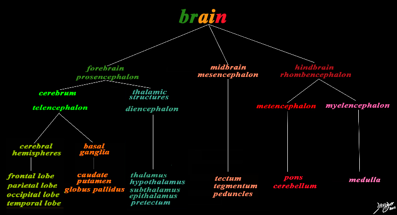
The basic and simplest classification of the brain into forebrain midbrain and hindbrain is shown in this diagram and advanced to a more complex tree using the embryological and evolutionary terminologies. The midbrain or mesencephalon and the Tectum is depicted in a salmon orange. The forebrain consists of the cerebrum also called the prosencephalon, which contains the more advanced form of the brain and the thalamic structures which contain more basic structures. The cerebrum (telencephalon) itself consists of two cerebral hemispheres and paired basal ganglial structures. Each cerebral hemisphere will have gray and white matter distributed in the frontal parietal temporal and occipital lobe, with the basal ganglia being part of the gray matter deep in the cerebral hemispheres. The most important thalamic structures arising from the diencephalons include the thalamus itself and the hypothalamus. The midbrain (mesencepaholon) consists of the tectum tegmentum and cerebral peduncles. The hindbrain has two major branch points based on the evolutionary development. The pons and cerebellum(part of the metencephalon) are grouped and the medulla (part of the myelencephalon is the second branch. Courtesy Ashley Davidoff MD Copyright 2010 All rights reserved 97686.8s |
|
Midbrain (mb) tectum (t) and Tegmentum (tg) Connecting the Forebrain with the Hindbrain |
|
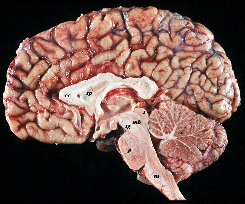
The midsagittal section view of brain reveals the distinctive shape position and character of the midline structures of the brain. The distinction between the character of the cerebral cortex which has a creamy color and the white matter exemplified by the corpus callosum (c) and septum pellucidum (sp) which are white, and the midbrain (mb) with the tegmentum anteriorly (tg) and the tectum (t) posteriorly. The pons (p) and medulla (m) which are off white are part of the hindbrain The cerebellum (c) aso part of the hindbrain is light salmon pink. The relative sizes of the forebrain, midbrain and hindbrain and their components are well appreciated in this section. Image Courtesy of Thomas W.Smith, MD; Department of Pathology, University of Massachusetts Medical School. 97805b03 |
|
Basic Parts of the Midbrain |
|
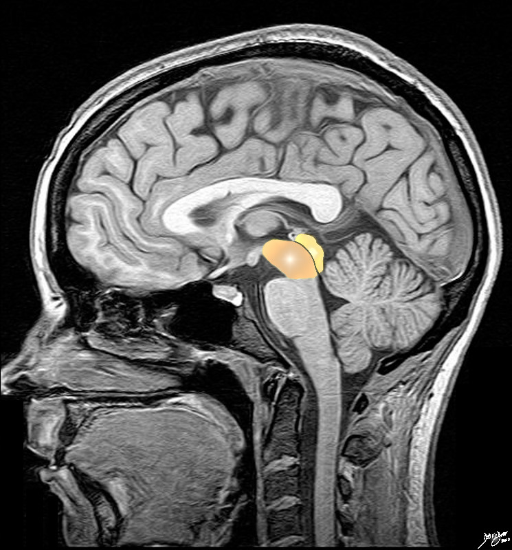
The midbrain consists of a larger anterior portion called the cerebral peduncles incorporated into the tegmentum or floor of the midbrain (orange ). The posterior portion is called tectum or the roof (yellow/orange) The tegmentum and tectum are separated by the aqueduct of Sylvius (black) Image provided by Philips Medical Systems Enhanced by Davidoff art Courtesy Ashley Davidoff MD 92141.3kb01bab02.8s |
|
Basic Divisions of the Midbrain Tegmentum and Tectum Cerebral Peduncles, Cerebral Crura and Colliculli |
|
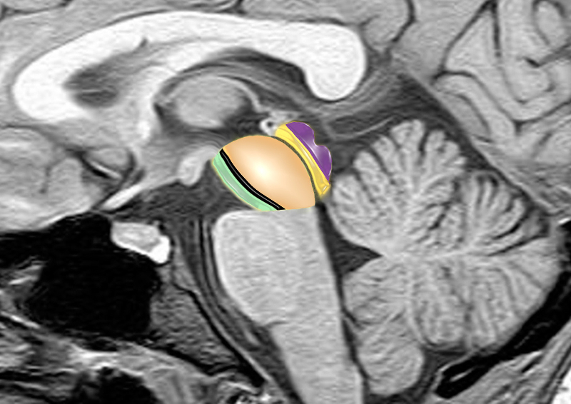
The midbrain consists of a larger anterior portion consisting of the cerebral peduncles, (green) the substantia nigra (black) and the tegmentum (light orange) that extends to the aqueduct (thin gray line) The posterior portion called tectum (orange) contains the colliculi (purple) which are the most posterior portions of the midbrain. brain anatomy neuroanatomy midbrain tegmentum colliculi tectum cerebral peduncle aqueduct of Sylvius conceptual diagram MRI principles Image provided by Philips Medical Systems Enhanced by Davidoff art Courtesy Ashley Davidoff MD 92141.3kb01ba02b02.8s |
|
Tectum and the Colliculi |
|
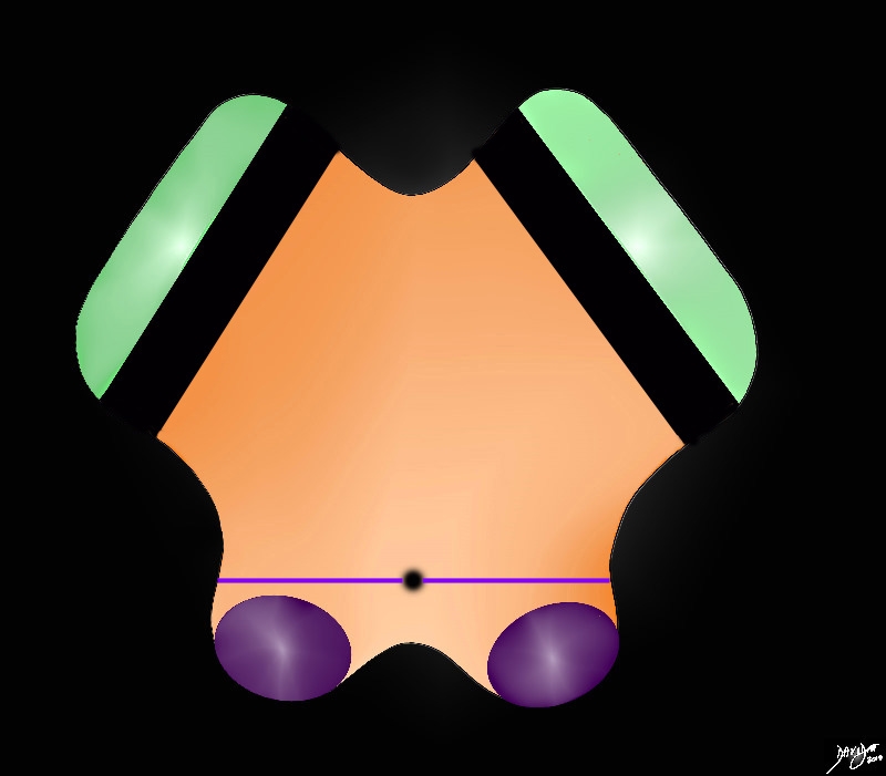
The anterior border iof the midbrain incorporates the cerbral peduncles(green), and the substantia nigra (black ? just posterior to the peduncles. Between the substantia nigra and the aqueduct is an area of the midbrain called the tegmentum (floor of the midbrain) The posterior end of the midbrain, posterior to the aquedct and behind the thin purple line is the tectum (roof) and it houses the colliculi (purple) . Davidoff art Courtesy Ashley Davidoff MD copyright 2010 all rights reserved 94074b09b05b.83s |
|
The Cerebral Crura and the Colliculi The Tegmentum and the Tectum |
|
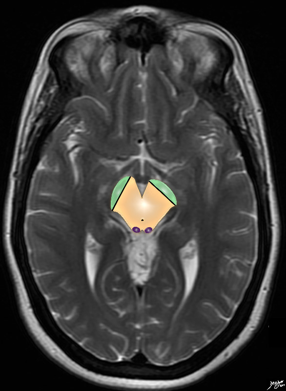
The T2 weighted MRI series focuses on the midbrain with characteristic shape and the small aqueduct of Sylvius posteriorly. The cerebral crura are now shown anteriorly (green) in the cerebral peduncles, and the colliculi are shown posteriorly in the tectum or roof posteriorly Courtesy Ashley Davidoff MD copyright 2010 all rights reserved 94081.4kb07.8s |
DOMElement Object
(
    [schemaTypeInfo] =>
    [tagName] => table
    [firstElementChild] => (object value omitted)
    [lastElementChild] => (object value omitted)
    [childElementCount] => 1
    [previousElementSibling] => (object value omitted)
    [nextElementSibling] =>
    [nodeName] => table
    [nodeValue] =>


The Cerebral Crura and the Colliculi
The Tegmentum and the Tectum


 The T2 weighted MRI series focuses on the midbrain with characteristic shape and the small aqueduct of Sylvius posteriorly. The cerebral crura are now shown anteriorly (green) in the cerebral peduncles, and the colliculi are shown posteriorly in the tectum or roof posteriorly
Courtesy Ashley Davidoff MD copyright 2010 all rights reserved 94081.4kb07.8s


    [nodeType] => 1
    [parentNode] => (object value omitted)
    [childNodes] => (object value omitted)
    [firstChild] => (object value omitted)
    [lastChild] => (object value omitted)
    [previousSibling] => (object value omitted)
    [nextSibling] => (object value omitted)
    [attributes] => (object value omitted)
    [ownerDocument] => (object value omitted)
    [namespaceURI] =>
    [prefix] =>
    [localName] => table
    [baseURI] =>
    [textContent] =>


The Cerebral Crura and the Colliculi
The Tegmentum and the Tectum


 The T2 weighted MRI series focuses on the midbrain with characteristic shape and the small aqueduct of Sylvius posteriorly. The cerebral crura are now shown anteriorly (green) in the cerebral peduncles, and the colliculi are shown posteriorly in the tectum or roof posteriorly
Courtesy Ashley Davidoff MD copyright 2010 all rights reserved 94081.4kb07.8s


)
DOMElement Object
(
    [schemaTypeInfo] =>
    [tagName] => td
    [firstElementChild] => (object value omitted)
    [lastElementChild] => (object value omitted)
    [childElementCount] => 2
    [previousElementSibling] =>
    [nextElementSibling] =>
    [nodeName] => td
    [nodeValue] =>
 The T2 weighted MRI series focuses on the midbrain with characteristic shape and the small aqueduct of Sylvius posteriorly. The cerebral crura are now shown anteriorly (green) in the cerebral peduncles, and the colliculi are shown posteriorly in the tectum or roof posteriorly
Courtesy Ashley Davidoff MD copyright 2010 all rights reserved 94081.4kb07.8s

    [nodeType] => 1
    [parentNode] => (object value omitted)
    [childNodes] => (object value omitted)
    [firstChild] => (object value omitted)
    [lastChild] => (object value omitted)
    [previousSibling] => (object value omitted)
    [nextSibling] => (object value omitted)
    [attributes] => (object value omitted)
    [ownerDocument] => (object value omitted)
    [namespaceURI] =>
    [prefix] =>
    [localName] => td
    [baseURI] =>
    [textContent] =>
 The T2 weighted MRI series focuses on the midbrain with characteristic shape and the small aqueduct of Sylvius posteriorly. The cerebral crura are now shown anteriorly (green) in the cerebral peduncles, and the colliculi are shown posteriorly in the tectum or roof posteriorly
Courtesy Ashley Davidoff MD copyright 2010 all rights reserved 94081.4kb07.8s

)
DOMElement Object
(
    [schemaTypeInfo] =>
    [tagName] => td
    [firstElementChild] => (object value omitted)
    [lastElementChild] => (object value omitted)
    [childElementCount] => 3
    [previousElementSibling] =>
    [nextElementSibling] =>
    [nodeName] => td
    [nodeValue] =>

The Cerebral Crura and the Colliculi
The Tegmentum and the Tectum
    [nodeType] => 1
    [parentNode] => (object value omitted)
    [childNodes] => (object value omitted)
    [firstChild] => (object value omitted)
    [lastChild] => (object value omitted)
    [previousSibling] => (object value omitted)
    [nextSibling] => (object value omitted)
    [attributes] => (object value omitted)
    [ownerDocument] => (object value omitted)
    [namespaceURI] =>
    [prefix] =>
    [localName] => td
    [baseURI] =>
    [textContent] =>

The Cerebral Crura and the Colliculi
The Tegmentum and the Tectum
)
DOMElement Object
(
    [schemaTypeInfo] =>
    [tagName] => table
    [firstElementChild] => (object value omitted)
    [lastElementChild] => (object value omitted)
    [childElementCount] => 1
    [previousElementSibling] => (object value omitted)
    [nextElementSibling] => (object value omitted)
    [nodeName] => table
    [nodeValue] =>


Tectum and the Colliculi


The anterior border iof the midbrain incorporates the cerbral peduncles(green), and the substantia nigra (black ? just posterior to the peduncles. Between the substantia nigra and the aqueduct is an area of the midbrain called the tegmentum (floor of the midbrain) The posterior end of the midbrain, posterior to the aquedct and behind the thin purple line is the tectum (roof)  and it houses the  colliculi (purple) .
Davidoff art Courtesy Ashley Davidoff MD copyright 2010 all rights reserved 94074b09b05b.83s


    [nodeType] => 1
    [parentNode] => (object value omitted)
    [childNodes] => (object value omitted)
    [firstChild] => (object value omitted)
    [lastChild] => (object value omitted)
    [previousSibling] => (object value omitted)
    [nextSibling] => (object value omitted)
    [attributes] => (object value omitted)
    [ownerDocument] => (object value omitted)
    [namespaceURI] =>
    [prefix] =>
    [localName] => table
    [baseURI] =>
    [textContent] =>


Tectum and the Colliculi


The anterior border iof the midbrain incorporates the cerbral peduncles(green), and the substantia nigra (black ? just posterior to the peduncles. Between the substantia nigra and the aqueduct is an area of the midbrain called the tegmentum (floor of the midbrain) The posterior end of the midbrain, posterior to the aquedct and behind the thin purple line is the tectum (roof)  and it houses the  colliculi (purple) .
Davidoff art Courtesy Ashley Davidoff MD copyright 2010 all rights reserved 94074b09b05b.83s


)
DOMElement Object
(
    [schemaTypeInfo] =>
    [tagName] => td
    [firstElementChild] => (object value omitted)
    [lastElementChild] => (object value omitted)
    [childElementCount] => 2
    [previousElementSibling] =>
    [nextElementSibling] =>
    [nodeName] => td
    [nodeValue] =>
The anterior border iof the midbrain incorporates the cerbral peduncles(green), and the substantia nigra (black ? just posterior to the peduncles. Between the substantia nigra and the aqueduct is an area of the midbrain called the tegmentum (floor of the midbrain) The posterior end of the midbrain, posterior to the aquedct and behind the thin purple line is the tectum (roof)  and it houses the  colliculi (purple) .
Davidoff art Courtesy Ashley Davidoff MD copyright 2010 all rights reserved 94074b09b05b.83s

    [nodeType] => 1
    [parentNode] => (object value omitted)
    [childNodes] => (object value omitted)
    [firstChild] => (object value omitted)
    [lastChild] => (object value omitted)
    [previousSibling] => (object value omitted)
    [nextSibling] => (object value omitted)
    [attributes] => (object value omitted)
    [ownerDocument] => (object value omitted)
    [namespaceURI] =>
    [prefix] =>
    [localName] => td
    [baseURI] =>
    [textContent] =>
The anterior border iof the midbrain incorporates the cerbral peduncles(green), and the substantia nigra (black ? just posterior to the peduncles. Between the substantia nigra and the aqueduct is an area of the midbrain called the tegmentum (floor of the midbrain) The posterior end of the midbrain, posterior to the aquedct and behind the thin purple line is the tectum (roof)  and it houses the  colliculi (purple) .
Davidoff art Courtesy Ashley Davidoff MD copyright 2010 all rights reserved 94074b09b05b.83s

)
DOMElement Object
(
    [schemaTypeInfo] =>
    [tagName] => td
    [firstElementChild] => (object value omitted)
    [lastElementChild] => (object value omitted)
    [childElementCount] => 2
    [previousElementSibling] =>
    [nextElementSibling] =>
    [nodeName] => td
    [nodeValue] =>

Tectum and the Colliculi
    [nodeType] => 1
    [parentNode] => (object value omitted)
    [childNodes] => (object value omitted)
    [firstChild] => (object value omitted)
    [lastChild] => (object value omitted)
    [previousSibling] => (object value omitted)
    [nextSibling] => (object value omitted)
    [attributes] => (object value omitted)
    [ownerDocument] => (object value omitted)
    [namespaceURI] =>
    [prefix] =>
    [localName] => td
    [baseURI] =>
    [textContent] =>

Tectum and the Colliculi
)
DOMElement Object
(
    [schemaTypeInfo] =>
    [tagName] => table
    [firstElementChild] => (object value omitted)
    [lastElementChild] => (object value omitted)
    [childElementCount] => 1
    [previousElementSibling] => (object value omitted)
    [nextElementSibling] => (object value omitted)
    [nodeName] => table
    [nodeValue] =>


Basic Divisions of the Midbrain
Tegmentum and Tectum
Cerebral Peduncles, Cerebral Crura and Colliculli


The midbrain consists of a larger anterior portion consisting of the cerebral peduncles, (green) the substantia nigra (black) and the tegmentum (light orange) that extends to the aqueduct (thin gray line) The posterior portion called tectum (orange) contains the colliculi (purple) which are the most posterior portions of the midbrain. brain anatomy neuroanatomy midbrain tegmentum colliculi tectum cerebral peduncle aqueduct of Sylvius conceptual diagram MRI principles
Image provided by Philips Medical Systems Enhanced by Davidoff art Courtesy Ashley Davidoff MD 92141.3kb01ba02b02.8s


    [nodeType] => 1
    [parentNode] => (object value omitted)
    [childNodes] => (object value omitted)
    [firstChild] => (object value omitted)
    [lastChild] => (object value omitted)
    [previousSibling] => (object value omitted)
    [nextSibling] => (object value omitted)
    [attributes] => (object value omitted)
    [ownerDocument] => (object value omitted)
    [namespaceURI] =>
    [prefix] =>
    [localName] => table
    [baseURI] =>
    [textContent] =>


Basic Divisions of the Midbrain
Tegmentum and Tectum
Cerebral Peduncles, Cerebral Crura and Colliculli


The midbrain consists of a larger anterior portion consisting of the cerebral peduncles, (green) the substantia nigra (black) and the tegmentum (light orange) that extends to the aqueduct (thin gray line) The posterior portion called tectum (orange) contains the colliculi (purple) which are the most posterior portions of the midbrain. brain anatomy neuroanatomy midbrain tegmentum colliculi tectum cerebral peduncle aqueduct of Sylvius conceptual diagram MRI principles
Image provided by Philips Medical Systems Enhanced by Davidoff art Courtesy Ashley Davidoff MD 92141.3kb01ba02b02.8s


)
DOMElement Object
(
    [schemaTypeInfo] =>
    [tagName] => td
    [firstElementChild] => (object value omitted)
    [lastElementChild] => (object value omitted)
    [childElementCount] => 2
    [previousElementSibling] =>
    [nextElementSibling] =>
    [nodeName] => td
    [nodeValue] =>
The midbrain consists of a larger anterior portion consisting of the cerebral peduncles, (green) the substantia nigra (black) and the tegmentum (light orange) that extends to the aqueduct (thin gray line) The posterior portion called tectum (orange) contains the colliculi (purple) which are the most posterior portions of the midbrain. brain anatomy neuroanatomy midbrain tegmentum colliculi tectum cerebral peduncle aqueduct of Sylvius conceptual diagram MRI principles
Image provided by Philips Medical Systems Enhanced by Davidoff art Courtesy Ashley Davidoff MD 92141.3kb01ba02b02.8s

    [nodeType] => 1
    [parentNode] => (object value omitted)
    [childNodes] => (object value omitted)
    [firstChild] => (object value omitted)
    [lastChild] => (object value omitted)
    [previousSibling] => (object value omitted)
    [nextSibling] => (object value omitted)
    [attributes] => (object value omitted)
    [ownerDocument] => (object value omitted)
    [namespaceURI] =>
    [prefix] =>
    [localName] => td
    [baseURI] =>
    [textContent] =>
The midbrain consists of a larger anterior portion consisting of the cerebral peduncles, (green) the substantia nigra (black) and the tegmentum (light orange) that extends to the aqueduct (thin gray line) The posterior portion called tectum (orange) contains the colliculi (purple) which are the most posterior portions of the midbrain. brain anatomy neuroanatomy midbrain tegmentum colliculi tectum cerebral peduncle aqueduct of Sylvius conceptual diagram MRI principles
Image provided by Philips Medical Systems Enhanced by Davidoff art Courtesy Ashley Davidoff MD 92141.3kb01ba02b02.8s

)
DOMElement Object
(
    [schemaTypeInfo] =>
    [tagName] => td
    [firstElementChild] => (object value omitted)
    [lastElementChild] => (object value omitted)
    [childElementCount] => 4
    [previousElementSibling] =>
    [nextElementSibling] =>
    [nodeName] => td
    [nodeValue] =>

Basic Divisions of the Midbrain
Tegmentum and Tectum
Cerebral Peduncles, Cerebral Crura and Colliculli

    [nodeType] => 1
    [parentNode] => (object value omitted)
    [childNodes] => (object value omitted)
    [firstChild] => (object value omitted)
    [lastChild] => (object value omitted)
    [previousSibling] => (object value omitted)
    [nextSibling] => (object value omitted)
    [attributes] => (object value omitted)
    [ownerDocument] => (object value omitted)
    [namespaceURI] =>
    [prefix] =>
    [localName] => td
    [baseURI] =>
    [textContent] =>

Basic Divisions of the Midbrain
Tegmentum and Tectum
Cerebral Peduncles, Cerebral Crura and Colliculli

)
DOMElement Object
(
    [schemaTypeInfo] =>
    [tagName] => table
    [firstElementChild] => (object value omitted)
    [lastElementChild] => (object value omitted)
    [childElementCount] => 1
    [previousElementSibling] => (object value omitted)
    [nextElementSibling] => (object value omitted)
    [nodeName] => table
    [nodeValue] =>


Basic Parts of the Midbrain


The midbrain consists of a larger anterior portion called the cerebral peduncles incorporated into the tegmentum or floor of the midbrain (orange ).  The  posterior portion is called tectum or the roof  (yellow/orange) The tegmentum and tectum are separated by the aqueduct of Sylvius (black)
Image provided by Philips Medical Systems Enhanced by Davidoff art Courtesy Ashley Davidoff MD 92141.3kb01bab02.8s


    [nodeType] => 1
    [parentNode] => (object value omitted)
    [childNodes] => (object value omitted)
    [firstChild] => (object value omitted)
    [lastChild] => (object value omitted)
    [previousSibling] => (object value omitted)
    [nextSibling] => (object value omitted)
    [attributes] => (object value omitted)
    [ownerDocument] => (object value omitted)
    [namespaceURI] =>
    [prefix] =>
    [localName] => table
    [baseURI] =>
    [textContent] =>


Basic Parts of the Midbrain


The midbrain consists of a larger anterior portion called the cerebral peduncles incorporated into the tegmentum or floor of the midbrain (orange ).  The  posterior portion is called tectum or the roof  (yellow/orange) The tegmentum and tectum are separated by the aqueduct of Sylvius (black)
Image provided by Philips Medical Systems Enhanced by Davidoff art Courtesy Ashley Davidoff MD 92141.3kb01bab02.8s


)
DOMElement Object
(
    [schemaTypeInfo] =>
    [tagName] => td
    [firstElementChild] => (object value omitted)
    [lastElementChild] => (object value omitted)
    [childElementCount] => 2
    [previousElementSibling] =>
    [nextElementSibling] =>
    [nodeName] => td
    [nodeValue] =>
The midbrain consists of a larger anterior portion called the cerebral peduncles incorporated into the tegmentum or floor of the midbrain (orange ).  The  posterior portion is called tectum or the roof  (yellow/orange) The tegmentum and tectum are separated by the aqueduct of Sylvius (black)
Image provided by Philips Medical Systems Enhanced by Davidoff art Courtesy Ashley Davidoff MD 92141.3kb01bab02.8s

    [nodeType] => 1
    [parentNode] => (object value omitted)
    [childNodes] => (object value omitted)
    [firstChild] => (object value omitted)
    [lastChild] => (object value omitted)
    [previousSibling] => (object value omitted)
    [nextSibling] => (object value omitted)
    [attributes] => (object value omitted)
    [ownerDocument] => (object value omitted)
    [namespaceURI] =>
    [prefix] =>
    [localName] => td
    [baseURI] =>
    [textContent] =>
The midbrain consists of a larger anterior portion called the cerebral peduncles incorporated into the tegmentum or floor of the midbrain (orange ).  The  posterior portion is called tectum or the roof  (yellow/orange) The tegmentum and tectum are separated by the aqueduct of Sylvius (black)
Image provided by Philips Medical Systems Enhanced by Davidoff art Courtesy Ashley Davidoff MD 92141.3kb01bab02.8s

)
DOMElement Object
(
    [schemaTypeInfo] =>
    [tagName] => td
    [firstElementChild] => (object value omitted)
    [lastElementChild] => (object value omitted)
    [childElementCount] => 2
    [previousElementSibling] =>
    [nextElementSibling] =>
    [nodeName] => td
    [nodeValue] =>

Basic Parts of the Midbrain
    [nodeType] => 1
    [parentNode] => (object value omitted)
    [childNodes] => (object value omitted)
    [firstChild] => (object value omitted)
    [lastChild] => (object value omitted)
    [previousSibling] => (object value omitted)
    [nextSibling] => (object value omitted)
    [attributes] => (object value omitted)
    [ownerDocument] => (object value omitted)
    [namespaceURI] =>
    [prefix] =>
    [localName] => td
    [baseURI] =>
    [textContent] =>

Basic Parts of the Midbrain
)
DOMElement Object
(
    [schemaTypeInfo] =>
    [tagName] => table
    [firstElementChild] => (object value omitted)
    [lastElementChild] => (object value omitted)
    [childElementCount] => 1
    [previousElementSibling] => (object value omitted)
    [nextElementSibling] => (object value omitted)
    [nodeName] => table
    [nodeValue] =>


Midbrain (mb) tectum (t) and Tegmentum (tg)
Connecting the Forebrain with the Hindbrain


The midsagittal section view of brain reveals the distinctive shape position and character of the midline structures of the brain. The distinction between the character of the cerebral cortex which has a creamy color and the white matter exemplified by the corpus callosum (c) and septum pellucidum (sp) which are white, and the midbrain (mb) with the tegmentum anteriorly (tg) and the tectum (t) posteriorly. The pons (p) and medulla (m) which are off white are part of the hindbrain The cerebellum (c) aso part of the hindbrain is light salmon pink. The relative sizes of the forebrain, midbrain and hindbrain and their components are well appreciated in this section.
Image Courtesy of Thomas W.Smith, MD; Department of Pathology, University of Massachusetts Medical School. 97805b03


    [nodeType] => 1
    [parentNode] => (object value omitted)
    [childNodes] => (object value omitted)
    [firstChild] => (object value omitted)
    [lastChild] => (object value omitted)
    [previousSibling] => (object value omitted)
    [nextSibling] => (object value omitted)
    [attributes] => (object value omitted)
    [ownerDocument] => (object value omitted)
    [namespaceURI] =>
    [prefix] =>
    [localName] => table
    [baseURI] =>
    [textContent] =>


Midbrain (mb) tectum (t) and Tegmentum (tg)
Connecting the Forebrain with the Hindbrain


The midsagittal section view of brain reveals the distinctive shape position and character of the midline structures of the brain. The distinction between the character of the cerebral cortex which has a creamy color and the white matter exemplified by the corpus callosum (c) and septum pellucidum (sp) which are white, and the midbrain (mb) with the tegmentum anteriorly (tg) and the tectum (t) posteriorly. The pons (p) and medulla (m) which are off white are part of the hindbrain The cerebellum (c) aso part of the hindbrain is light salmon pink. The relative sizes of the forebrain, midbrain and hindbrain and their components are well appreciated in this section.
Image Courtesy of Thomas W.Smith, MD; Department of Pathology, University of Massachusetts Medical School. 97805b03


)
DOMElement Object
(
    [schemaTypeInfo] =>
    [tagName] => td
    [firstElementChild] => (object value omitted)
    [lastElementChild] => (object value omitted)
    [childElementCount] => 2
    [previousElementSibling] =>
    [nextElementSibling] =>
    [nodeName] => td
    [nodeValue] =>
The midsagittal section view of brain reveals the distinctive shape position and character of the midline structures of the brain. The distinction between the character of the cerebral cortex which has a creamy color and the white matter exemplified by the corpus callosum (c) and septum pellucidum (sp) which are white, and the midbrain (mb) with the tegmentum anteriorly (tg) and the tectum (t) posteriorly. The pons (p) and medulla (m) which are off white are part of the hindbrain The cerebellum (c) aso part of the hindbrain is light salmon pink. The relative sizes of the forebrain, midbrain and hindbrain and their components are well appreciated in this section.
Image Courtesy of Thomas W.Smith, MD; Department of Pathology, University of Massachusetts Medical School. 97805b03

    [nodeType] => 1
    [parentNode] => (object value omitted)
    [childNodes] => (object value omitted)
    [firstChild] => (object value omitted)
    [lastChild] => (object value omitted)
    [previousSibling] => (object value omitted)
    [nextSibling] => (object value omitted)
    [attributes] => (object value omitted)
    [ownerDocument] => (object value omitted)
    [namespaceURI] =>
    [prefix] =>
    [localName] => td
    [baseURI] =>
    [textContent] =>
The midsagittal section view of brain reveals the distinctive shape position and character of the midline structures of the brain. The distinction between the character of the cerebral cortex which has a creamy color and the white matter exemplified by the corpus callosum (c) and septum pellucidum (sp) which are white, and the midbrain (mb) with the tegmentum anteriorly (tg) and the tectum (t) posteriorly. The pons (p) and medulla (m) which are off white are part of the hindbrain The cerebellum (c) aso part of the hindbrain is light salmon pink. The relative sizes of the forebrain, midbrain and hindbrain and their components are well appreciated in this section.
Image Courtesy of Thomas W.Smith, MD; Department of Pathology, University of Massachusetts Medical School. 97805b03

)
DOMElement Object
(
    [schemaTypeInfo] =>
    [tagName] => td
    [firstElementChild] => (object value omitted)
    [lastElementChild] => (object value omitted)
    [childElementCount] => 3
    [previousElementSibling] =>
    [nextElementSibling] =>
    [nodeName] => td
    [nodeValue] =>

Midbrain (mb) tectum (t) and Tegmentum (tg)
Connecting the Forebrain with the Hindbrain
    [nodeType] => 1
    [parentNode] => (object value omitted)
    [childNodes] => (object value omitted)
    [firstChild] => (object value omitted)
    [lastChild] => (object value omitted)
    [previousSibling] => (object value omitted)
    [nextSibling] => (object value omitted)
    [attributes] => (object value omitted)
    [ownerDocument] => (object value omitted)
    [namespaceURI] =>
    [prefix] =>
    [localName] => td
    [baseURI] =>
    [textContent] =>

Midbrain (mb) tectum (t) and Tegmentum (tg)
Connecting the Forebrain with the Hindbrain
)
DOMElement Object
(
    [schemaTypeInfo] =>
    [tagName] => table
    [firstElementChild] => (object value omitted)
    [lastElementChild] => (object value omitted)
    [childElementCount] => 1
    [previousElementSibling] => (object value omitted)
    [nextElementSibling] => (object value omitted)
    [nodeName] => table
    [nodeValue] =>


The Tectum (mesencephalon) in Context
Part of the Mesencepaolon


The basic and simplest classification of the brain into forebrain midbrain and hindbrain is shown in this diagram and advanced to a more complex tree using the embryological and evolutionary terminologies.
The midbrain or mesencephalon and the Tectum is depicted in a salmon orange.
The forebrain consists of the cerebrum also called the prosencephalon, which contains the more advanced form of the brain and the thalamic structures which contain more basic structures. The cerebrum (telencephalon) itself consists of two cerebral hemispheres and paired basal ganglial structures. Each cerebral hemisphere will have gray and white matter distributed in the frontal parietal temporal and occipital lobe, with the basal ganglia being part of the gray matter deep in the cerebral hemispheres. The most important thalamic structures arising from the diencephalons include the thalamus itself and the hypothalamus. The midbrain (mesencepaholon) consists of the tectum tegmentum and cerebral peduncles. The hindbrain has two major branch points based on the evolutionary development. The pons and cerebellum(part of the metencephalon) are grouped and the medulla (part of the myelencephalon is the second branch.
Courtesy Ashley Davidoff MD Copyright 2010 All rights reserved 97686.8s


    [nodeType] => 1
    [parentNode] => (object value omitted)
    [childNodes] => (object value omitted)
    [firstChild] => (object value omitted)
    [lastChild] => (object value omitted)
    [previousSibling] => (object value omitted)
    [nextSibling] => (object value omitted)
    [attributes] => (object value omitted)
    [ownerDocument] => (object value omitted)
    [namespaceURI] =>
    [prefix] =>
    [localName] => table
    [baseURI] =>
    [textContent] =>


The Tectum (mesencephalon) in Context
Part of the Mesencepaolon


The basic and simplest classification of the brain into forebrain midbrain and hindbrain is shown in this diagram and advanced to a more complex tree using the embryological and evolutionary terminologies.
The midbrain or mesencephalon and the Tectum is depicted in a salmon orange.
The forebrain consists of the cerebrum also called the prosencephalon, which contains the more advanced form of the brain and the thalamic structures which contain more basic structures. The cerebrum (telencephalon) itself consists of two cerebral hemispheres and paired basal ganglial structures. Each cerebral hemisphere will have gray and white matter distributed in the frontal parietal temporal and occipital lobe, with the basal ganglia being part of the gray matter deep in the cerebral hemispheres. The most important thalamic structures arising from the diencephalons include the thalamus itself and the hypothalamus. The midbrain (mesencepaholon) consists of the tectum tegmentum and cerebral peduncles. The hindbrain has two major branch points based on the evolutionary development. The pons and cerebellum(part of the metencephalon) are grouped and the medulla (part of the myelencephalon is the second branch.
Courtesy Ashley Davidoff MD Copyright 2010 All rights reserved 97686.8s


)
DOMElement Object
(
    [schemaTypeInfo] =>
    [tagName] => td
    [firstElementChild] => (object value omitted)
    [lastElementChild] => (object value omitted)
    [childElementCount] => 4
    [previousElementSibling] =>
    [nextElementSibling] =>
    [nodeName] => td
    [nodeValue] =>
The basic and simplest classification of the brain into forebrain midbrain and hindbrain is shown in this diagram and advanced to a more complex tree using the embryological and evolutionary terminologies.
The midbrain or mesencephalon and the Tectum is depicted in a salmon orange.
The forebrain consists of the cerebrum also called the prosencephalon, which contains the more advanced form of the brain and the thalamic structures which contain more basic structures. The cerebrum (telencephalon) itself consists of two cerebral hemispheres and paired basal ganglial structures. Each cerebral hemisphere will have gray and white matter distributed in the frontal parietal temporal and occipital lobe, with the basal ganglia being part of the gray matter deep in the cerebral hemispheres. The most important thalamic structures arising from the diencephalons include the thalamus itself and the hypothalamus. The midbrain (mesencepaholon) consists of the tectum tegmentum and cerebral peduncles. The hindbrain has two major branch points based on the evolutionary development. The pons and cerebellum(part of the metencephalon) are grouped and the medulla (part of the myelencephalon is the second branch.
Courtesy Ashley Davidoff MD Copyright 2010 All rights reserved 97686.8s

    [nodeType] => 1
    [parentNode] => (object value omitted)
    [childNodes] => (object value omitted)
    [firstChild] => (object value omitted)
    [lastChild] => (object value omitted)
    [previousSibling] => (object value omitted)
    [nextSibling] => (object value omitted)
    [attributes] => (object value omitted)
    [ownerDocument] => (object value omitted)
    [namespaceURI] =>
    [prefix] =>
    [localName] => td
    [baseURI] =>
    [textContent] =>
The basic and simplest classification of the brain into forebrain midbrain and hindbrain is shown in this diagram and advanced to a more complex tree using the embryological and evolutionary terminologies.
The midbrain or mesencephalon and the Tectum is depicted in a salmon orange.
The forebrain consists of the cerebrum also called the prosencephalon, which contains the more advanced form of the brain and the thalamic structures which contain more basic structures. The cerebrum (telencephalon) itself consists of two cerebral hemispheres and paired basal ganglial structures. Each cerebral hemisphere will have gray and white matter distributed in the frontal parietal temporal and occipital lobe, with the basal ganglia being part of the gray matter deep in the cerebral hemispheres. The most important thalamic structures arising from the diencephalons include the thalamus itself and the hypothalamus. The midbrain (mesencepaholon) consists of the tectum tegmentum and cerebral peduncles. The hindbrain has two major branch points based on the evolutionary development. The pons and cerebellum(part of the metencephalon) are grouped and the medulla (part of the myelencephalon is the second branch.
Courtesy Ashley Davidoff MD Copyright 2010 All rights reserved 97686.8s

)
DOMElement Object
(
    [schemaTypeInfo] =>
    [tagName] => td
    [firstElementChild] => (object value omitted)
    [lastElementChild] => (object value omitted)
    [childElementCount] => 3
    [previousElementSibling] =>
    [nextElementSibling] =>
    [nodeName] => td
    [nodeValue] =>

The Tectum (mesencephalon) in Context
Part of the Mesencepaolon
    [nodeType] => 1
    [parentNode] => (object value omitted)
    [childNodes] => (object value omitted)
    [firstChild] => (object value omitted)
    [lastChild] => (object value omitted)
    [previousSibling] => (object value omitted)
    [nextSibling] => (object value omitted)
    [attributes] => (object value omitted)
    [ownerDocument] => (object value omitted)
    [namespaceURI] =>
    [prefix] =>
    [localName] => td
    [baseURI] =>
    [textContent] =>

The Tectum (mesencephalon) in Context
Part of the Mesencepaolon
)

