Sumit Karia MD Ashley Davidoff MD
The Common Vein Copyright 2010
Introduction
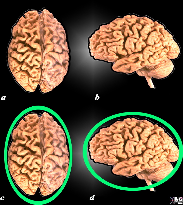
Overall, when viewed from the outside, and from above, the brain has an oval shape with the long axis oriented in the anteroposterior direction. The broader part of the oval is posterior and the narrower part is anterior.
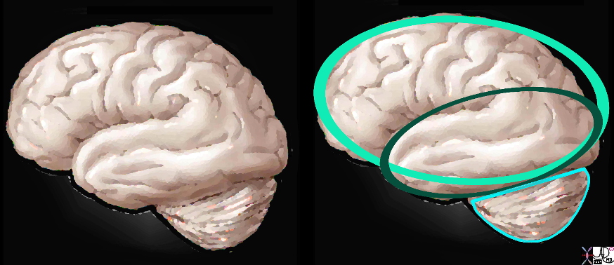
When viewed from the side, its overall shape is still oval but the temporal lobe and the cerebellum start to add complexity to the shape.
Basic Shapes of the Surface
As one explores the surface of the brain the external shape becomes more complex by virtue of the ridges in the brain surface. The ridges and clefts – gyri and sulci have characteristic shape and position and each named according to structure and function.
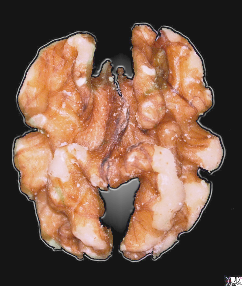
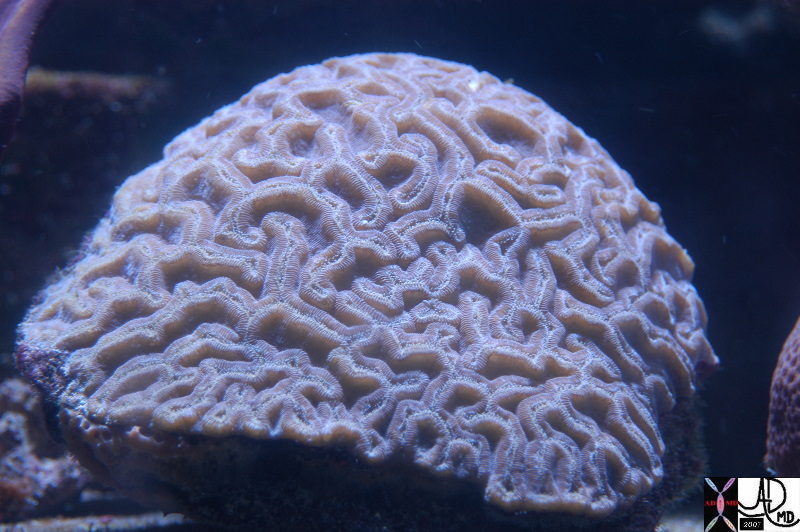
There are not many other structures that look like the brain in nature but there are two that have similar convolutional appearance including the walnut as described above and brain coral.
|
Form and Colors reminiscent of the Brain |
|
This image of a walnut has a few features that are reminiscent of a brain, including the symmetric hemispheric components, the pinkish color, and the undulations of the surface. However in reality it is about 1.8cms long, and only 5-8mms high, and is hard. Courtesy Ashley Davidoff MD copyright 2010 all rights reserved 100286pb.93s |
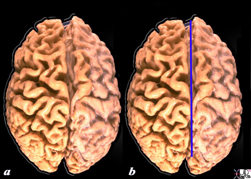
Looking at the brain from above, it has, in its midline, a deep fissure that divides the organ in 2 halves called hemispheres. The external surface is also marked by serpiginous ridges and furrows called the gyri and sulci.
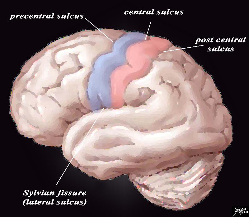
The gyri and fissures have characteristic form and position though there is variation among individuals. As we advance into the structure we will explore the detail of the gyri and sulci. As a a start we will exemplify the most central of the sulci and gyri in the diagram below.
|
Shapes on the Top |
|
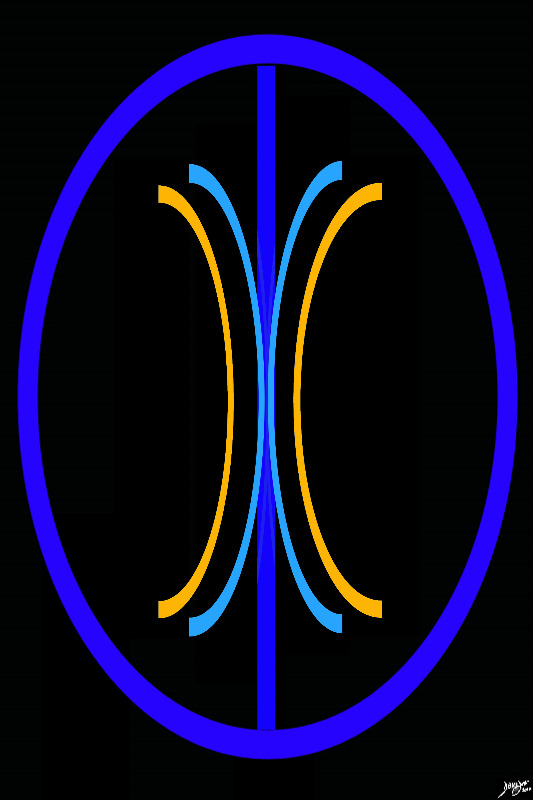
When the external surface is explored from above a deep midline fissure (blue line) divides the oval of the brain into two hemispheres. The deep cleft is called the interhemishpheric fissure In addition, the serpiginous folds and crevices that characterize the outside surface of the brain start to become apparrent as one explores the shape of the brain in more depth. It becomes obvious that it is more than a mere oval. Source unknown Image modified by Davidoff 52981b05d05 |
The brain conforms to the shape of the bony shape of the calvarium. The superior aspect of the brain is convex, and lies in close association with the outer bony layer of the cranium, while in the anterior cranial fossa and middle cranial fossa its inferior part is flatter as it abuts on the inferior portions of the bony vault.
|
The Basal Ganglia of the Forebrain in Sagittal Projection Recurring C- Shaped Pattern |
|
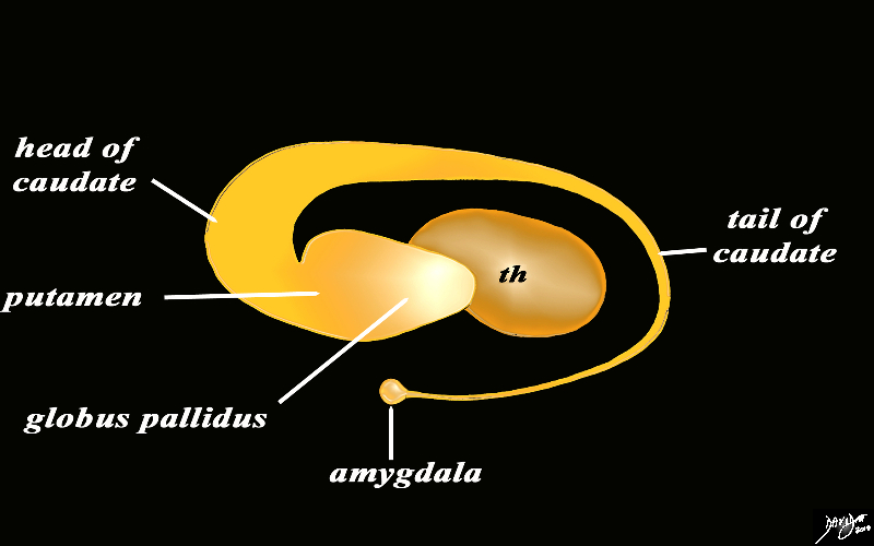
The basal ganglia lie both in the forebrain and in the midbrain. In the forebrain they are distributed in paraventricular fashion almost in an inverted C fashion. In the conceptual diagrams we have colored the inverted ?C? that abuts the ventricular system orange, and this includes the thalamus and the basal ganglia. The basal ganglia that lie in the forebrain include the globus pallidus, putamen, head of the caudate nucleus, tail of the caudate nucleus and the amygdala. The amygdala appears to be part of the limbic system and the basal ganglia. Courtesy Ashley Davidoff MD Copyright 2010 All rights reserved 93907d13b05dL3.8s |
|
Basal Ganglia in the Forebrain Lie within the The Orange Paraventricular Layer
|
|
The paraventricular orange layer contains the basal ganglia and the thalamus. The paraventricular structures in general conform to the axis of the lateral ventricles superiorly. In this instance we will highlight the components of the basal ganglia that lie along this plane. Courtesy Ashley DAvidoff MD Copyright 2010 all rights reserved 93914.3ka04b01.8s |
|
Forebrain Basal Ganglia in Axial Projection |
|
The basal ganglia in the forebrain in the axial projection are seen as a continuous inverted C as well. In this diagram their relationship to the ventricles distributed in paraventricular fashion is exemplified. In the conceptual diagrams we have colored the inverted ?C? that abuts the ventricular system orange, and this includes the putamen, globus pallidus, head of the caudate nucleus, tail of the caudate nucleus and the amygdala. The amygdala appears to be part of the limbic system and the basal ganglia. Courtesy Ashley Davidoff MD Copyright 2010 All rights reserved 93914.3ka04b01e05.8s |
The human brain is contained within the calvarium. In its normal anatomical position, the normal directional terminology does not apply simply to the brain. The connection between the brainstem and cerebrum results in an angle of about 100 degrees. This angle is called the cephalic flexure and it is responsible for the change in directional terminology. For example, the inferior surface of the cerebrum is also its ventral surface, where as the ventral surface of the spinal cord is the anterior surface. When referring to position in the cerebrum it is more convenient to refer with terminology such as superior/inferior/anterior/posterior.
The hemispheres have a form of a triangular prism, with 2 extremities, 3 faces and 3 borders. The anterior extremity is called the frontal, the posterior is called occipital.
Human brain folds are a highly evolved trait. Only mammals have evolved this structural advance and amongst them only cats, dogs, monkeys, dolphins and humans. Other animals and mammals like rats are still have flat, unfolded brains.
|
Axial Images from Vertex to Skull Base |
|
These axial T1 weighted images taken sequentially fronm the vertex of the brain (top left) progressively through the forebrain and anterior cranial fossa, (approximately first two rows) through the midbrain (3rd row) and then hindbrain (posterior fossa)reveal the initial oval shape of the brain with progressive shortening in the AP projection to the butterfly shape of the cerebellum in the posterior fossa. Courtesy Philips Medical Systems 92127.8s |
DOMElement Object
(
    [schemaTypeInfo] =>
    [tagName] => table
    [firstElementChild] => (object value omitted)
    [lastElementChild] => (object value omitted)
    [childElementCount] => 1
    [previousElementSibling] => (object value omitted)
    [nextElementSibling] =>
    [nodeName] => table
    [nodeValue] =>


Axial Images from Vertex to Skull Base


These axial T1 weighted images taken sequentially fronm the vertex of the brain (top left) progressively through the forebrain and anterior cranial fossa, (approximately first two rows) through the midbrain (3rd row) and then hindbrain (posterior fossa)reveal the initial oval shape of the brain with progressive shortening in the AP projection to the butterfly shape of the cerebellum in the posterior fossa.
Courtesy Philips Medical Systems 92127.8s


    [nodeType] => 1
    [parentNode] => (object value omitted)
    [childNodes] => (object value omitted)
    [firstChild] => (object value omitted)
    [lastChild] => (object value omitted)
    [previousSibling] => (object value omitted)
    [nextSibling] => (object value omitted)
    [attributes] => (object value omitted)
    [ownerDocument] => (object value omitted)
    [namespaceURI] =>
    [prefix] =>
    [localName] => table
    [baseURI] =>
    [textContent] =>


Axial Images from Vertex to Skull Base


These axial T1 weighted images taken sequentially fronm the vertex of the brain (top left) progressively through the forebrain and anterior cranial fossa, (approximately first two rows) through the midbrain (3rd row) and then hindbrain (posterior fossa)reveal the initial oval shape of the brain with progressive shortening in the AP projection to the butterfly shape of the cerebellum in the posterior fossa.
Courtesy Philips Medical Systems 92127.8s


)
DOMElement Object
(
    [schemaTypeInfo] =>
    [tagName] => td
    [firstElementChild] => (object value omitted)
    [lastElementChild] => (object value omitted)
    [childElementCount] => 2
    [previousElementSibling] =>
    [nextElementSibling] =>
    [nodeName] => td
    [nodeValue] =>
These axial T1 weighted images taken sequentially fronm the vertex of the brain (top left) progressively through the forebrain and anterior cranial fossa, (approximately first two rows) through the midbrain (3rd row) and then hindbrain (posterior fossa)reveal the initial oval shape of the brain with progressive shortening in the AP projection to the butterfly shape of the cerebellum in the posterior fossa.
Courtesy Philips Medical Systems 92127.8s

    [nodeType] => 1
    [parentNode] => (object value omitted)
    [childNodes] => (object value omitted)
    [firstChild] => (object value omitted)
    [lastChild] => (object value omitted)
    [previousSibling] => (object value omitted)
    [nextSibling] => (object value omitted)
    [attributes] => (object value omitted)
    [ownerDocument] => (object value omitted)
    [namespaceURI] =>
    [prefix] =>
    [localName] => td
    [baseURI] =>
    [textContent] =>
These axial T1 weighted images taken sequentially fronm the vertex of the brain (top left) progressively through the forebrain and anterior cranial fossa, (approximately first two rows) through the midbrain (3rd row) and then hindbrain (posterior fossa)reveal the initial oval shape of the brain with progressive shortening in the AP projection to the butterfly shape of the cerebellum in the posterior fossa.
Courtesy Philips Medical Systems 92127.8s

)
DOMElement Object
(
    [schemaTypeInfo] =>
    [tagName] => td
    [firstElementChild] => (object value omitted)
    [lastElementChild] => (object value omitted)
    [childElementCount] => 2
    [previousElementSibling] =>
    [nextElementSibling] =>
    [nodeName] => td
    [nodeValue] =>

Axial Images from Vertex to Skull Base

    [nodeType] => 1
    [parentNode] => (object value omitted)
    [childNodes] => (object value omitted)
    [firstChild] => (object value omitted)
    [lastChild] => (object value omitted)
    [previousSibling] => (object value omitted)
    [nextSibling] => (object value omitted)
    [attributes] => (object value omitted)
    [ownerDocument] => (object value omitted)
    [namespaceURI] =>
    [prefix] =>
    [localName] => td
    [baseURI] =>
    [textContent] =>

Axial Images from Vertex to Skull Base

)
DOMElement Object
(
    [schemaTypeInfo] =>
    [tagName] => table
    [firstElementChild] => (object value omitted)
    [lastElementChild] => (object value omitted)
    [childElementCount] => 1
    [previousElementSibling] => (object value omitted)
    [nextElementSibling] => (object value omitted)
    [nodeName] => table
    [nodeValue] =>


Forebrain Basal Ganglia in Axial Projection


The basal ganglia in the forebrain in the axial projection are seen as a continuous inverted C as well. In this diagram their relationship to the ventricles distributed in paraventricular fashion is exemplified. In the conceptual diagrams we have colored the inverted ?C? that abuts the ventricular system orange, and this includes the putamen, globus pallidus, head of the caudate nucleus, tail of the caudate nucleus and the amygdala. The amygdala appears to be part of the limbic system and the basal ganglia.
Courtesy Ashley Davidoff MD Copyright 2010 All rights reserved 93914.3ka04b01e05.8s


    [nodeType] => 1
    [parentNode] => (object value omitted)
    [childNodes] => (object value omitted)
    [firstChild] => (object value omitted)
    [lastChild] => (object value omitted)
    [previousSibling] => (object value omitted)
    [nextSibling] => (object value omitted)
    [attributes] => (object value omitted)
    [ownerDocument] => (object value omitted)
    [namespaceURI] =>
    [prefix] =>
    [localName] => table
    [baseURI] =>
    [textContent] =>


Forebrain Basal Ganglia in Axial Projection


The basal ganglia in the forebrain in the axial projection are seen as a continuous inverted C as well. In this diagram their relationship to the ventricles distributed in paraventricular fashion is exemplified. In the conceptual diagrams we have colored the inverted ?C? that abuts the ventricular system orange, and this includes the putamen, globus pallidus, head of the caudate nucleus, tail of the caudate nucleus and the amygdala. The amygdala appears to be part of the limbic system and the basal ganglia.
Courtesy Ashley Davidoff MD Copyright 2010 All rights reserved 93914.3ka04b01e05.8s


)
DOMElement Object
(
    [schemaTypeInfo] =>
    [tagName] => td
    [firstElementChild] => (object value omitted)
    [lastElementChild] => (object value omitted)
    [childElementCount] => 2
    [previousElementSibling] =>
    [nextElementSibling] =>
    [nodeName] => td
    [nodeValue] =>
The basal ganglia in the forebrain in the axial projection are seen as a continuous inverted C as well. In this diagram their relationship to the ventricles distributed in paraventricular fashion is exemplified. In the conceptual diagrams we have colored the inverted ?C? that abuts the ventricular system orange, and this includes the putamen, globus pallidus, head of the caudate nucleus, tail of the caudate nucleus and the amygdala. The amygdala appears to be part of the limbic system and the basal ganglia.
Courtesy Ashley Davidoff MD Copyright 2010 All rights reserved 93914.3ka04b01e05.8s

    [nodeType] => 1
    [parentNode] => (object value omitted)
    [childNodes] => (object value omitted)
    [firstChild] => (object value omitted)
    [lastChild] => (object value omitted)
    [previousSibling] => (object value omitted)
    [nextSibling] => (object value omitted)
    [attributes] => (object value omitted)
    [ownerDocument] => (object value omitted)
    [namespaceURI] =>
    [prefix] =>
    [localName] => td
    [baseURI] =>
    [textContent] =>
The basal ganglia in the forebrain in the axial projection are seen as a continuous inverted C as well. In this diagram their relationship to the ventricles distributed in paraventricular fashion is exemplified. In the conceptual diagrams we have colored the inverted ?C? that abuts the ventricular system orange, and this includes the putamen, globus pallidus, head of the caudate nucleus, tail of the caudate nucleus and the amygdala. The amygdala appears to be part of the limbic system and the basal ganglia.
Courtesy Ashley Davidoff MD Copyright 2010 All rights reserved 93914.3ka04b01e05.8s

)
DOMElement Object
(
    [schemaTypeInfo] =>
    [tagName] => td
    [firstElementChild] => (object value omitted)
    [lastElementChild] => (object value omitted)
    [childElementCount] => 2
    [previousElementSibling] =>
    [nextElementSibling] =>
    [nodeName] => td
    [nodeValue] =>

Forebrain Basal Ganglia in Axial Projection
    [nodeType] => 1
    [parentNode] => (object value omitted)
    [childNodes] => (object value omitted)
    [firstChild] => (object value omitted)
    [lastChild] => (object value omitted)
    [previousSibling] => (object value omitted)
    [nextSibling] => (object value omitted)
    [attributes] => (object value omitted)
    [ownerDocument] => (object value omitted)
    [namespaceURI] =>
    [prefix] =>
    [localName] => td
    [baseURI] =>
    [textContent] =>

Forebrain Basal Ganglia in Axial Projection
)
DOMElement Object
(
    [schemaTypeInfo] =>
    [tagName] => table
    [firstElementChild] => (object value omitted)
    [lastElementChild] => (object value omitted)
    [childElementCount] => 1
    [previousElementSibling] => (object value omitted)
    [nextElementSibling] => (object value omitted)
    [nodeName] => table
    [nodeValue] =>


Basal Ganglia in the Forebrain Lie within the The Orange Paraventricular Layer


The paraventricular orange layer contains the basal ganglia and the thalamus.  The paraventricular structures in general conform to the axis of the lateral ventricles superiorly.  In this instance we will highlight the components of the basal ganglia that lie along this plane.
Courtesy Ashley DAvidoff MD Copyright 2010 all rights reserved 93914.3ka04b01.8s


    [nodeType] => 1
    [parentNode] => (object value omitted)
    [childNodes] => (object value omitted)
    [firstChild] => (object value omitted)
    [lastChild] => (object value omitted)
    [previousSibling] => (object value omitted)
    [nextSibling] => (object value omitted)
    [attributes] => (object value omitted)
    [ownerDocument] => (object value omitted)
    [namespaceURI] =>
    [prefix] =>
    [localName] => table
    [baseURI] =>
    [textContent] =>


Basal Ganglia in the Forebrain Lie within the The Orange Paraventricular Layer


The paraventricular orange layer contains the basal ganglia and the thalamus.  The paraventricular structures in general conform to the axis of the lateral ventricles superiorly.  In this instance we will highlight the components of the basal ganglia that lie along this plane.
Courtesy Ashley DAvidoff MD Copyright 2010 all rights reserved 93914.3ka04b01.8s


)
DOMElement Object
(
    [schemaTypeInfo] =>
    [tagName] => td
    [firstElementChild] => (object value omitted)
    [lastElementChild] => (object value omitted)
    [childElementCount] => 2
    [previousElementSibling] =>
    [nextElementSibling] =>
    [nodeName] => td
    [nodeValue] =>
The paraventricular orange layer contains the basal ganglia and the thalamus.  The paraventricular structures in general conform to the axis of the lateral ventricles superiorly.  In this instance we will highlight the components of the basal ganglia that lie along this plane.
Courtesy Ashley DAvidoff MD Copyright 2010 all rights reserved 93914.3ka04b01.8s

    [nodeType] => 1
    [parentNode] => (object value omitted)
    [childNodes] => (object value omitted)
    [firstChild] => (object value omitted)
    [lastChild] => (object value omitted)
    [previousSibling] => (object value omitted)
    [nextSibling] => (object value omitted)
    [attributes] => (object value omitted)
    [ownerDocument] => (object value omitted)
    [namespaceURI] =>
    [prefix] =>
    [localName] => td
    [baseURI] =>
    [textContent] =>
The paraventricular orange layer contains the basal ganglia and the thalamus.  The paraventricular structures in general conform to the axis of the lateral ventricles superiorly.  In this instance we will highlight the components of the basal ganglia that lie along this plane.
Courtesy Ashley DAvidoff MD Copyright 2010 all rights reserved 93914.3ka04b01.8s

)
DOMElement Object
(
    [schemaTypeInfo] =>
    [tagName] => td
    [firstElementChild] => (object value omitted)
    [lastElementChild] => (object value omitted)
    [childElementCount] => 3
    [previousElementSibling] =>
    [nextElementSibling] =>
    [nodeName] => td
    [nodeValue] =>

Basal Ganglia in the Forebrain Lie within the The Orange Paraventricular Layer


    [nodeType] => 1
    [parentNode] => (object value omitted)
    [childNodes] => (object value omitted)
    [firstChild] => (object value omitted)
    [lastChild] => (object value omitted)
    [previousSibling] => (object value omitted)
    [nextSibling] => (object value omitted)
    [attributes] => (object value omitted)
    [ownerDocument] => (object value omitted)
    [namespaceURI] =>
    [prefix] =>
    [localName] => td
    [baseURI] =>
    [textContent] =>

Basal Ganglia in the Forebrain Lie within the The Orange Paraventricular Layer


)
DOMElement Object
(
    [schemaTypeInfo] =>
    [tagName] => table
    [firstElementChild] => (object value omitted)
    [lastElementChild] => (object value omitted)
    [childElementCount] => 1
    [previousElementSibling] => (object value omitted)
    [nextElementSibling] => (object value omitted)
    [nodeName] => table
    [nodeValue] =>


The Basal Ganglia of the Forebrain in Sagittal Projection
Recurring C- Shaped Pattern


The basal ganglia lie both in the forebrain and in the midbrain. In the forebrain they are distributed in paraventricular fashion almost in an inverted C fashion. In the conceptual diagrams we have colored the inverted ?C? that abuts the ventricular system orange, and this includes the thalamus and the basal ganglia. The basal ganglia that lie in the forebrain include the globus pallidus, putamen, head of the caudate nucleus, tail of the caudate nucleus and the amygdala. The amygdala appears to be part of the limbic system and the basal ganglia.
Courtesy Ashley Davidoff MD Copyright 2010 All rights reserved 93907d13b05dL3.8s


    [nodeType] => 1
    [parentNode] => (object value omitted)
    [childNodes] => (object value omitted)
    [firstChild] => (object value omitted)
    [lastChild] => (object value omitted)
    [previousSibling] => (object value omitted)
    [nextSibling] => (object value omitted)
    [attributes] => (object value omitted)
    [ownerDocument] => (object value omitted)
    [namespaceURI] =>
    [prefix] =>
    [localName] => table
    [baseURI] =>
    [textContent] =>


The Basal Ganglia of the Forebrain in Sagittal Projection
Recurring C- Shaped Pattern


The basal ganglia lie both in the forebrain and in the midbrain. In the forebrain they are distributed in paraventricular fashion almost in an inverted C fashion. In the conceptual diagrams we have colored the inverted ?C? that abuts the ventricular system orange, and this includes the thalamus and the basal ganglia. The basal ganglia that lie in the forebrain include the globus pallidus, putamen, head of the caudate nucleus, tail of the caudate nucleus and the amygdala. The amygdala appears to be part of the limbic system and the basal ganglia.
Courtesy Ashley Davidoff MD Copyright 2010 All rights reserved 93907d13b05dL3.8s


)
DOMElement Object
(
    [schemaTypeInfo] =>
    [tagName] => td
    [firstElementChild] => (object value omitted)
    [lastElementChild] => (object value omitted)
    [childElementCount] => 2
    [previousElementSibling] =>
    [nextElementSibling] =>
    [nodeName] => td
    [nodeValue] =>
The basal ganglia lie both in the forebrain and in the midbrain. In the forebrain they are distributed in paraventricular fashion almost in an inverted C fashion. In the conceptual diagrams we have colored the inverted ?C? that abuts the ventricular system orange, and this includes the thalamus and the basal ganglia. The basal ganglia that lie in the forebrain include the globus pallidus, putamen, head of the caudate nucleus, tail of the caudate nucleus and the amygdala. The amygdala appears to be part of the limbic system and the basal ganglia.
Courtesy Ashley Davidoff MD Copyright 2010 All rights reserved 93907d13b05dL3.8s

    [nodeType] => 1
    [parentNode] => (object value omitted)
    [childNodes] => (object value omitted)
    [firstChild] => (object value omitted)
    [lastChild] => (object value omitted)
    [previousSibling] => (object value omitted)
    [nextSibling] => (object value omitted)
    [attributes] => (object value omitted)
    [ownerDocument] => (object value omitted)
    [namespaceURI] =>
    [prefix] =>
    [localName] => td
    [baseURI] =>
    [textContent] =>
The basal ganglia lie both in the forebrain and in the midbrain. In the forebrain they are distributed in paraventricular fashion almost in an inverted C fashion. In the conceptual diagrams we have colored the inverted ?C? that abuts the ventricular system orange, and this includes the thalamus and the basal ganglia. The basal ganglia that lie in the forebrain include the globus pallidus, putamen, head of the caudate nucleus, tail of the caudate nucleus and the amygdala. The amygdala appears to be part of the limbic system and the basal ganglia.
Courtesy Ashley Davidoff MD Copyright 2010 All rights reserved 93907d13b05dL3.8s

)
DOMElement Object
(
    [schemaTypeInfo] =>
    [tagName] => td
    [firstElementChild] => (object value omitted)
    [lastElementChild] => (object value omitted)
    [childElementCount] => 3
    [previousElementSibling] =>
    [nextElementSibling] =>
    [nodeName] => td
    [nodeValue] =>

The Basal Ganglia of the Forebrain in Sagittal Projection
Recurring C- Shaped Pattern
    [nodeType] => 1
    [parentNode] => (object value omitted)
    [childNodes] => (object value omitted)
    [firstChild] => (object value omitted)
    [lastChild] => (object value omitted)
    [previousSibling] => (object value omitted)
    [nextSibling] => (object value omitted)
    [attributes] => (object value omitted)
    [ownerDocument] => (object value omitted)
    [namespaceURI] =>
    [prefix] =>
    [localName] => td
    [baseURI] =>
    [textContent] =>

The Basal Ganglia of the Forebrain in Sagittal Projection
Recurring C- Shaped Pattern
)
DOMElement Object
(
    [schemaTypeInfo] =>
    [tagName] => table
    [firstElementChild] => (object value omitted)
    [lastElementChild] => (object value omitted)
    [childElementCount] => 1
    [previousElementSibling] => (object value omitted)
    [nextElementSibling] => (object value omitted)
    [nodeName] => table
    [nodeValue] =>


The Inner Orange C- Ring


The orange c shaped ring lies lateral and to some extent inferior to the lateral ventricles typified by the light blue ring and consists of the thalamus and basal ganglia including the globus pallidus, putamen, head of the caudate nucleus, tail of the caudate nucleus and the amydala. This diagram infers the concept of recurring inverted C shaped structures that are layered both from deep to superficial but also to some extent medial to lateral.
Courtesy Ashley Davidoff MD Copyright 2010 All rights reserved 93907d13b05b02.8s


    [nodeType] => 1
    [parentNode] => (object value omitted)
    [childNodes] => (object value omitted)
    [firstChild] => (object value omitted)
    [lastChild] => (object value omitted)
    [previousSibling] => (object value omitted)
    [nextSibling] => (object value omitted)
    [attributes] => (object value omitted)
    [ownerDocument] => (object value omitted)
    [namespaceURI] =>
    [prefix] =>
    [localName] => table
    [baseURI] =>
    [textContent] =>


The Inner Orange C- Ring


The orange c shaped ring lies lateral and to some extent inferior to the lateral ventricles typified by the light blue ring and consists of the thalamus and basal ganglia including the globus pallidus, putamen, head of the caudate nucleus, tail of the caudate nucleus and the amydala. This diagram infers the concept of recurring inverted C shaped structures that are layered both from deep to superficial but also to some extent medial to lateral.
Courtesy Ashley Davidoff MD Copyright 2010 All rights reserved 93907d13b05b02.8s


)
DOMElement Object
(
    [schemaTypeInfo] =>
    [tagName] => td
    [firstElementChild] => (object value omitted)
    [lastElementChild] => (object value omitted)
    [childElementCount] => 2
    [previousElementSibling] =>
    [nextElementSibling] =>
    [nodeName] => td
    [nodeValue] =>
The orange c shaped ring lies lateral and to some extent inferior to the lateral ventricles typified by the light blue ring and consists of the thalamus and basal ganglia including the globus pallidus, putamen, head of the caudate nucleus, tail of the caudate nucleus and the amydala. This diagram infers the concept of recurring inverted C shaped structures that are layered both from deep to superficial but also to some extent medial to lateral.
Courtesy Ashley Davidoff MD Copyright 2010 All rights reserved 93907d13b05b02.8s

    [nodeType] => 1
    [parentNode] => (object value omitted)
    [childNodes] => (object value omitted)
    [firstChild] => (object value omitted)
    [lastChild] => (object value omitted)
    [previousSibling] => (object value omitted)
    [nextSibling] => (object value omitted)
    [attributes] => (object value omitted)
    [ownerDocument] => (object value omitted)
    [namespaceURI] =>
    [prefix] =>
    [localName] => td
    [baseURI] =>
    [textContent] =>
The orange c shaped ring lies lateral and to some extent inferior to the lateral ventricles typified by the light blue ring and consists of the thalamus and basal ganglia including the globus pallidus, putamen, head of the caudate nucleus, tail of the caudate nucleus and the amydala. This diagram infers the concept of recurring inverted C shaped structures that are layered both from deep to superficial but also to some extent medial to lateral.
Courtesy Ashley Davidoff MD Copyright 2010 All rights reserved 93907d13b05b02.8s

)
DOMElement Object
(
    [schemaTypeInfo] =>
    [tagName] => td
    [firstElementChild] => (object value omitted)
    [lastElementChild] => (object value omitted)
    [childElementCount] => 2
    [previousElementSibling] =>
    [nextElementSibling] =>
    [nodeName] => td
    [nodeValue] =>
The Inner Orange C- Ring
    [nodeType] => 1
    [parentNode] => (object value omitted)
    [childNodes] => (object value omitted)
    [firstChild] => (object value omitted)
    [lastChild] => (object value omitted)
    [previousSibling] => (object value omitted)
    [nextSibling] => (object value omitted)
    [attributes] => (object value omitted)
    [ownerDocument] => (object value omitted)
    [namespaceURI] =>
    [prefix] =>
    [localName] => td
    [baseURI] =>
    [textContent] =>
The Inner Orange C- Ring
)
DOMElement Object
(
    [schemaTypeInfo] =>
    [tagName] => table
    [firstElementChild] => (object value omitted)
    [lastElementChild] => (object value omitted)
    [childElementCount] => 1
    [previousElementSibling] => (object value omitted)
    [nextElementSibling] => (object value omitted)
    [nodeName] => table
    [nodeValue] =>


The Inner Light Blue Layer – The Ventricles


In the diagram that describes the series of inverted “C” the ventricles are the deepest and most midline of the structures In this diagram it is demonstrated as the innermost teal blue ring. The frontal horns are superior and anterior and the temporal horns are the most inferior and anterior.
Courtesy Ashley Davidoff MD copyright 2010 all rights reserved 93890b01b05b1.8s


    [nodeType] => 1
    [parentNode] => (object value omitted)
    [childNodes] => (object value omitted)
    [firstChild] => (object value omitted)
    [lastChild] => (object value omitted)
    [previousSibling] => (object value omitted)
    [nextSibling] => (object value omitted)
    [attributes] => (object value omitted)
    [ownerDocument] => (object value omitted)
    [namespaceURI] =>
    [prefix] =>
    [localName] => table
    [baseURI] =>
    [textContent] =>


The Inner Light Blue Layer – The Ventricles


In the diagram that describes the series of inverted “C” the ventricles are the deepest and most midline of the structures In this diagram it is demonstrated as the innermost teal blue ring. The frontal horns are superior and anterior and the temporal horns are the most inferior and anterior.
Courtesy Ashley Davidoff MD copyright 2010 all rights reserved 93890b01b05b1.8s


)
DOMElement Object
(
    [schemaTypeInfo] =>
    [tagName] => td
    [firstElementChild] => (object value omitted)
    [lastElementChild] => (object value omitted)
    [childElementCount] => 2
    [previousElementSibling] =>
    [nextElementSibling] =>
    [nodeName] => td
    [nodeValue] =>
In the diagram that describes the series of inverted “C” the ventricles are the deepest and most midline of the structures In this diagram it is demonstrated as the innermost teal blue ring. The frontal horns are superior and anterior and the temporal horns are the most inferior and anterior.
Courtesy Ashley Davidoff MD copyright 2010 all rights reserved 93890b01b05b1.8s

    [nodeType] => 1
    [parentNode] => (object value omitted)
    [childNodes] => (object value omitted)
    [firstChild] => (object value omitted)
    [lastChild] => (object value omitted)
    [previousSibling] => (object value omitted)
    [nextSibling] => (object value omitted)
    [attributes] => (object value omitted)
    [ownerDocument] => (object value omitted)
    [namespaceURI] =>
    [prefix] =>
    [localName] => td
    [baseURI] =>
    [textContent] =>
In the diagram that describes the series of inverted “C” the ventricles are the deepest and most midline of the structures In this diagram it is demonstrated as the innermost teal blue ring. The frontal horns are superior and anterior and the temporal horns are the most inferior and anterior.
Courtesy Ashley Davidoff MD copyright 2010 all rights reserved 93890b01b05b1.8s

)
DOMElement Object
(
    [schemaTypeInfo] =>
    [tagName] => td
    [firstElementChild] => (object value omitted)
    [lastElementChild] => (object value omitted)
    [childElementCount] => 2
    [previousElementSibling] =>
    [nextElementSibling] =>
    [nodeName] => td
    [nodeValue] =>
The Inner Light Blue Layer – The Ventricles
    [nodeType] => 1
    [parentNode] => (object value omitted)
    [childNodes] => (object value omitted)
    [firstChild] => (object value omitted)
    [lastChild] => (object value omitted)
    [previousSibling] => (object value omitted)
    [nextSibling] => (object value omitted)
    [attributes] => (object value omitted)
    [ownerDocument] => (object value omitted)
    [namespaceURI] =>
    [prefix] =>
    [localName] => td
    [baseURI] =>
    [textContent] =>
The Inner Light Blue Layer – The Ventricles
)
DOMElement Object
(
    [schemaTypeInfo] =>
    [tagName] => table
    [firstElementChild] => (object value omitted)
    [lastElementChild] => (object value omitted)
    [childElementCount] => 1
    [previousElementSibling] => (object value omitted)
    [nextElementSibling] => (object value omitted)
    [nodeName] => table
    [nodeValue] =>


The Inverted C Shapes of the Forebrain


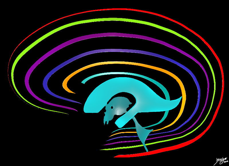
The forebrain has most of its components aligned in a series of inverted c- shaped rings starting from the outer cortex and advancing through a series of smaller inner rings with each intimately connected to the others. The thalamus appears diagramatically as the centre of these rings as seen from the sagittal view The outer and largest ring is the cerebral cortex, followed consecutively by the green ring which is the cingulate gyrus superiorly and the parahippocampal gyrus inferiorly. The red ring represents the distribution of the main portion of the anterior cerebral artery. Next is the yellow c which is the indusium griseum superiorly and the hippocampus inferiorly. This is followed by a the corpus callosum (purple) which enables the white ring of white matter to connect between hemispheres. The next ring is the thin pin kring whichrepresents the fornix superiorly and the fimbria inferiorly. The innermost ring (light blue) represents the lateral horns of the ventricular system The navy blue arrowheaded structure is the septum pellucidum
Courtesy Ashley Davidoff copyright 2010 all rights reserved 93907d14.8s


    [nodeType] => 1
    [parentNode] => (object value omitted)
    [childNodes] => (object value omitted)
    [firstChild] => (object value omitted)
    [lastChild] => (object value omitted)
    [previousSibling] => (object value omitted)
    [nextSibling] => (object value omitted)
    [attributes] => (object value omitted)
    [ownerDocument] => (object value omitted)
    [namespaceURI] =>
    [prefix] =>
    [localName] => table
    [baseURI] =>
    [textContent] =>


The Inverted C Shapes of the Forebrain


The forebrain has most of its components aligned in a series of inverted c- shaped rings starting from the outer cortex and advancing through a series of smaller inner rings with each intimately connected to the others. The thalamus appears diagramatically as the centre of these rings as seen from the sagittal view The outer and largest ring is the cerebral cortex, followed consecutively by the green ring which is the cingulate gyrus superiorly and the parahippocampal gyrus inferiorly. The red ring represents the distribution of the main portion of the anterior cerebral artery. Next is the yellow c which is the indusium griseum superiorly and the hippocampus inferiorly. This is followed by a the corpus callosum (purple) which enables the white ring of white matter to connect between hemispheres. The next ring is the thin pin kring whichrepresents the fornix superiorly and the fimbria inferiorly. The innermost ring (light blue) represents the lateral horns of the ventricular system The navy blue arrowheaded structure is the septum pellucidum
Courtesy Ashley Davidoff copyright 2010 all rights reserved 93907d14.8s


)
DOMElement Object
(
    [schemaTypeInfo] =>
    [tagName] => td
    [firstElementChild] => (object value omitted)
    [lastElementChild] => (object value omitted)
    [childElementCount] => 2
    [previousElementSibling] =>
    [nextElementSibling] =>
    [nodeName] => td
    [nodeValue] =>
The forebrain has most of its components aligned in a series of inverted c- shaped rings starting from the outer cortex and advancing through a series of smaller inner rings with each intimately connected to the others. The thalamus appears diagramatically as the centre of these rings as seen from the sagittal view The outer and largest ring is the cerebral cortex, followed consecutively by the green ring which is the cingulate gyrus superiorly and the parahippocampal gyrus inferiorly. The red ring represents the distribution of the main portion of the anterior cerebral artery. Next is the yellow c which is the indusium griseum superiorly and the hippocampus inferiorly. This is followed by a the corpus callosum (purple) which enables the white ring of white matter to connect between hemispheres. The next ring is the thin pin kring whichrepresents the fornix superiorly and the fimbria inferiorly. The innermost ring (light blue) represents the lateral horns of the ventricular system The navy blue arrowheaded structure is the septum pellucidum
Courtesy Ashley Davidoff copyright 2010 all rights reserved 93907d14.8s

    [nodeType] => 1
    [parentNode] => (object value omitted)
    [childNodes] => (object value omitted)
    [firstChild] => (object value omitted)
    [lastChild] => (object value omitted)
    [previousSibling] => (object value omitted)
    [nextSibling] => (object value omitted)
    [attributes] => (object value omitted)
    [ownerDocument] => (object value omitted)
    [namespaceURI] =>
    [prefix] =>
    [localName] => td
    [baseURI] =>
    [textContent] =>
The forebrain has most of its components aligned in a series of inverted c- shaped rings starting from the outer cortex and advancing through a series of smaller inner rings with each intimately connected to the others. The thalamus appears diagramatically as the centre of these rings as seen from the sagittal view The outer and largest ring is the cerebral cortex, followed consecutively by the green ring which is the cingulate gyrus superiorly and the parahippocampal gyrus inferiorly. The red ring represents the distribution of the main portion of the anterior cerebral artery. Next is the yellow c which is the indusium griseum superiorly and the hippocampus inferiorly. This is followed by a the corpus callosum (purple) which enables the white ring of white matter to connect between hemispheres. The next ring is the thin pin kring whichrepresents the fornix superiorly and the fimbria inferiorly. The innermost ring (light blue) represents the lateral horns of the ventricular system The navy blue arrowheaded structure is the septum pellucidum
Courtesy Ashley Davidoff copyright 2010 all rights reserved 93907d14.8s

)
DOMElement Object
(
    [schemaTypeInfo] =>
    [tagName] => td
    [firstElementChild] => (object value omitted)
    [lastElementChild] => (object value omitted)
    [childElementCount] => 2
    [previousElementSibling] =>
    [nextElementSibling] =>
    [nodeName] => td
    [nodeValue] =>
The Inverted C Shapes of the Forebrain
    [nodeType] => 1
    [parentNode] => (object value omitted)
    [childNodes] => (object value omitted)
    [firstChild] => (object value omitted)
    [lastChild] => (object value omitted)
    [previousSibling] => (object value omitted)
    [nextSibling] => (object value omitted)
    [attributes] => (object value omitted)
    [ownerDocument] => (object value omitted)
    [namespaceURI] =>
    [prefix] =>
    [localName] => td
    [baseURI] =>
    [textContent] =>
The Inverted C Shapes of the Forebrain
)
DOMElement Object
(
    [schemaTypeInfo] =>
    [tagName] => table
    [firstElementChild] => (object value omitted)
    [lastElementChild] => (object value omitted)
    [childElementCount] => 1
    [previousElementSibling] => (object value omitted)
    [nextElementSibling] => (object value omitted)
    [nodeName] => table
    [nodeValue] =>


Sagittal Midline Image
The Forebrain is Shaped like the Pancreas
Through the Interhemispheric Fissure


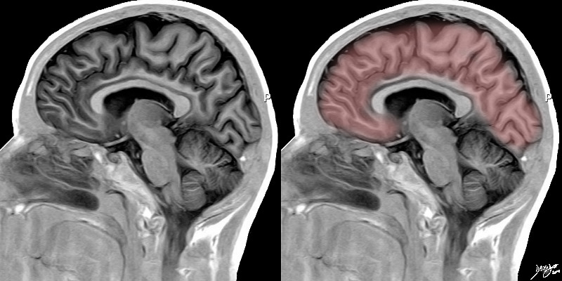
When the brain is cut through the middle fissure and divided into twio hemispheres, and then viewed by a inversion recovery MRI sequence the shape of the forebrain is reminiscent of the shape of the pancreas, almost comma shaped
Courtesy Ashley Davidoff MD Images provided by Philips Medical Systems Artistically rendered by Davidoff  92170c02.8s


    [nodeType] => 1
    [parentNode] => (object value omitted)
    [childNodes] => (object value omitted)
    [firstChild] => (object value omitted)
    [lastChild] => (object value omitted)
    [previousSibling] => (object value omitted)
    [nextSibling] => (object value omitted)
    [attributes] => (object value omitted)
    [ownerDocument] => (object value omitted)
    [namespaceURI] =>
    [prefix] =>
    [localName] => table
    [baseURI] =>
    [textContent] =>


Sagittal Midline Image
The Forebrain is Shaped like the Pancreas
Through the Interhemispheric Fissure


When the brain is cut through the middle fissure and divided into twio hemispheres, and then viewed by a inversion recovery MRI sequence the shape of the forebrain is reminiscent of the shape of the pancreas, almost comma shaped
Courtesy Ashley Davidoff MD Images provided by Philips Medical Systems Artistically rendered by Davidoff  92170c02.8s


)
DOMElement Object
(
    [schemaTypeInfo] =>
    [tagName] => td
    [firstElementChild] => (object value omitted)
    [lastElementChild] => (object value omitted)
    [childElementCount] => 2
    [previousElementSibling] =>
    [nextElementSibling] =>
    [nodeName] => td
    [nodeValue] =>
When the brain is cut through the middle fissure and divided into twio hemispheres, and then viewed by a inversion recovery MRI sequence the shape of the forebrain is reminiscent of the shape of the pancreas, almost comma shaped
Courtesy Ashley Davidoff MD Images provided by Philips Medical Systems Artistically rendered by Davidoff  92170c02.8s

    [nodeType] => 1
    [parentNode] => (object value omitted)
    [childNodes] => (object value omitted)
    [firstChild] => (object value omitted)
    [lastChild] => (object value omitted)
    [previousSibling] => (object value omitted)
    [nextSibling] => (object value omitted)
    [attributes] => (object value omitted)
    [ownerDocument] => (object value omitted)
    [namespaceURI] =>
    [prefix] =>
    [localName] => td
    [baseURI] =>
    [textContent] =>
When the brain is cut through the middle fissure and divided into twio hemispheres, and then viewed by a inversion recovery MRI sequence the shape of the forebrain is reminiscent of the shape of the pancreas, almost comma shaped
Courtesy Ashley Davidoff MD Images provided by Philips Medical Systems Artistically rendered by Davidoff  92170c02.8s

)
DOMElement Object
(
    [schemaTypeInfo] =>
    [tagName] => td
    [firstElementChild] => (object value omitted)
    [lastElementChild] => (object value omitted)
    [childElementCount] => 4
    [previousElementSibling] =>
    [nextElementSibling] =>
    [nodeName] => td
    [nodeValue] =>
Sagittal Midline Image
The Forebrain is Shaped like the Pancreas
Through the Interhemispheric Fissure

    [nodeType] => 1
    [parentNode] => (object value omitted)
    [childNodes] => (object value omitted)
    [firstChild] => (object value omitted)
    [lastChild] => (object value omitted)
    [previousSibling] => (object value omitted)
    [nextSibling] => (object value omitted)
    [attributes] => (object value omitted)
    [ownerDocument] => (object value omitted)
    [namespaceURI] =>
    [prefix] =>
    [localName] => td
    [baseURI] =>
    [textContent] =>
Sagittal Midline Image
The Forebrain is Shaped like the Pancreas
Through the Interhemispheric Fissure

)
DOMElement Object
(
    [schemaTypeInfo] =>
    [tagName] => table
    [firstElementChild] => (object value omitted)
    [lastElementChild] => (object value omitted)
    [childElementCount] => 1
    [previousElementSibling] => (object value omitted)
    [nextElementSibling] => (object value omitted)
    [nodeName] => table
    [nodeValue] =>


Coronal View
The Interhemispheric Fissure


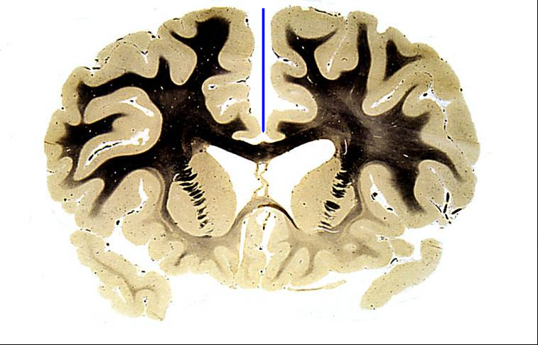
The coronal section of a normal anatomic section of the brain shows the interhemispheric fissure (blue line) and in particular the depth to which it extends into the brain.
Courtesy Elisa Flower MD Department of Radiology Boston Medical Center and Courtesy of Department Anatomy Boston University School of Medicine 97104b02


    [nodeType] => 1
    [parentNode] => (object value omitted)
    [childNodes] => (object value omitted)
    [firstChild] => (object value omitted)
    [lastChild] => (object value omitted)
    [previousSibling] => (object value omitted)
    [nextSibling] => (object value omitted)
    [attributes] => (object value omitted)
    [ownerDocument] => (object value omitted)
    [namespaceURI] =>
    [prefix] =>
    [localName] => table
    [baseURI] =>
    [textContent] =>


Coronal View
The Interhemispheric Fissure


The coronal section of a normal anatomic section of the brain shows the interhemispheric fissure (blue line) and in particular the depth to which it extends into the brain.
Courtesy Elisa Flower MD Department of Radiology Boston Medical Center and Courtesy of Department Anatomy Boston University School of Medicine 97104b02


)
DOMElement Object
(
    [schemaTypeInfo] =>
    [tagName] => td
    [firstElementChild] => (object value omitted)
    [lastElementChild] => (object value omitted)
    [childElementCount] => 2
    [previousElementSibling] =>
    [nextElementSibling] =>
    [nodeName] => td
    [nodeValue] =>
The coronal section of a normal anatomic section of the brain shows the interhemispheric fissure (blue line) and in particular the depth to which it extends into the brain.
Courtesy Elisa Flower MD Department of Radiology Boston Medical Center and Courtesy of Department Anatomy Boston University School of Medicine 97104b02

    [nodeType] => 1
    [parentNode] => (object value omitted)
    [childNodes] => (object value omitted)
    [firstChild] => (object value omitted)
    [lastChild] => (object value omitted)
    [previousSibling] => (object value omitted)
    [nextSibling] => (object value omitted)
    [attributes] => (object value omitted)
    [ownerDocument] => (object value omitted)
    [namespaceURI] =>
    [prefix] =>
    [localName] => td
    [baseURI] =>
    [textContent] =>
The coronal section of a normal anatomic section of the brain shows the interhemispheric fissure (blue line) and in particular the depth to which it extends into the brain.
Courtesy Elisa Flower MD Department of Radiology Boston Medical Center and Courtesy of Department Anatomy Boston University School of Medicine 97104b02

)
DOMElement Object
(
    [schemaTypeInfo] =>
    [tagName] => td
    [firstElementChild] => (object value omitted)
    [lastElementChild] => (object value omitted)
    [childElementCount] => 3
    [previousElementSibling] =>
    [nextElementSibling] =>
    [nodeName] => td
    [nodeValue] =>
Coronal View
The Interhemispheric Fissure
    [nodeType] => 1
    [parentNode] => (object value omitted)
    [childNodes] => (object value omitted)
    [firstChild] => (object value omitted)
    [lastChild] => (object value omitted)
    [previousSibling] => (object value omitted)
    [nextSibling] => (object value omitted)
    [attributes] => (object value omitted)
    [ownerDocument] => (object value omitted)
    [namespaceURI] =>
    [prefix] =>
    [localName] => td
    [baseURI] =>
    [textContent] =>
Coronal View
The Interhemispheric Fissure
)
DOMElement Object
(
    [schemaTypeInfo] =>
    [tagName] => table
    [firstElementChild] => (object value omitted)
    [lastElementChild] => (object value omitted)
    [childElementCount] => 1
    [previousElementSibling] => (object value omitted)
    [nextElementSibling] => (object value omitted)
    [nodeName] => table
    [nodeValue] =>


T2 weighted MRI
The Interhemispheric Fissure


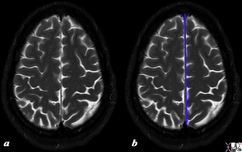
The T2 weighted MR taken a few cms from the vertex demonstrates the overall ovoid shape of the brain, the interhemishperic fissure, the sulci filled with fluid (white) enabling visualization of the gyri (black)
Courtesy Ashley DAvidoff MD copyright 2010 all rights reserved 90720c01.8s


    [nodeType] => 1
    [parentNode] => (object value omitted)
    [childNodes] => (object value omitted)
    [firstChild] => (object value omitted)
    [lastChild] => (object value omitted)
    [previousSibling] => (object value omitted)
    [nextSibling] => (object value omitted)
    [attributes] => (object value omitted)
    [ownerDocument] => (object value omitted)
    [namespaceURI] =>
    [prefix] =>
    [localName] => table
    [baseURI] =>
    [textContent] =>


T2 weighted MRI
The Interhemispheric Fissure


The T2 weighted MR taken a few cms from the vertex demonstrates the overall ovoid shape of the brain, the interhemishperic fissure, the sulci filled with fluid (white) enabling visualization of the gyri (black)
Courtesy Ashley DAvidoff MD copyright 2010 all rights reserved 90720c01.8s


)
DOMElement Object
(
    [schemaTypeInfo] =>
    [tagName] => td
    [firstElementChild] => (object value omitted)
    [lastElementChild] => (object value omitted)
    [childElementCount] => 2
    [previousElementSibling] =>
    [nextElementSibling] =>
    [nodeName] => td
    [nodeValue] =>
The T2 weighted MR taken a few cms from the vertex demonstrates the overall ovoid shape of the brain, the interhemishperic fissure, the sulci filled with fluid (white) enabling visualization of the gyri (black)
Courtesy Ashley DAvidoff MD copyright 2010 all rights reserved 90720c01.8s

    [nodeType] => 1
    [parentNode] => (object value omitted)
    [childNodes] => (object value omitted)
    [firstChild] => (object value omitted)
    [lastChild] => (object value omitted)
    [previousSibling] => (object value omitted)
    [nextSibling] => (object value omitted)
    [attributes] => (object value omitted)
    [ownerDocument] => (object value omitted)
    [namespaceURI] =>
    [prefix] =>
    [localName] => td
    [baseURI] =>
    [textContent] =>
The T2 weighted MR taken a few cms from the vertex demonstrates the overall ovoid shape of the brain, the interhemishperic fissure, the sulci filled with fluid (white) enabling visualization of the gyri (black)
Courtesy Ashley DAvidoff MD copyright 2010 all rights reserved 90720c01.8s

)
DOMElement Object
(
    [schemaTypeInfo] =>
    [tagName] => td
    [firstElementChild] => (object value omitted)
    [lastElementChild] => (object value omitted)
    [childElementCount] => 3
    [previousElementSibling] =>
    [nextElementSibling] =>
    [nodeName] => td
    [nodeValue] =>
T2 weighted MRI
The Interhemispheric Fissure

    [nodeType] => 1
    [parentNode] => (object value omitted)
    [childNodes] => (object value omitted)
    [firstChild] => (object value omitted)
    [lastChild] => (object value omitted)
    [previousSibling] => (object value omitted)
    [nextSibling] => (object value omitted)
    [attributes] => (object value omitted)
    [ownerDocument] => (object value omitted)
    [namespaceURI] =>
    [prefix] =>
    [localName] => td
    [baseURI] =>
    [textContent] =>
T2 weighted MRI
The Interhemispheric Fissure

)
DOMElement Object
(
    [schemaTypeInfo] =>
    [tagName] => table
    [firstElementChild] => (object value omitted)
    [lastElementChild] => (object value omitted)
    [childElementCount] => 1
    [previousElementSibling] => (object value omitted)
    [nextElementSibling] => (object value omitted)
    [nodeName] => table
    [nodeValue] =>


Shapes on the Top


When the external surface is explored from above a deep midline fissure (blue line) divides the oval of the brain into two hemispheres. The deep cleft is called the interhemishpheric fissure In addition, the serpiginous folds and crevices that characterize the outside surface of the brain start to become apparrent as one explores the shape of the brain in more depth. It becomes obvious that it is more than a mere oval.
 Source unknown Image modified by Davidoff 52981b05d05


    [nodeType] => 1
    [parentNode] => (object value omitted)
    [childNodes] => (object value omitted)
    [firstChild] => (object value omitted)
    [lastChild] => (object value omitted)
    [previousSibling] => (object value omitted)
    [nextSibling] => (object value omitted)
    [attributes] => (object value omitted)
    [ownerDocument] => (object value omitted)
    [namespaceURI] =>
    [prefix] =>
    [localName] => table
    [baseURI] =>
    [textContent] =>


Shapes on the Top


When the external surface is explored from above a deep midline fissure (blue line) divides the oval of the brain into two hemispheres. The deep cleft is called the interhemishpheric fissure In addition, the serpiginous folds and crevices that characterize the outside surface of the brain start to become apparrent as one explores the shape of the brain in more depth. It becomes obvious that it is more than a mere oval.
 Source unknown Image modified by Davidoff 52981b05d05


)
DOMElement Object
(
    [schemaTypeInfo] =>
    [tagName] => td
    [firstElementChild] => (object value omitted)
    [lastElementChild] => (object value omitted)
    [childElementCount] => 2
    [previousElementSibling] =>
    [nextElementSibling] =>
    [nodeName] => td
    [nodeValue] =>
When the external surface is explored from above a deep midline fissure (blue line) divides the oval of the brain into two hemispheres. The deep cleft is called the interhemishpheric fissure In addition, the serpiginous folds and crevices that characterize the outside surface of the brain start to become apparrent as one explores the shape of the brain in more depth. It becomes obvious that it is more than a mere oval.
 Source unknown Image modified by Davidoff 52981b05d05

    [nodeType] => 1
    [parentNode] => (object value omitted)
    [childNodes] => (object value omitted)
    [firstChild] => (object value omitted)
    [lastChild] => (object value omitted)
    [previousSibling] => (object value omitted)
    [nextSibling] => (object value omitted)
    [attributes] => (object value omitted)
    [ownerDocument] => (object value omitted)
    [namespaceURI] =>
    [prefix] =>
    [localName] => td
    [baseURI] =>
    [textContent] =>
When the external surface is explored from above a deep midline fissure (blue line) divides the oval of the brain into two hemispheres. The deep cleft is called the interhemishpheric fissure In addition, the serpiginous folds and crevices that characterize the outside surface of the brain start to become apparrent as one explores the shape of the brain in more depth. It becomes obvious that it is more than a mere oval.
 Source unknown Image modified by Davidoff 52981b05d05

)
DOMElement Object
(
    [schemaTypeInfo] =>
    [tagName] => td
    [firstElementChild] => (object value omitted)
    [lastElementChild] => (object value omitted)
    [childElementCount] => 2
    [previousElementSibling] =>
    [nextElementSibling] =>
    [nodeName] => td
    [nodeValue] =>

Shapes on the Top

    [nodeType] => 1
    [parentNode] => (object value omitted)
    [childNodes] => (object value omitted)
    [firstChild] => (object value omitted)
    [lastChild] => (object value omitted)
    [previousSibling] => (object value omitted)
    [nextSibling] => (object value omitted)
    [attributes] => (object value omitted)
    [ownerDocument] => (object value omitted)
    [namespaceURI] =>
    [prefix] =>
    [localName] => td
    [baseURI] =>
    [textContent] =>

Shapes on the Top

)
DOMElement Object
(
    [schemaTypeInfo] =>
    [tagName] => table
    [firstElementChild] => (object value omitted)
    [lastElementChild] => (object value omitted)
    [childElementCount] => 1
    [previousElementSibling] => (object value omitted)
    [nextElementSibling] => (object value omitted)
    [nodeName] => table
    [nodeValue] =>


Introduction to the Central Sulci and Gyri
Characteristic Location and Shape


The lateral view of the brain shows the 4 important central sulci (aka fissures), 3 of which are vertically oriented and 1 of which is horizontal. The vertical sulci include the central sulcus, precentral sulcus and post central sulcus. The central sulcus is important because it separates the frontal lobe from the parietal lobe. The post central sulcus is important because together with the central sulcus it forms the borders of the post central gyrus (pink) which is the sensory cortex. The precentral sulcus together with the central sulcus forms the borders post central gyrus (blue) which is the motor cortex. The Sylvian fissure or lateral sulcus runs horizontally and separates the temporal lobe below from the frontal lobe and parietal lobe above.
Courtesy Ashley Davidoff copyright 2010 all rights reserved 83029b01b01b02g01L.8s


    [nodeType] => 1
    [parentNode] => (object value omitted)
    [childNodes] => (object value omitted)
    [firstChild] => (object value omitted)
    [lastChild] => (object value omitted)
    [previousSibling] => (object value omitted)
    [nextSibling] => (object value omitted)
    [attributes] => (object value omitted)
    [ownerDocument] => (object value omitted)
    [namespaceURI] =>
    [prefix] =>
    [localName] => table
    [baseURI] =>
    [textContent] =>


Introduction to the Central Sulci and Gyri
Characteristic Location and Shape


The lateral view of the brain shows the 4 important central sulci (aka fissures), 3 of which are vertically oriented and 1 of which is horizontal. The vertical sulci include the central sulcus, precentral sulcus and post central sulcus. The central sulcus is important because it separates the frontal lobe from the parietal lobe. The post central sulcus is important because together with the central sulcus it forms the borders of the post central gyrus (pink) which is the sensory cortex. The precentral sulcus together with the central sulcus forms the borders post central gyrus (blue) which is the motor cortex. The Sylvian fissure or lateral sulcus runs horizontally and separates the temporal lobe below from the frontal lobe and parietal lobe above.
Courtesy Ashley Davidoff copyright 2010 all rights reserved 83029b01b01b02g01L.8s


)
DOMElement Object
(
    [schemaTypeInfo] =>
    [tagName] => td
    [firstElementChild] => (object value omitted)
    [lastElementChild] => (object value omitted)
    [childElementCount] => 2
    [previousElementSibling] =>
    [nextElementSibling] =>
    [nodeName] => td
    [nodeValue] =>
The lateral view of the brain shows the 4 important central sulci (aka fissures), 3 of which are vertically oriented and 1 of which is horizontal. The vertical sulci include the central sulcus, precentral sulcus and post central sulcus. The central sulcus is important because it separates the frontal lobe from the parietal lobe. The post central sulcus is important because together with the central sulcus it forms the borders of the post central gyrus (pink) which is the sensory cortex. The precentral sulcus together with the central sulcus forms the borders post central gyrus (blue) which is the motor cortex. The Sylvian fissure or lateral sulcus runs horizontally and separates the temporal lobe below from the frontal lobe and parietal lobe above.
Courtesy Ashley Davidoff copyright 2010 all rights reserved 83029b01b01b02g01L.8s
    [nodeType] => 1
    [parentNode] => (object value omitted)
    [childNodes] => (object value omitted)
    [firstChild] => (object value omitted)
    [lastChild] => (object value omitted)
    [previousSibling] => (object value omitted)
    [nextSibling] => (object value omitted)
    [attributes] => (object value omitted)
    [ownerDocument] => (object value omitted)
    [namespaceURI] =>
    [prefix] =>
    [localName] => td
    [baseURI] =>
    [textContent] =>
The lateral view of the brain shows the 4 important central sulci (aka fissures), 3 of which are vertically oriented and 1 of which is horizontal. The vertical sulci include the central sulcus, precentral sulcus and post central sulcus. The central sulcus is important because it separates the frontal lobe from the parietal lobe. The post central sulcus is important because together with the central sulcus it forms the borders of the post central gyrus (pink) which is the sensory cortex. The precentral sulcus together with the central sulcus forms the borders post central gyrus (blue) which is the motor cortex. The Sylvian fissure or lateral sulcus runs horizontally and separates the temporal lobe below from the frontal lobe and parietal lobe above.
Courtesy Ashley Davidoff copyright 2010 all rights reserved 83029b01b01b02g01L.8s
)
DOMElement Object
(
    [schemaTypeInfo] =>
    [tagName] => td
    [firstElementChild] => (object value omitted)
    [lastElementChild] => (object value omitted)
    [childElementCount] => 3
    [previousElementSibling] =>
    [nextElementSibling] =>
    [nodeName] => td
    [nodeValue] =>
Introduction to the Central Sulci and Gyri
Characteristic Location and Shape
    [nodeType] => 1
    [parentNode] => (object value omitted)
    [childNodes] => (object value omitted)
    [firstChild] => (object value omitted)
    [lastChild] => (object value omitted)
    [previousSibling] => (object value omitted)
    [nextSibling] => (object value omitted)
    [attributes] => (object value omitted)
    [ownerDocument] => (object value omitted)
    [namespaceURI] =>
    [prefix] =>
    [localName] => td
    [baseURI] =>
    [textContent] =>
Introduction to the Central Sulci and Gyri
Characteristic Location and Shape
)
DOMElement Object
(
    [schemaTypeInfo] =>
    [tagName] => table
    [firstElementChild] => (object value omitted)
    [lastElementChild] => (object value omitted)
    [childElementCount] => 1
    [previousElementSibling] => (object value omitted)
    [nextElementSibling] => (object value omitted)
    [nodeName] => table
    [nodeValue] =>


Brain Coral


The brain coral
83018.800 brain cerebral coral Bahamas water animalsulcus sulci gyrus gyri gray matter anatomy character parts white matter TCV the common vein applied biology Davidoff photography


    [nodeType] => 1
    [parentNode] => (object value omitted)
    [childNodes] => (object value omitted)
    [firstChild] => (object value omitted)
    [lastChild] => (object value omitted)
    [previousSibling] => (object value omitted)
    [nextSibling] => (object value omitted)
    [attributes] => (object value omitted)
    [ownerDocument] => (object value omitted)
    [namespaceURI] =>
    [prefix] =>
    [localName] => table
    [baseURI] =>
    [textContent] =>


Brain Coral


The brain coral
83018.800 brain cerebral coral Bahamas water animalsulcus sulci gyrus gyri gray matter anatomy character parts white matter TCV the common vein applied biology Davidoff photography


)
DOMElement Object
(
    [schemaTypeInfo] =>
    [tagName] => td
    [firstElementChild] => (object value omitted)
    [lastElementChild] => (object value omitted)
    [childElementCount] => 2
    [previousElementSibling] =>
    [nextElementSibling] =>
    [nodeName] => td
    [nodeValue] =>
The brain coral
83018.800 brain cerebral coral Bahamas water animalsulcus sulci gyrus gyri gray matter anatomy character parts white matter TCV the common vein applied biology Davidoff photography

    [nodeType] => 1
    [parentNode] => (object value omitted)
    [childNodes] => (object value omitted)
    [firstChild] => (object value omitted)
    [lastChild] => (object value omitted)
    [previousSibling] => (object value omitted)
    [nextSibling] => (object value omitted)
    [attributes] => (object value omitted)
    [ownerDocument] => (object value omitted)
    [namespaceURI] =>
    [prefix] =>
    [localName] => td
    [baseURI] =>
    [textContent] =>
The brain coral
83018.800 brain cerebral coral Bahamas water animalsulcus sulci gyrus gyri gray matter anatomy character parts white matter TCV the common vein applied biology Davidoff photography

)
DOMElement Object
(
    [schemaTypeInfo] =>
    [tagName] => td
    [firstElementChild] => (object value omitted)
    [lastElementChild] => (object value omitted)
    [childElementCount] => 2
    [previousElementSibling] =>
    [nextElementSibling] =>
    [nodeName] => td
    [nodeValue] =>
Brain Coral
    [nodeType] => 1
    [parentNode] => (object value omitted)
    [childNodes] => (object value omitted)
    [firstChild] => (object value omitted)
    [lastChild] => (object value omitted)
    [previousSibling] => (object value omitted)
    [nextSibling] => (object value omitted)
    [attributes] => (object value omitted)
    [ownerDocument] => (object value omitted)
    [namespaceURI] =>
    [prefix] =>
    [localName] => td
    [baseURI] =>
    [textContent] =>
Brain Coral
)
DOMElement Object
(
    [schemaTypeInfo] =>
    [tagName] => table
    [firstElementChild] => (object value omitted)
    [lastElementChild] => (object value omitted)
    [childElementCount] => 1
    [previousElementSibling] => (object value omitted)
    [nextElementSibling] => (object value omitted)
    [nodeName] => table
    [nodeValue] =>


Form and Colors reminiscent of the Brain


This image of a walnut has a few features that are reminiscent of a brain, including the symmetric hemispheric components, the pinkish color, and the undulations of the surface. However in reality it is about 1.8cms long, and only 5-8mms high, and is hard.
Courtesy Ashley Davidoff MD copyright 2010 all rights reserved 100286pb.93s


    [nodeType] => 1
    [parentNode] => (object value omitted)
    [childNodes] => (object value omitted)
    [firstChild] => (object value omitted)
    [lastChild] => (object value omitted)
    [previousSibling] => (object value omitted)
    [nextSibling] => (object value omitted)
    [attributes] => (object value omitted)
    [ownerDocument] => (object value omitted)
    [namespaceURI] =>
    [prefix] =>
    [localName] => table
    [baseURI] =>
    [textContent] =>


Form and Colors reminiscent of the Brain


This image of a walnut has a few features that are reminiscent of a brain, including the symmetric hemispheric components, the pinkish color, and the undulations of the surface. However in reality it is about 1.8cms long, and only 5-8mms high, and is hard.
Courtesy Ashley Davidoff MD copyright 2010 all rights reserved 100286pb.93s


)
DOMElement Object
(
    [schemaTypeInfo] =>
    [tagName] => td
    [firstElementChild] => (object value omitted)
    [lastElementChild] => (object value omitted)
    [childElementCount] => 2
    [previousElementSibling] =>
    [nextElementSibling] =>
    [nodeName] => td
    [nodeValue] =>
This image of a walnut has a few features that are reminiscent of a brain, including the symmetric hemispheric components, the pinkish color, and the undulations of the surface. However in reality it is about 1.8cms long, and only 5-8mms high, and is hard.
Courtesy Ashley Davidoff MD copyright 2010 all rights reserved 100286pb.93s

    [nodeType] => 1
    [parentNode] => (object value omitted)
    [childNodes] => (object value omitted)
    [firstChild] => (object value omitted)
    [lastChild] => (object value omitted)
    [previousSibling] => (object value omitted)
    [nextSibling] => (object value omitted)
    [attributes] => (object value omitted)
    [ownerDocument] => (object value omitted)
    [namespaceURI] =>
    [prefix] =>
    [localName] => td
    [baseURI] =>
    [textContent] =>
This image of a walnut has a few features that are reminiscent of a brain, including the symmetric hemispheric components, the pinkish color, and the undulations of the surface. However in reality it is about 1.8cms long, and only 5-8mms high, and is hard.
Courtesy Ashley Davidoff MD copyright 2010 all rights reserved 100286pb.93s

)
DOMElement Object
(
    [schemaTypeInfo] =>
    [tagName] => td
    [firstElementChild] => (object value omitted)
    [lastElementChild] => (object value omitted)
    [childElementCount] => 2
    [previousElementSibling] =>
    [nextElementSibling] =>
    [nodeName] => td
    [nodeValue] =>

Form and Colors reminiscent of the Brain
    [nodeType] => 1
    [parentNode] => (object value omitted)
    [childNodes] => (object value omitted)
    [firstChild] => (object value omitted)
    [lastChild] => (object value omitted)
    [previousSibling] => (object value omitted)
    [nextSibling] => (object value omitted)
    [attributes] => (object value omitted)
    [ownerDocument] => (object value omitted)
    [namespaceURI] =>
    [prefix] =>
    [localName] => td
    [baseURI] =>
    [textContent] =>

Form and Colors reminiscent of the Brain
)
DOMElement Object
(
    [schemaTypeInfo] =>
    [tagName] => table
    [firstElementChild] => (object value omitted)
    [lastElementChild] => (object value omitted)
    [childElementCount] => 1
    [previousElementSibling] => (object value omitted)
    [nextElementSibling] => (object value omitted)
    [nodeName] => table
    [nodeValue] =>


Basic Shapes
Lateral Projection


When the external surface of the brain is viewed from its lateral aspect, , the ovoid shape of the conglomerate frontal parietal, occipital and part of the temporal lobe is still maintained. The part of the temporal lobe that projects into the middle cranial fossa adds a second oval directed slightly inferiorly. The medulla adds a new dimension having the shape somewhere between a half a sphere and the bottom of a top.
Davidoff art All Rights reserved copyright 2010 83029d03c01.9s


    [nodeType] => 1
    [parentNode] => (object value omitted)
    [childNodes] => (object value omitted)
    [firstChild] => (object value omitted)
    [lastChild] => (object value omitted)
    [previousSibling] => (object value omitted)
    [nextSibling] => (object value omitted)
    [attributes] => (object value omitted)
    [ownerDocument] => (object value omitted)
    [namespaceURI] =>
    [prefix] =>
    [localName] => table
    [baseURI] =>
    [textContent] =>


Basic Shapes
Lateral Projection


When the external surface of the brain is viewed from its lateral aspect, , the ovoid shape of the conglomerate frontal parietal, occipital and part of the temporal lobe is still maintained. The part of the temporal lobe that projects into the middle cranial fossa adds a second oval directed slightly inferiorly. The medulla adds a new dimension having the shape somewhere between a half a sphere and the bottom of a top.
Davidoff art All Rights reserved copyright 2010 83029d03c01.9s


)
DOMElement Object
(
    [schemaTypeInfo] =>
    [tagName] => td
    [firstElementChild] => (object value omitted)
    [lastElementChild] => (object value omitted)
    [childElementCount] => 2
    [previousElementSibling] =>
    [nextElementSibling] =>
    [nodeName] => td
    [nodeValue] =>
When the external surface of the brain is viewed from its lateral aspect, , the ovoid shape of the conglomerate frontal parietal, occipital and part of the temporal lobe is still maintained. The part of the temporal lobe that projects into the middle cranial fossa adds a second oval directed slightly inferiorly. The medulla adds a new dimension having the shape somewhere between a half a sphere and the bottom of a top.
Davidoff art All Rights reserved copyright 2010 83029d03c01.9s

    [nodeType] => 1
    [parentNode] => (object value omitted)
    [childNodes] => (object value omitted)
    [firstChild] => (object value omitted)
    [lastChild] => (object value omitted)
    [previousSibling] => (object value omitted)
    [nextSibling] => (object value omitted)
    [attributes] => (object value omitted)
    [ownerDocument] => (object value omitted)
    [namespaceURI] =>
    [prefix] =>
    [localName] => td
    [baseURI] =>
    [textContent] =>
When the external surface of the brain is viewed from its lateral aspect, , the ovoid shape of the conglomerate frontal parietal, occipital and part of the temporal lobe is still maintained. The part of the temporal lobe that projects into the middle cranial fossa adds a second oval directed slightly inferiorly. The medulla adds a new dimension having the shape somewhere between a half a sphere and the bottom of a top.
Davidoff art All Rights reserved copyright 2010 83029d03c01.9s

)
DOMElement Object
(
    [schemaTypeInfo] =>
    [tagName] => td
    [firstElementChild] => (object value omitted)
    [lastElementChild] => (object value omitted)
    [childElementCount] => 3
    [previousElementSibling] =>
    [nextElementSibling] =>
    [nodeName] => td
    [nodeValue] =>
Basic Shapes
Lateral Projection

    [nodeType] => 1
    [parentNode] => (object value omitted)
    [childNodes] => (object value omitted)
    [firstChild] => (object value omitted)
    [lastChild] => (object value omitted)
    [previousSibling] => (object value omitted)
    [nextSibling] => (object value omitted)
    [attributes] => (object value omitted)
    [ownerDocument] => (object value omitted)
    [namespaceURI] =>
    [prefix] =>
    [localName] => td
    [baseURI] =>
    [textContent] =>
Basic Shapes
Lateral Projection

)
DOMElement Object
(
    [schemaTypeInfo] =>
    [tagName] => table
    [firstElementChild] => (object value omitted)
    [lastElementChild] => (object value omitted)
    [childElementCount] => 1
    [previousElementSibling] => (object value omitted)
    [nextElementSibling] => (object value omitted)
    [nodeName] => table
    [nodeValue] =>


General Shape of the Brain
External Surface


The ovoid shape is easily appreciated when the external surface of the brain is viewed from above, and although it in general it has an ovoid shape when viewed from the side, more complex shapes start to appear, as the temporal and cerebellum start to appear.
source unknown modified by Davidoff 52981b05.8sd01


    [nodeType] => 1
    [parentNode] => (object value omitted)
    [childNodes] => (object value omitted)
    [firstChild] => (object value omitted)
    [lastChild] => (object value omitted)
    [previousSibling] => (object value omitted)
    [nextSibling] => (object value omitted)
    [attributes] => (object value omitted)
    [ownerDocument] => (object value omitted)
    [namespaceURI] =>
    [prefix] =>
    [localName] => table
    [baseURI] =>
    [textContent] =>


General Shape of the Brain
External Surface


The ovoid shape is easily appreciated when the external surface of the brain is viewed from above, and although it in general it has an ovoid shape when viewed from the side, more complex shapes start to appear, as the temporal and cerebellum start to appear.
source unknown modified by Davidoff 52981b05.8sd01


)
DOMElement Object
(
    [schemaTypeInfo] =>
    [tagName] => td
    [firstElementChild] => (object value omitted)
    [lastElementChild] => (object value omitted)
    [childElementCount] => 2
    [previousElementSibling] =>
    [nextElementSibling] =>
    [nodeName] => td
    [nodeValue] =>
The ovoid shape is easily appreciated when the external surface of the brain is viewed from above, and although it in general it has an ovoid shape when viewed from the side, more complex shapes start to appear, as the temporal and cerebellum start to appear.
source unknown modified by Davidoff 52981b05.8sd01

    [nodeType] => 1
    [parentNode] => (object value omitted)
    [childNodes] => (object value omitted)
    [firstChild] => (object value omitted)
    [lastChild] => (object value omitted)
    [previousSibling] => (object value omitted)
    [nextSibling] => (object value omitted)
    [attributes] => (object value omitted)
    [ownerDocument] => (object value omitted)
    [namespaceURI] =>
    [prefix] =>
    [localName] => td
    [baseURI] =>
    [textContent] =>
The ovoid shape is easily appreciated when the external surface of the brain is viewed from above, and although it in general it has an ovoid shape when viewed from the side, more complex shapes start to appear, as the temporal and cerebellum start to appear.
source unknown modified by Davidoff 52981b05.8sd01

)
DOMElement Object
(
    [schemaTypeInfo] =>
    [tagName] => td
    [firstElementChild] => (object value omitted)
    [lastElementChild] => (object value omitted)
    [childElementCount] => 3
    [previousElementSibling] =>
    [nextElementSibling] =>
    [nodeName] => td
    [nodeValue] =>
General Shape of the Brain
External Surface

    [nodeType] => 1
    [parentNode] => (object value omitted)
    [childNodes] => (object value omitted)
    [firstChild] => (object value omitted)
    [lastChild] => (object value omitted)
    [previousSibling] => (object value omitted)
    [nextSibling] => (object value omitted)
    [attributes] => (object value omitted)
    [ownerDocument] => (object value omitted)
    [namespaceURI] =>
    [prefix] =>
    [localName] => td
    [baseURI] =>
    [textContent] =>
General Shape of the Brain
External Surface

)

