Sumit Karia MD Ashley Davidoff MD
The Common Vein Copyright 2010
Introduction
There are many places to start the study of the brain, but we have decided to start with the classical lateral view, and to define the main vectors in this view, and thereafter superimpose the vectors on a brain specimen.
|
The Basic Structure of the Brain from its LAteral Aspect The Brain at its Simplest |
|
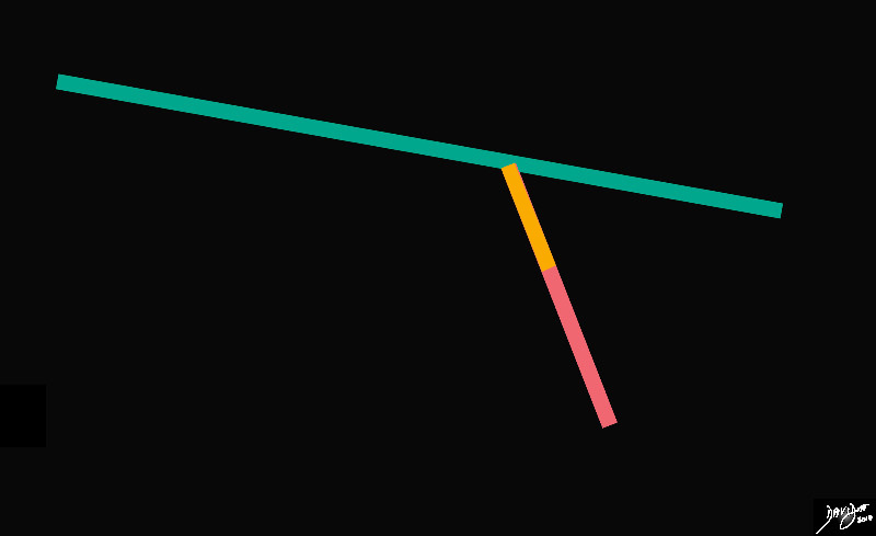
The stick diagram divides the brain into a relatively long almost horizontal component, and a relatively short almost vertical component. Anatomically the horizontal component is called the forebrain, and the vertical component will make up the midbrain (orange) and hind brain (salmon) and extend ino the spinal cord Davidoff art Courtesy Ashley Davidoff MD copyright 2010 all rights reserved 93881b02.81s |
|
The Sagital Conceptual Vectors Overlying an External Lateral View of the Brain |
|
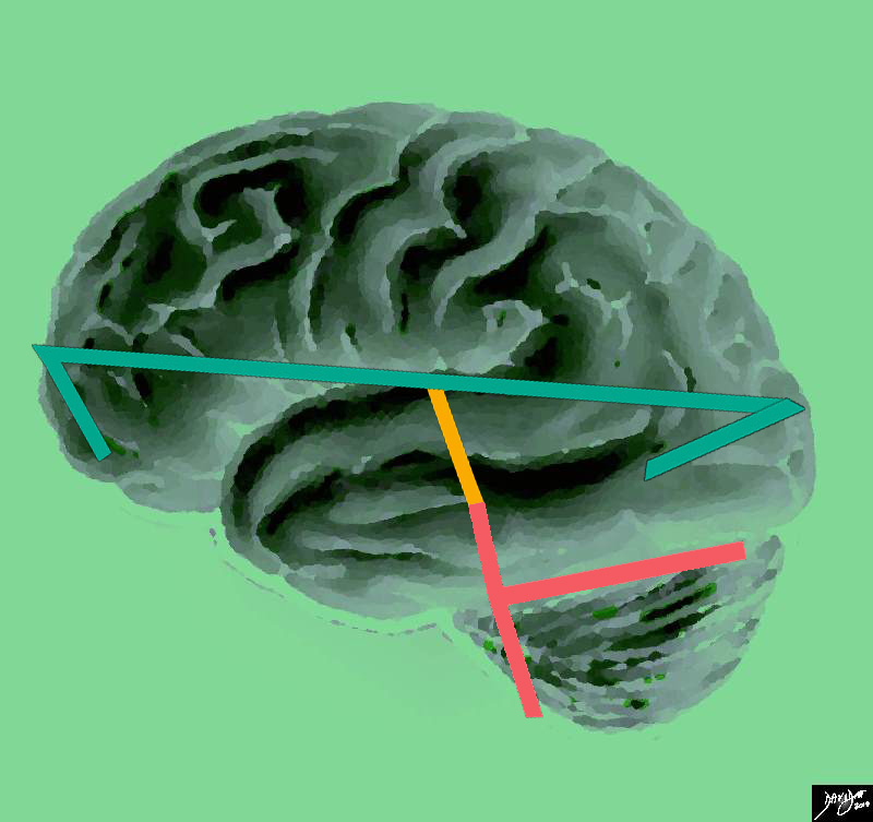
The vectors of the brain from a side view are superimposed on a specimen of the brain, showing a long anteroposterior vector of the forebrain (light green) and a shorter more vertical vector consisting of the midbrain (orange) and the hind brain (salmon pink) Davidoff Art courtesy Ashley Davidoff copyright 2010 all rights reserved 83029e04b01.85s |
|
Artistic rendition Reflecting the Concept of Inverted C -Shaped Rings |
|
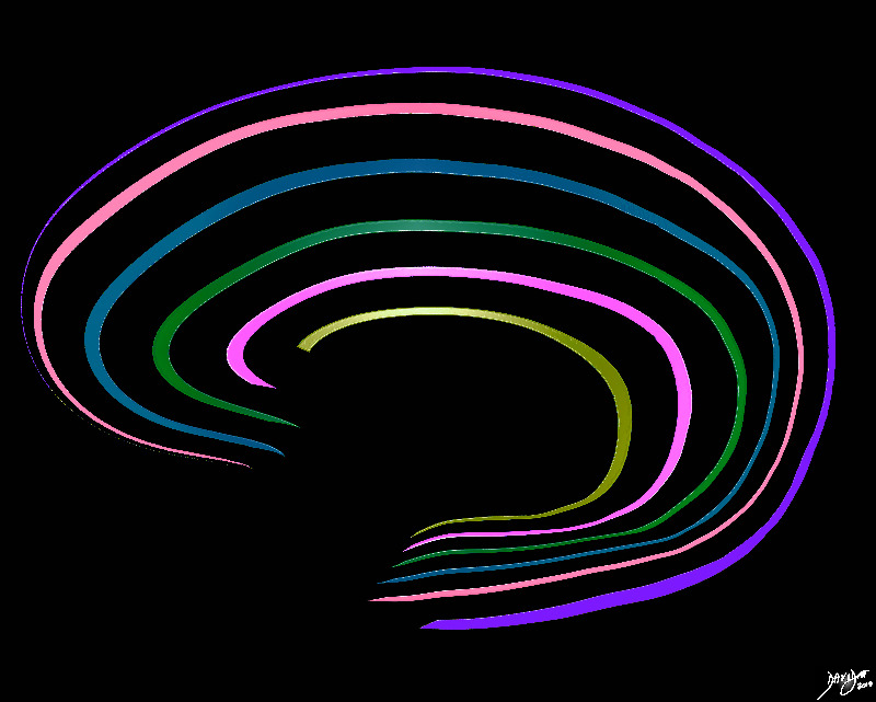
The shape of the brain in general is an ovoid. As one goes beyond the surface in the sagittal plane, the brain can be viewed as a series of inverted c shaped structures. Davidoff art Courtesy Ashley Davidoff MD copyright 2010 all rights reserved 93890b01b06.8 |
|
The Forebrain A series of C rings
|
|
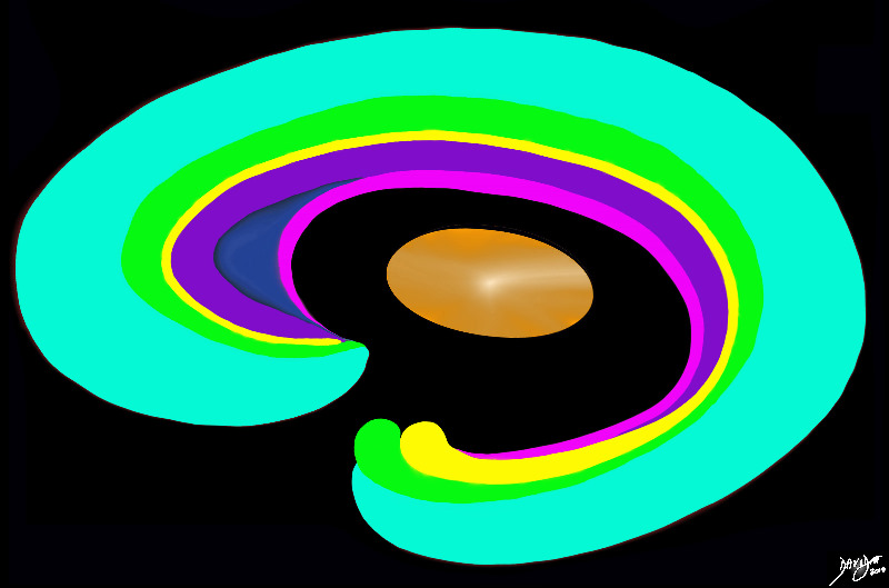
The forebrain has most of its components aligned in a series of c-shaped rings starting from the outer cortex and advancing through a series of smaller inner rings with each intimately connected to the others. The thalamus apeears diagramatically as the center of these rings as seen from the sagittal view Davidoff art Courtesy Ashley Davidoff copyright 2010 all rights reserved 93907b01.8s |
|
Conceptual Framework of the Hindbrain |
|
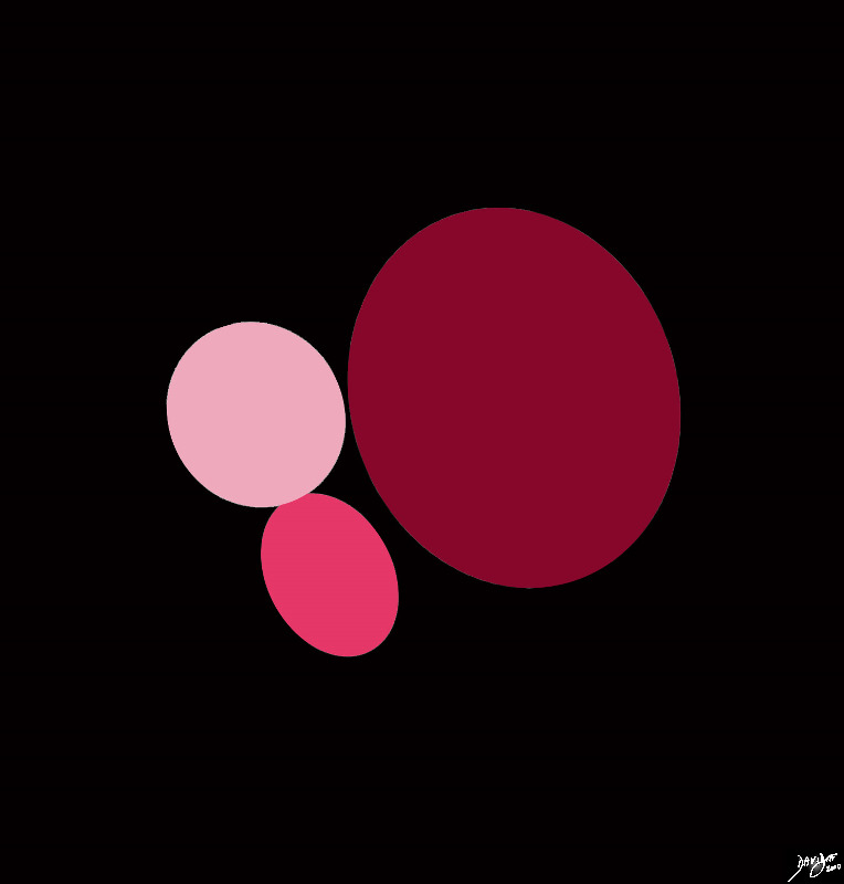
The hindbrain conceptually consists of 3 ovoids The pons is anterior and superior (light pink), the medulla is smaller and is anterior and inferior, and the cerebellum is the largest and is posterior. Courtesy Ashley Davidoff MD Copyright 2010 all rights reserved 92141.3kd03b03b01.8s |
|
The Concept In Vivo |
|
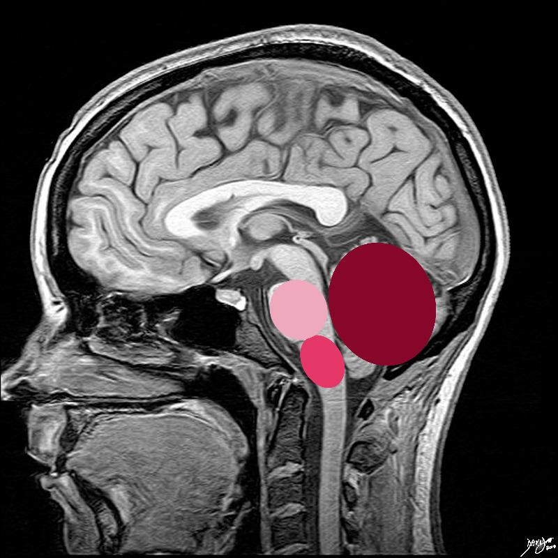
The 3 ovoids are situated in the posterior cranial fossa the pons is anterior and superior (light pink), the medulla is smaller and is anterior and inferior, and the cerebellum is the largest and is posterior. Courtesy Ashley Davidoff MD Copyright 2010 all rights reserved 92141.3kd03b03b.8s |
DOMElement Object
(
    [schemaTypeInfo] =>
    [tagName] => table
    [firstElementChild] => (object value omitted)
    [lastElementChild] => (object value omitted)
    [childElementCount] => 1
    [previousElementSibling] => (object value omitted)
    [nextElementSibling] =>
    [nodeName] => table
    [nodeValue] =>


The Concept In Vivo


The 3 ovoids are situated in the posterior cranial fossa the pons is anterior and superior (light pink), the medulla is smaller and is anterior and inferior, and the cerebellum is the largest and is posterior.
Courtesy Ashley Davidoff MD Copyright 2010 all rights reserved 92141.3kd03b03b.8s


    [nodeType] => 1
    [parentNode] => (object value omitted)
    [childNodes] => (object value omitted)
    [firstChild] => (object value omitted)
    [lastChild] => (object value omitted)
    [previousSibling] => (object value omitted)
    [nextSibling] => (object value omitted)
    [attributes] => (object value omitted)
    [ownerDocument] => (object value omitted)
    [namespaceURI] =>
    [prefix] =>
    [localName] => table
    [baseURI] =>
    [textContent] =>


The Concept In Vivo


The 3 ovoids are situated in the posterior cranial fossa the pons is anterior and superior (light pink), the medulla is smaller and is anterior and inferior, and the cerebellum is the largest and is posterior.
Courtesy Ashley Davidoff MD Copyright 2010 all rights reserved 92141.3kd03b03b.8s


)
DOMElement Object
(
    [schemaTypeInfo] =>
    [tagName] => td
    [firstElementChild] => (object value omitted)
    [lastElementChild] => (object value omitted)
    [childElementCount] => 2
    [previousElementSibling] =>
    [nextElementSibling] =>
    [nodeName] => td
    [nodeValue] =>
The 3 ovoids are situated in the posterior cranial fossa the pons is anterior and superior (light pink), the medulla is smaller and is anterior and inferior, and the cerebellum is the largest and is posterior.
Courtesy Ashley Davidoff MD Copyright 2010 all rights reserved 92141.3kd03b03b.8s

    [nodeType] => 1
    [parentNode] => (object value omitted)
    [childNodes] => (object value omitted)
    [firstChild] => (object value omitted)
    [lastChild] => (object value omitted)
    [previousSibling] => (object value omitted)
    [nextSibling] => (object value omitted)
    [attributes] => (object value omitted)
    [ownerDocument] => (object value omitted)
    [namespaceURI] =>
    [prefix] =>
    [localName] => td
    [baseURI] =>
    [textContent] =>
The 3 ovoids are situated in the posterior cranial fossa the pons is anterior and superior (light pink), the medulla is smaller and is anterior and inferior, and the cerebellum is the largest and is posterior.
Courtesy Ashley Davidoff MD Copyright 2010 all rights reserved 92141.3kd03b03b.8s

)
DOMElement Object
(
    [schemaTypeInfo] =>
    [tagName] => td
    [firstElementChild] => (object value omitted)
    [lastElementChild] => (object value omitted)
    [childElementCount] => 2
    [previousElementSibling] =>
    [nextElementSibling] =>
    [nodeName] => td
    [nodeValue] =>

The Concept In Vivo
    [nodeType] => 1
    [parentNode] => (object value omitted)
    [childNodes] => (object value omitted)
    [firstChild] => (object value omitted)
    [lastChild] => (object value omitted)
    [previousSibling] => (object value omitted)
    [nextSibling] => (object value omitted)
    [attributes] => (object value omitted)
    [ownerDocument] => (object value omitted)
    [namespaceURI] =>
    [prefix] =>
    [localName] => td
    [baseURI] =>
    [textContent] =>

The Concept In Vivo
)
DOMElement Object
(
    [schemaTypeInfo] =>
    [tagName] => table
    [firstElementChild] => (object value omitted)
    [lastElementChild] => (object value omitted)
    [childElementCount] => 1
    [previousElementSibling] => (object value omitted)
    [nextElementSibling] => (object value omitted)
    [nodeName] => table
    [nodeValue] =>


Conceptual Framework of the Hindbrain


The hindbrain conceptually consists of 3 ovoids The pons is anterior and superior (light pink), the medulla is smaller and is anterior and inferior, and the cerebellum is the largest and is posterior.
Courtesy Ashley Davidoff MD Copyright 2010 all rights reserved 92141.3kd03b03b01.8s


    [nodeType] => 1
    [parentNode] => (object value omitted)
    [childNodes] => (object value omitted)
    [firstChild] => (object value omitted)
    [lastChild] => (object value omitted)
    [previousSibling] => (object value omitted)
    [nextSibling] => (object value omitted)
    [attributes] => (object value omitted)
    [ownerDocument] => (object value omitted)
    [namespaceURI] =>
    [prefix] =>
    [localName] => table
    [baseURI] =>
    [textContent] =>


Conceptual Framework of the Hindbrain


The hindbrain conceptually consists of 3 ovoids The pons is anterior and superior (light pink), the medulla is smaller and is anterior and inferior, and the cerebellum is the largest and is posterior.
Courtesy Ashley Davidoff MD Copyright 2010 all rights reserved 92141.3kd03b03b01.8s


)
DOMElement Object
(
    [schemaTypeInfo] =>
    [tagName] => td
    [firstElementChild] => (object value omitted)
    [lastElementChild] => (object value omitted)
    [childElementCount] => 2
    [previousElementSibling] =>
    [nextElementSibling] =>
    [nodeName] => td
    [nodeValue] =>
The hindbrain conceptually consists of 3 ovoids The pons is anterior and superior (light pink), the medulla is smaller and is anterior and inferior, and the cerebellum is the largest and is posterior.
Courtesy Ashley Davidoff MD Copyright 2010 all rights reserved 92141.3kd03b03b01.8s

    [nodeType] => 1
    [parentNode] => (object value omitted)
    [childNodes] => (object value omitted)
    [firstChild] => (object value omitted)
    [lastChild] => (object value omitted)
    [previousSibling] => (object value omitted)
    [nextSibling] => (object value omitted)
    [attributes] => (object value omitted)
    [ownerDocument] => (object value omitted)
    [namespaceURI] =>
    [prefix] =>
    [localName] => td
    [baseURI] =>
    [textContent] =>
The hindbrain conceptually consists of 3 ovoids The pons is anterior and superior (light pink), the medulla is smaller and is anterior and inferior, and the cerebellum is the largest and is posterior.
Courtesy Ashley Davidoff MD Copyright 2010 all rights reserved 92141.3kd03b03b01.8s

)
DOMElement Object
(
    [schemaTypeInfo] =>
    [tagName] => td
    [firstElementChild] => (object value omitted)
    [lastElementChild] => (object value omitted)
    [childElementCount] => 2
    [previousElementSibling] =>
    [nextElementSibling] =>
    [nodeName] => td
    [nodeValue] =>

Conceptual Framework of the Hindbrain
    [nodeType] => 1
    [parentNode] => (object value omitted)
    [childNodes] => (object value omitted)
    [firstChild] => (object value omitted)
    [lastChild] => (object value omitted)
    [previousSibling] => (object value omitted)
    [nextSibling] => (object value omitted)
    [attributes] => (object value omitted)
    [ownerDocument] => (object value omitted)
    [namespaceURI] =>
    [prefix] =>
    [localName] => td
    [baseURI] =>
    [textContent] =>

Conceptual Framework of the Hindbrain
)
DOMElement Object
(
    [schemaTypeInfo] =>
    [tagName] => table
    [firstElementChild] => (object value omitted)
    [lastElementChild] => (object value omitted)
    [childElementCount] => 1
    [previousElementSibling] => (object value omitted)
    [nextElementSibling] => (object value omitted)
    [nodeName] => table
    [nodeValue] =>


MRI of the Midbrain


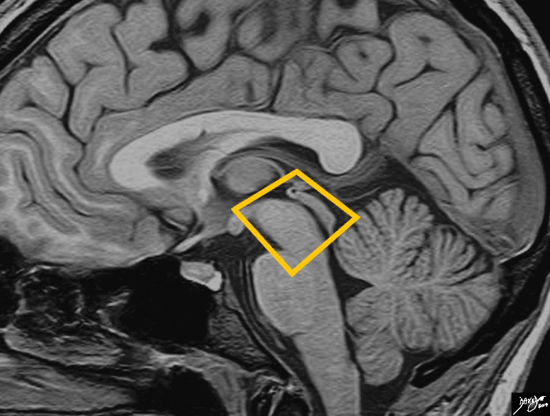
The midbrain is an almost rectangular structure that bridges the forebrain and hindbrain. It is the smallest of the 3 major components of the brain
Davidoff art Image Courtesy Philips Medical System copyright 2010 92141.3kb03.81s


    [nodeType] => 1
    [parentNode] => (object value omitted)
    [childNodes] => (object value omitted)
    [firstChild] => (object value omitted)
    [lastChild] => (object value omitted)
    [previousSibling] => (object value omitted)
    [nextSibling] => (object value omitted)
    [attributes] => (object value omitted)
    [ownerDocument] => (object value omitted)
    [namespaceURI] =>
    [prefix] =>
    [localName] => table
    [baseURI] =>
    [textContent] =>


MRI of the Midbrain


The midbrain is an almost rectangular structure that bridges the forebrain and hindbrain. It is the smallest of the 3 major components of the brain
Davidoff art Image Courtesy Philips Medical System copyright 2010 92141.3kb03.81s


)
DOMElement Object
(
    [schemaTypeInfo] =>
    [tagName] => td
    [firstElementChild] => (object value omitted)
    [lastElementChild] => (object value omitted)
    [childElementCount] => 2
    [previousElementSibling] =>
    [nextElementSibling] =>
    [nodeName] => td
    [nodeValue] =>
The midbrain is an almost rectangular structure that bridges the forebrain and hindbrain. It is the smallest of the 3 major components of the brain
Davidoff art Image Courtesy Philips Medical System copyright 2010 92141.3kb03.81s

    [nodeType] => 1
    [parentNode] => (object value omitted)
    [childNodes] => (object value omitted)
    [firstChild] => (object value omitted)
    [lastChild] => (object value omitted)
    [previousSibling] => (object value omitted)
    [nextSibling] => (object value omitted)
    [attributes] => (object value omitted)
    [ownerDocument] => (object value omitted)
    [namespaceURI] =>
    [prefix] =>
    [localName] => td
    [baseURI] =>
    [textContent] =>
The midbrain is an almost rectangular structure that bridges the forebrain and hindbrain. It is the smallest of the 3 major components of the brain
Davidoff art Image Courtesy Philips Medical System copyright 2010 92141.3kb03.81s

)
DOMElement Object
(
    [schemaTypeInfo] =>
    [tagName] => td
    [firstElementChild] => (object value omitted)
    [lastElementChild] => (object value omitted)
    [childElementCount] => 2
    [previousElementSibling] =>
    [nextElementSibling] =>
    [nodeName] => td
    [nodeValue] =>
MRI of the Midbrain
    [nodeType] => 1
    [parentNode] => (object value omitted)
    [childNodes] => (object value omitted)
    [firstChild] => (object value omitted)
    [lastChild] => (object value omitted)
    [previousSibling] => (object value omitted)
    [nextSibling] => (object value omitted)
    [attributes] => (object value omitted)
    [ownerDocument] => (object value omitted)
    [namespaceURI] =>
    [prefix] =>
    [localName] => td
    [baseURI] =>
    [textContent] =>
MRI of the Midbrain
)
DOMElement Object
(
    [schemaTypeInfo] =>
    [tagName] => table
    [firstElementChild] => (object value omitted)
    [lastElementChild] => (object value omitted)
    [childElementCount] => 1
    [previousElementSibling] => (object value omitted)
    [nextElementSibling] => (object value omitted)
    [nodeName] => table
    [nodeValue] =>


The Forebrain
A series of C rings


The forebrain has most of its components aligned in a series of c-shaped rings starting from the outer cortex and advancing through a series of smaller inner rings with each intimately connected to the others. The thalamus apeears diagramatically as the center of these rings as seen from the sagittal view
Davidoff art Courtesy Ashley Davidoff copyright 2010 all rights reserved 93907b01.8s


    [nodeType] => 1
    [parentNode] => (object value omitted)
    [childNodes] => (object value omitted)
    [firstChild] => (object value omitted)
    [lastChild] => (object value omitted)
    [previousSibling] => (object value omitted)
    [nextSibling] => (object value omitted)
    [attributes] => (object value omitted)
    [ownerDocument] => (object value omitted)
    [namespaceURI] =>
    [prefix] =>
    [localName] => table
    [baseURI] =>
    [textContent] =>


The Forebrain
A series of C rings


The forebrain has most of its components aligned in a series of c-shaped rings starting from the outer cortex and advancing through a series of smaller inner rings with each intimately connected to the others. The thalamus apeears diagramatically as the center of these rings as seen from the sagittal view
Davidoff art Courtesy Ashley Davidoff copyright 2010 all rights reserved 93907b01.8s


)
DOMElement Object
(
    [schemaTypeInfo] =>
    [tagName] => td
    [firstElementChild] => (object value omitted)
    [lastElementChild] => (object value omitted)
    [childElementCount] => 2
    [previousElementSibling] =>
    [nextElementSibling] =>
    [nodeName] => td
    [nodeValue] =>
The forebrain has most of its components aligned in a series of c-shaped rings starting from the outer cortex and advancing through a series of smaller inner rings with each intimately connected to the others. The thalamus apeears diagramatically as the center of these rings as seen from the sagittal view
Davidoff art Courtesy Ashley Davidoff copyright 2010 all rights reserved 93907b01.8s

    [nodeType] => 1
    [parentNode] => (object value omitted)
    [childNodes] => (object value omitted)
    [firstChild] => (object value omitted)
    [lastChild] => (object value omitted)
    [previousSibling] => (object value omitted)
    [nextSibling] => (object value omitted)
    [attributes] => (object value omitted)
    [ownerDocument] => (object value omitted)
    [namespaceURI] =>
    [prefix] =>
    [localName] => td
    [baseURI] =>
    [textContent] =>
The forebrain has most of its components aligned in a series of c-shaped rings starting from the outer cortex and advancing through a series of smaller inner rings with each intimately connected to the others. The thalamus apeears diagramatically as the center of these rings as seen from the sagittal view
Davidoff art Courtesy Ashley Davidoff copyright 2010 all rights reserved 93907b01.8s

)
DOMElement Object
(
    [schemaTypeInfo] =>
    [tagName] => td
    [firstElementChild] => (object value omitted)
    [lastElementChild] => (object value omitted)
    [childElementCount] => 4
    [previousElementSibling] =>
    [nextElementSibling] =>
    [nodeName] => td
    [nodeValue] =>

The Forebrain
A series of C rings


    [nodeType] => 1
    [parentNode] => (object value omitted)
    [childNodes] => (object value omitted)
    [firstChild] => (object value omitted)
    [lastChild] => (object value omitted)
    [previousSibling] => (object value omitted)
    [nextSibling] => (object value omitted)
    [attributes] => (object value omitted)
    [ownerDocument] => (object value omitted)
    [namespaceURI] =>
    [prefix] =>
    [localName] => td
    [baseURI] =>
    [textContent] =>

The Forebrain
A series of C rings


)
DOMElement Object
(
    [schemaTypeInfo] =>
    [tagName] => table
    [firstElementChild] => (object value omitted)
    [lastElementChild] => (object value omitted)
    [childElementCount] => 1
    [previousElementSibling] => (object value omitted)
    [nextElementSibling] => (object value omitted)
    [nodeName] => table
    [nodeValue] =>


Artistic rendition Reflecting the Concept of Inverted C -Shaped Rings


The shape of the brain in general is an ovoid. As one goes beyond the surface in the sagittal plane, the brain can be viewed as a series of inverted c shaped structures.
Davidoff art Courtesy Ashley Davidoff MD copyright 2010 all rights reserved 93890b01b06.8


    [nodeType] => 1
    [parentNode] => (object value omitted)
    [childNodes] => (object value omitted)
    [firstChild] => (object value omitted)
    [lastChild] => (object value omitted)
    [previousSibling] => (object value omitted)
    [nextSibling] => (object value omitted)
    [attributes] => (object value omitted)
    [ownerDocument] => (object value omitted)
    [namespaceURI] =>
    [prefix] =>
    [localName] => table
    [baseURI] =>
    [textContent] =>


Artistic rendition Reflecting the Concept of Inverted C -Shaped Rings


The shape of the brain in general is an ovoid. As one goes beyond the surface in the sagittal plane, the brain can be viewed as a series of inverted c shaped structures.
Davidoff art Courtesy Ashley Davidoff MD copyright 2010 all rights reserved 93890b01b06.8


)
DOMElement Object
(
    [schemaTypeInfo] =>
    [tagName] => td
    [firstElementChild] => (object value omitted)
    [lastElementChild] => (object value omitted)
    [childElementCount] => 2
    [previousElementSibling] =>
    [nextElementSibling] =>
    [nodeName] => td
    [nodeValue] =>
The shape of the brain in general is an ovoid. As one goes beyond the surface in the sagittal plane, the brain can be viewed as a series of inverted c shaped structures.
Davidoff art Courtesy Ashley Davidoff MD copyright 2010 all rights reserved 93890b01b06.8

    [nodeType] => 1
    [parentNode] => (object value omitted)
    [childNodes] => (object value omitted)
    [firstChild] => (object value omitted)
    [lastChild] => (object value omitted)
    [previousSibling] => (object value omitted)
    [nextSibling] => (object value omitted)
    [attributes] => (object value omitted)
    [ownerDocument] => (object value omitted)
    [namespaceURI] =>
    [prefix] =>
    [localName] => td
    [baseURI] =>
    [textContent] =>
The shape of the brain in general is an ovoid. As one goes beyond the surface in the sagittal plane, the brain can be viewed as a series of inverted c shaped structures.
Davidoff art Courtesy Ashley Davidoff MD copyright 2010 all rights reserved 93890b01b06.8

)
DOMElement Object
(
    [schemaTypeInfo] =>
    [tagName] => td
    [firstElementChild] => (object value omitted)
    [lastElementChild] => (object value omitted)
    [childElementCount] => 2
    [previousElementSibling] =>
    [nextElementSibling] =>
    [nodeName] => td
    [nodeValue] =>

Artistic rendition Reflecting the Concept of Inverted C -Shaped Rings
    [nodeType] => 1
    [parentNode] => (object value omitted)
    [childNodes] => (object value omitted)
    [firstChild] => (object value omitted)
    [lastChild] => (object value omitted)
    [previousSibling] => (object value omitted)
    [nextSibling] => (object value omitted)
    [attributes] => (object value omitted)
    [ownerDocument] => (object value omitted)
    [namespaceURI] =>
    [prefix] =>
    [localName] => td
    [baseURI] =>
    [textContent] =>

Artistic rendition Reflecting the Concept of Inverted C -Shaped Rings
)
DOMElement Object
(
    [schemaTypeInfo] =>
    [tagName] => table
    [firstElementChild] => (object value omitted)
    [lastElementChild] => (object value omitted)
    [childElementCount] => 1
    [previousElementSibling] => (object value omitted)
    [nextElementSibling] => (object value omitted)
    [nodeName] => table
    [nodeValue] =>


Major Parts of the Brain


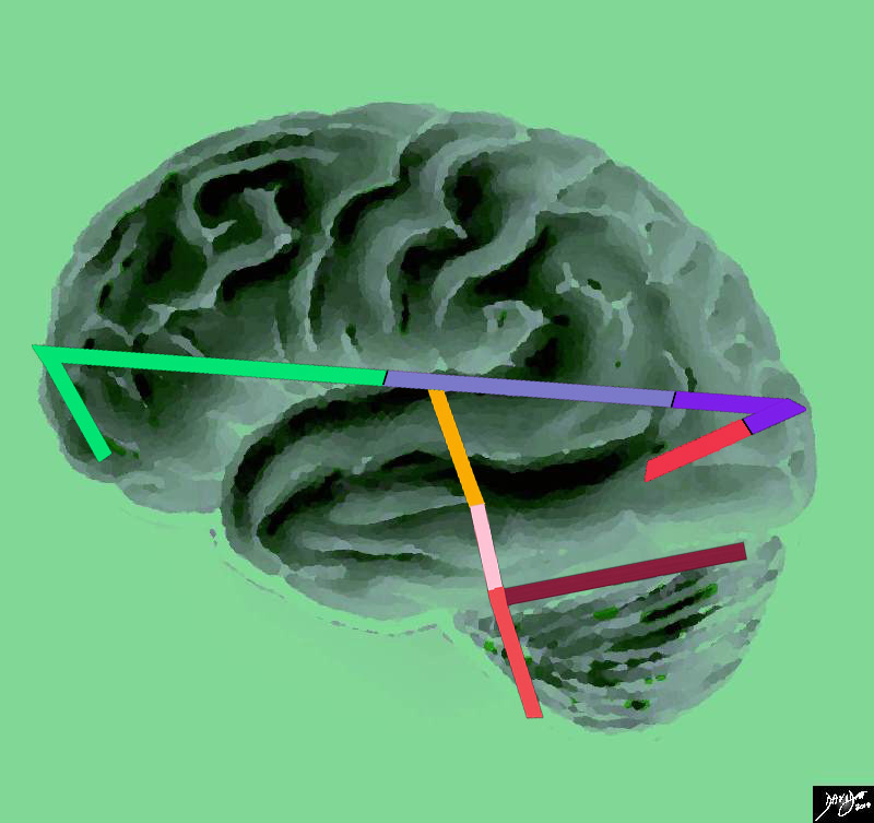
This artistic rendition of the brain reflects the vectors of the major parts of the brain with the stick diagram overlaid on a sagittal external view of the brain. In the stick diagram, the forebrain has now been divided into the frontal lobe (bright green), parietal lobe (light mauve) occipital lobe (purple) and temporal lobe (red). The midbrain is represented in orange, and the hind brain consists of the pons (pink) medulla (salmon) and the cerebellum (maroon)
Courtesy Ashley Davidoff copyright 2010 all rights reserved 83029e04.83s


    [nodeType] => 1
    [parentNode] => (object value omitted)
    [childNodes] => (object value omitted)
    [firstChild] => (object value omitted)
    [lastChild] => (object value omitted)
    [previousSibling] => (object value omitted)
    [nextSibling] => (object value omitted)
    [attributes] => (object value omitted)
    [ownerDocument] => (object value omitted)
    [namespaceURI] =>
    [prefix] =>
    [localName] => table
    [baseURI] =>
    [textContent] =>


Major Parts of the Brain


This artistic rendition of the brain reflects the vectors of the major parts of the brain with the stick diagram overlaid on a sagittal external view of the brain. In the stick diagram, the forebrain has now been divided into the frontal lobe (bright green), parietal lobe (light mauve) occipital lobe (purple) and temporal lobe (red). The midbrain is represented in orange, and the hind brain consists of the pons (pink) medulla (salmon) and the cerebellum (maroon)
Courtesy Ashley Davidoff copyright 2010 all rights reserved 83029e04.83s


)
DOMElement Object
(
    [schemaTypeInfo] =>
    [tagName] => td
    [firstElementChild] => (object value omitted)
    [lastElementChild] => (object value omitted)
    [childElementCount] => 2
    [previousElementSibling] =>
    [nextElementSibling] =>
    [nodeName] => td
    [nodeValue] =>
This artistic rendition of the brain reflects the vectors of the major parts of the brain with the stick diagram overlaid on a sagittal external view of the brain. In the stick diagram, the forebrain has now been divided into the frontal lobe (bright green), parietal lobe (light mauve) occipital lobe (purple) and temporal lobe (red). The midbrain is represented in orange, and the hind brain consists of the pons (pink) medulla (salmon) and the cerebellum (maroon)
Courtesy Ashley Davidoff copyright 2010 all rights reserved 83029e04.83s

    [nodeType] => 1
    [parentNode] => (object value omitted)
    [childNodes] => (object value omitted)
    [firstChild] => (object value omitted)
    [lastChild] => (object value omitted)
    [previousSibling] => (object value omitted)
    [nextSibling] => (object value omitted)
    [attributes] => (object value omitted)
    [ownerDocument] => (object value omitted)
    [namespaceURI] =>
    [prefix] =>
    [localName] => td
    [baseURI] =>
    [textContent] =>
This artistic rendition of the brain reflects the vectors of the major parts of the brain with the stick diagram overlaid on a sagittal external view of the brain. In the stick diagram, the forebrain has now been divided into the frontal lobe (bright green), parietal lobe (light mauve) occipital lobe (purple) and temporal lobe (red). The midbrain is represented in orange, and the hind brain consists of the pons (pink) medulla (salmon) and the cerebellum (maroon)
Courtesy Ashley Davidoff copyright 2010 all rights reserved 83029e04.83s

)
DOMElement Object
(
    [schemaTypeInfo] =>
    [tagName] => td
    [firstElementChild] => (object value omitted)
    [lastElementChild] => (object value omitted)
    [childElementCount] => 2
    [previousElementSibling] =>
    [nextElementSibling] =>
    [nodeName] => td
    [nodeValue] =>
Major Parts of the Brain
    [nodeType] => 1
    [parentNode] => (object value omitted)
    [childNodes] => (object value omitted)
    [firstChild] => (object value omitted)
    [lastChild] => (object value omitted)
    [previousSibling] => (object value omitted)
    [nextSibling] => (object value omitted)
    [attributes] => (object value omitted)
    [ownerDocument] => (object value omitted)
    [namespaceURI] =>
    [prefix] =>
    [localName] => td
    [baseURI] =>
    [textContent] =>
Major Parts of the Brain
)
DOMElement Object
(
    [schemaTypeInfo] =>
    [tagName] => table
    [firstElementChild] => (object value omitted)
    [lastElementChild] => (object value omitted)
    [childElementCount] => 1
    [previousElementSibling] => (object value omitted)
    [nextElementSibling] => (object value omitted)
    [nodeName] => table
    [nodeValue] =>


The Sagital Conceptual Vectors Overlying an External Lateral View of the Brain


The vectors of the brain from a side view are superimposed on a specimen of the brain, showing a long anteroposterior vector of the forebrain (light green) and a shorter more vertical vector consisting of the midbrain (orange) and the hind brain (salmon pink)
Davidoff Art courtesy Ashley Davidoff copyright 2010 all rights reserved 83029e04b01.85s


    [nodeType] => 1
    [parentNode] => (object value omitted)
    [childNodes] => (object value omitted)
    [firstChild] => (object value omitted)
    [lastChild] => (object value omitted)
    [previousSibling] => (object value omitted)
    [nextSibling] => (object value omitted)
    [attributes] => (object value omitted)
    [ownerDocument] => (object value omitted)
    [namespaceURI] =>
    [prefix] =>
    [localName] => table
    [baseURI] =>
    [textContent] =>


The Sagital Conceptual Vectors Overlying an External Lateral View of the Brain


The vectors of the brain from a side view are superimposed on a specimen of the brain, showing a long anteroposterior vector of the forebrain (light green) and a shorter more vertical vector consisting of the midbrain (orange) and the hind brain (salmon pink)
Davidoff Art courtesy Ashley Davidoff copyright 2010 all rights reserved 83029e04b01.85s


)
DOMElement Object
(
    [schemaTypeInfo] =>
    [tagName] => td
    [firstElementChild] => (object value omitted)
    [lastElementChild] => (object value omitted)
    [childElementCount] => 2
    [previousElementSibling] =>
    [nextElementSibling] =>
    [nodeName] => td
    [nodeValue] =>
The vectors of the brain from a side view are superimposed on a specimen of the brain, showing a long anteroposterior vector of the forebrain (light green) and a shorter more vertical vector consisting of the midbrain (orange) and the hind brain (salmon pink)
Davidoff Art courtesy Ashley Davidoff copyright 2010 all rights reserved 83029e04b01.85s

    [nodeType] => 1
    [parentNode] => (object value omitted)
    [childNodes] => (object value omitted)
    [firstChild] => (object value omitted)
    [lastChild] => (object value omitted)
    [previousSibling] => (object value omitted)
    [nextSibling] => (object value omitted)
    [attributes] => (object value omitted)
    [ownerDocument] => (object value omitted)
    [namespaceURI] =>
    [prefix] =>
    [localName] => td
    [baseURI] =>
    [textContent] =>
The vectors of the brain from a side view are superimposed on a specimen of the brain, showing a long anteroposterior vector of the forebrain (light green) and a shorter more vertical vector consisting of the midbrain (orange) and the hind brain (salmon pink)
Davidoff Art courtesy Ashley Davidoff copyright 2010 all rights reserved 83029e04b01.85s

)
DOMElement Object
(
    [schemaTypeInfo] =>
    [tagName] => td
    [firstElementChild] => (object value omitted)
    [lastElementChild] => (object value omitted)
    [childElementCount] => 2
    [previousElementSibling] =>
    [nextElementSibling] =>
    [nodeName] => td
    [nodeValue] =>

The Sagital Conceptual Vectors Overlying an External Lateral View of the Brain
    [nodeType] => 1
    [parentNode] => (object value omitted)
    [childNodes] => (object value omitted)
    [firstChild] => (object value omitted)
    [lastChild] => (object value omitted)
    [previousSibling] => (object value omitted)
    [nextSibling] => (object value omitted)
    [attributes] => (object value omitted)
    [ownerDocument] => (object value omitted)
    [namespaceURI] =>
    [prefix] =>
    [localName] => td
    [baseURI] =>
    [textContent] =>

The Sagital Conceptual Vectors Overlying an External Lateral View of the Brain
)
DOMElement Object
(
    [schemaTypeInfo] =>
    [tagName] => table
    [firstElementChild] => (object value omitted)
    [lastElementChild] => (object value omitted)
    [childElementCount] => 1
    [previousElementSibling] => (object value omitted)
    [nextElementSibling] => (object value omitted)
    [nodeName] => table
    [nodeValue] =>


The Basic Structure of the Brain from its LAteral Aspect
The Brain at its Simplest


 The stick diagram divides the brain into a relatively long almost horizontal component, and a relatively short almost vertical component. Anatomically the horizontal component is called the forebrain, and the vertical component will make up the midbrain (orange) and hind brain (salmon) and extend ino the spinal cord
Davidoff art Courtesy Ashley Davidoff MD copyright 2010 all rights reserved 93881b02.81s


    [nodeType] => 1
    [parentNode] => (object value omitted)
    [childNodes] => (object value omitted)
    [firstChild] => (object value omitted)
    [lastChild] => (object value omitted)
    [previousSibling] => (object value omitted)
    [nextSibling] => (object value omitted)
    [attributes] => (object value omitted)
    [ownerDocument] => (object value omitted)
    [namespaceURI] =>
    [prefix] =>
    [localName] => table
    [baseURI] =>
    [textContent] =>


The Basic Structure of the Brain from its LAteral Aspect
The Brain at its Simplest


 The stick diagram divides the brain into a relatively long almost horizontal component, and a relatively short almost vertical component. Anatomically the horizontal component is called the forebrain, and the vertical component will make up the midbrain (orange) and hind brain (salmon) and extend ino the spinal cord
Davidoff art Courtesy Ashley Davidoff MD copyright 2010 all rights reserved 93881b02.81s


)
DOMElement Object
(
    [schemaTypeInfo] =>
    [tagName] => td
    [firstElementChild] => (object value omitted)
    [lastElementChild] => (object value omitted)
    [childElementCount] => 2
    [previousElementSibling] =>
    [nextElementSibling] =>
    [nodeName] => td
    [nodeValue] =>
 The stick diagram divides the brain into a relatively long almost horizontal component, and a relatively short almost vertical component. Anatomically the horizontal component is called the forebrain, and the vertical component will make up the midbrain (orange) and hind brain (salmon) and extend ino the spinal cord
Davidoff art Courtesy Ashley Davidoff MD copyright 2010 all rights reserved 93881b02.81s

    [nodeType] => 1
    [parentNode] => (object value omitted)
    [childNodes] => (object value omitted)
    [firstChild] => (object value omitted)
    [lastChild] => (object value omitted)
    [previousSibling] => (object value omitted)
    [nextSibling] => (object value omitted)
    [attributes] => (object value omitted)
    [ownerDocument] => (object value omitted)
    [namespaceURI] =>
    [prefix] =>
    [localName] => td
    [baseURI] =>
    [textContent] =>
 The stick diagram divides the brain into a relatively long almost horizontal component, and a relatively short almost vertical component. Anatomically the horizontal component is called the forebrain, and the vertical component will make up the midbrain (orange) and hind brain (salmon) and extend ino the spinal cord
Davidoff art Courtesy Ashley Davidoff MD copyright 2010 all rights reserved 93881b02.81s

)
DOMElement Object
(
    [schemaTypeInfo] =>
    [tagName] => td
    [firstElementChild] => (object value omitted)
    [lastElementChild] => (object value omitted)
    [childElementCount] => 3
    [previousElementSibling] =>
    [nextElementSibling] =>
    [nodeName] => td
    [nodeValue] =>

The Basic Structure of the Brain from its LAteral Aspect
The Brain at its Simplest

    [nodeType] => 1
    [parentNode] => (object value omitted)
    [childNodes] => (object value omitted)
    [firstChild] => (object value omitted)
    [lastChild] => (object value omitted)
    [previousSibling] => (object value omitted)
    [nextSibling] => (object value omitted)
    [attributes] => (object value omitted)
    [ownerDocument] => (object value omitted)
    [namespaceURI] =>
    [prefix] =>
    [localName] => td
    [baseURI] =>
    [textContent] =>

The Basic Structure of the Brain from its LAteral Aspect
The Brain at its Simplest

)

