Sumit Karia MD Ashley Davidoff MD
The Common Vein Copyright 2010
Introduction
The brain is both structurally and functionally the crowning glory of the body. It is the most superior organ in the body.
It is enclosed in the cavity of the bony cranium and occupies almost all the space provided by the cranium.
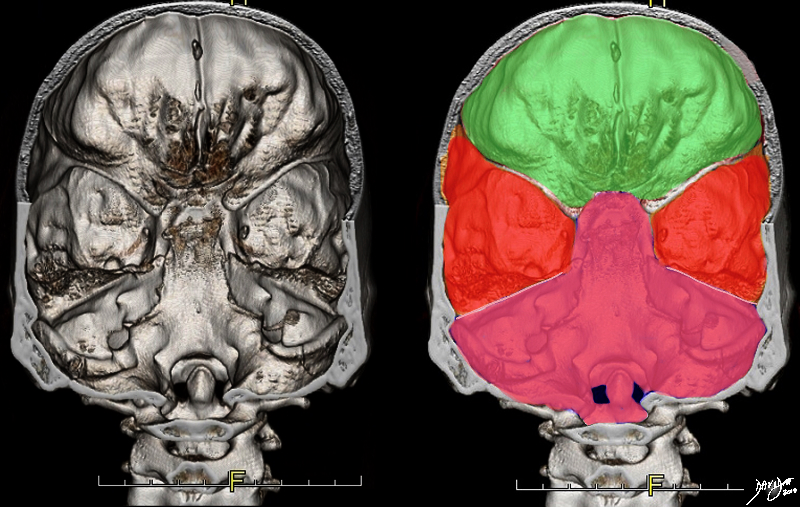
It is positioned between the three fossae; anterior cranial fossa, middle cranial fossa and posterior cranial fossa. The anterior cranial fossa houses the frontal lobe, and the middle cranial fossa houses the temporal lobe and the upper part of the midbrain above the tentorium, while the posterior cranial fossa houses the lower part of the midbrain and all the hindbrain.
|
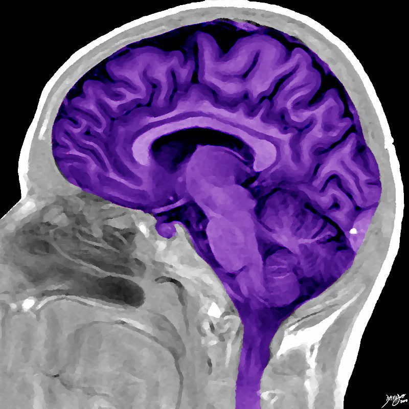
The Cranial Position of the Brain Snug in the Calvarium |
|
This sagittal MRI of the brain has ben artistically rendered, and reflects more detail of the anatomy of the forebrain, midbrain and hindbrain within the cranium. Image Courtesy Philips medical Systems Davidoff art 92170b06b01.8s |
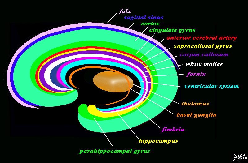
As dicussed in the conceptual organization of the brain there is a positional format. The ventricular suystem (light blue ring) is central, as is the corpus callosum that lies superior to the lateral ventricles. The anterior cerebral circulation lies superior to the corpus callosum. The centrally positioned falx and sagittal sinuses are superior to the artery.
Positional relationships in the Sagittal Plane
Lateral to these central structures the basal ganglia (the orange ring) are positioned
Positional Relationships in the Axial Plane
|
Concepts in Positional Relationships of the Brain in Axial Projection Gray Matter on the Outside |
|
The anchoring positional concept of the brain in the axial plane is reflected as two cerebral hemispheres with a series of structures reminiscent of backward and downward facing “C’s” symetrically positioned around the center. The most medial and central are the ventricular system (light blue) and the corpus callosum (purple) which lies superior and midline. The orange circle is paramedian and consists of the thalamus and basal ganglia. On the periphery of the brain is the gray matter, which umbrella all the other components of the brain. Davidoff art Courtesy Ashley Davidoff copyright 2010 all rights reserved 93914.3ka12.8s |
The Concept of Supratentorial and Infratentorial – The Coronal Plane
Position in Disease
Crossing the Midline
|
Glioblastome Multiforme |
|
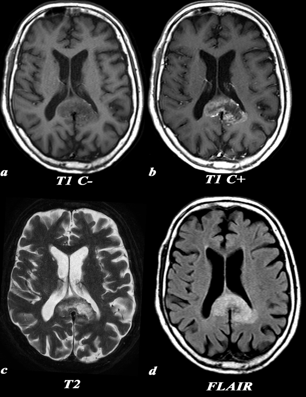
This 74 year old male presented with memory impairment and confusion. A previously performed head CT had shown a space occupying lesion in the splenium of the corpus callosum. MRI: (a,b,c,d) T1 weighted axial images before contrast (a) demonstrates a low signal in the enlarged splenium of the corpus callosum. The post contrast T1 weighted image (b) in the axial plane demonstrates the peripheral and irregular enhancement of the mass. This configuration is classic and often called a ?butterfly? glioma. Remember that due to the infiltrative nature of the mass, the extent of actual tumor involvement is larger than what is demonstrated by imaging on the post contrast images. This lesion which is hyperintense on T2 (c), enlarges the splenium, crosses the midline and involves the medial aspect of the left parietal lobe. Similar findings are identified on the FLAIR image. The extension into the parietal lobe is best seen on the FLAIR image These findings are consistent with a diagnosis of glioblastoma multiforme. Included in the differential diagnosis for lesions crossing the midline is primary lymphoma of the brain Image Courtesy Elisa Flower MD and Asim Mian MD 97647c01.8 |
DOMElement Object
(
    [schemaTypeInfo] =>
    [tagName] => table
    [firstElementChild] => (object value omitted)
    [lastElementChild] => (object value omitted)
    [childElementCount] => 1
    [previousElementSibling] => (object value omitted)
    [nextElementSibling] =>
    [nodeName] => table
    [nodeValue] =>


Glioblastome Multiforme


This 74 year old male presented with memory impairment and confusion. A previously performed head CT had shown a space occupying lesion in the splenium of the corpus callosum.
MRI: (a,b,c,d) T1 weighted axial images before contrast (a) demonstrates a low signal in the enlarged splenium of the corpus callosum. The post contrast T1 weighted image (b) in the axial plane demonstrates the peripheral and irregular enhancement of the mass. This configuration is classic and often called a ?butterfly? glioma.
Remember that due to the infiltrative nature of the mass, the extent of actual tumor involvement is larger than what is demonstrated by imaging on the post contrast images. This lesion which is hyperintense on T2 (c), enlarges the splenium, crosses the midline and involves the medial aspect of the left parietal lobe. Similar findings are identified on the FLAIR image. The extension into the parietal lobe is best seen on the FLAIR image
These findings are consistent with a diagnosis of glioblastoma multiforme. Included in the differential diagnosis for lesions crossing the midline is primary lymphoma of the brain
Image Courtesy Elisa Flower MD and Asim Mian MD 97647c01.8


    [nodeType] => 1
    [parentNode] => (object value omitted)
    [childNodes] => (object value omitted)
    [firstChild] => (object value omitted)
    [lastChild] => (object value omitted)
    [previousSibling] => (object value omitted)
    [nextSibling] => (object value omitted)
    [attributes] => (object value omitted)
    [ownerDocument] => (object value omitted)
    [namespaceURI] =>
    [prefix] =>
    [localName] => table
    [baseURI] =>
    [textContent] =>


Glioblastome Multiforme


This 74 year old male presented with memory impairment and confusion. A previously performed head CT had shown a space occupying lesion in the splenium of the corpus callosum.
MRI: (a,b,c,d) T1 weighted axial images before contrast (a) demonstrates a low signal in the enlarged splenium of the corpus callosum. The post contrast T1 weighted image (b) in the axial plane demonstrates the peripheral and irregular enhancement of the mass. This configuration is classic and often called a ?butterfly? glioma.
Remember that due to the infiltrative nature of the mass, the extent of actual tumor involvement is larger than what is demonstrated by imaging on the post contrast images. This lesion which is hyperintense on T2 (c), enlarges the splenium, crosses the midline and involves the medial aspect of the left parietal lobe. Similar findings are identified on the FLAIR image. The extension into the parietal lobe is best seen on the FLAIR image
These findings are consistent with a diagnosis of glioblastoma multiforme. Included in the differential diagnosis for lesions crossing the midline is primary lymphoma of the brain
Image Courtesy Elisa Flower MD and Asim Mian MD 97647c01.8


)
DOMElement Object
(
    [schemaTypeInfo] =>
    [tagName] => td
    [firstElementChild] => (object value omitted)
    [lastElementChild] => (object value omitted)
    [childElementCount] => 5
    [previousElementSibling] =>
    [nextElementSibling] =>
    [nodeName] => td
    [nodeValue] =>
This 74 year old male presented with memory impairment and confusion. A previously performed head CT had shown a space occupying lesion in the splenium of the corpus callosum.
MRI: (a,b,c,d) T1 weighted axial images before contrast (a) demonstrates a low signal in the enlarged splenium of the corpus callosum. The post contrast T1 weighted image (b) in the axial plane demonstrates the peripheral and irregular enhancement of the mass. This configuration is classic and often called a ?butterfly? glioma.
Remember that due to the infiltrative nature of the mass, the extent of actual tumor involvement is larger than what is demonstrated by imaging on the post contrast images. This lesion which is hyperintense on T2 (c), enlarges the splenium, crosses the midline and involves the medial aspect of the left parietal lobe. Similar findings are identified on the FLAIR image. The extension into the parietal lobe is best seen on the FLAIR image
These findings are consistent with a diagnosis of glioblastoma multiforme. Included in the differential diagnosis for lesions crossing the midline is primary lymphoma of the brain
Image Courtesy Elisa Flower MD and Asim Mian MD 97647c01.8

    [nodeType] => 1
    [parentNode] => (object value omitted)
    [childNodes] => (object value omitted)
    [firstChild] => (object value omitted)
    [lastChild] => (object value omitted)
    [previousSibling] => (object value omitted)
    [nextSibling] => (object value omitted)
    [attributes] => (object value omitted)
    [ownerDocument] => (object value omitted)
    [namespaceURI] =>
    [prefix] =>
    [localName] => td
    [baseURI] =>
    [textContent] =>
This 74 year old male presented with memory impairment and confusion. A previously performed head CT had shown a space occupying lesion in the splenium of the corpus callosum.
MRI: (a,b,c,d) T1 weighted axial images before contrast (a) demonstrates a low signal in the enlarged splenium of the corpus callosum. The post contrast T1 weighted image (b) in the axial plane demonstrates the peripheral and irregular enhancement of the mass. This configuration is classic and often called a ?butterfly? glioma.
Remember that due to the infiltrative nature of the mass, the extent of actual tumor involvement is larger than what is demonstrated by imaging on the post contrast images. This lesion which is hyperintense on T2 (c), enlarges the splenium, crosses the midline and involves the medial aspect of the left parietal lobe. Similar findings are identified on the FLAIR image. The extension into the parietal lobe is best seen on the FLAIR image
These findings are consistent with a diagnosis of glioblastoma multiforme. Included in the differential diagnosis for lesions crossing the midline is primary lymphoma of the brain
Image Courtesy Elisa Flower MD and Asim Mian MD 97647c01.8

)
DOMElement Object
(
    [schemaTypeInfo] =>
    [tagName] => td
    [firstElementChild] => (object value omitted)
    [lastElementChild] => (object value omitted)
    [childElementCount] => 2
    [previousElementSibling] =>
    [nextElementSibling] =>
    [nodeName] => td
    [nodeValue] =>

Glioblastome Multiforme
    [nodeType] => 1
    [parentNode] => (object value omitted)
    [childNodes] => (object value omitted)
    [firstChild] => (object value omitted)
    [lastChild] => (object value omitted)
    [previousSibling] => (object value omitted)
    [nextSibling] => (object value omitted)
    [attributes] => (object value omitted)
    [ownerDocument] => (object value omitted)
    [namespaceURI] =>
    [prefix] =>
    [localName] => td
    [baseURI] =>
    [textContent] =>

Glioblastome Multiforme
)
DOMElement Object
(
    [schemaTypeInfo] =>
    [tagName] => table
    [firstElementChild] => (object value omitted)
    [lastElementChild] => (object value omitted)
    [childElementCount] => 1
    [previousElementSibling] => (object value omitted)
    [nextElementSibling] => (object value omitted)
    [nodeName] => table
    [nodeValue] =>


Supratentorial and Infratentorial Structures


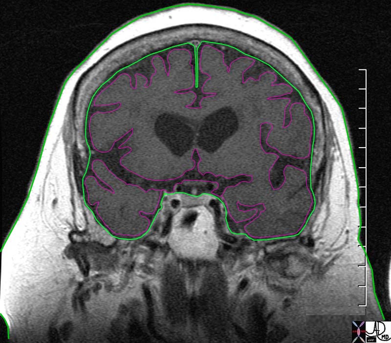
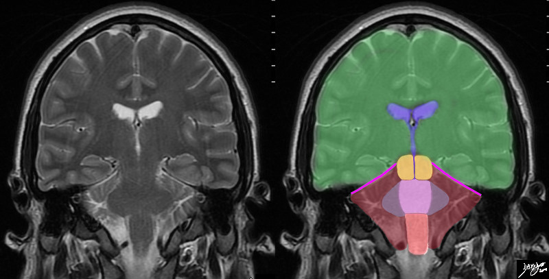
In this T2 weighted coronal MRI image the distinction between the supratentorial and infratentorial structures is made apparent by the bright pink tentorium that acts as a roof of the posterior fossa. The forebrain (green) midbrain (orange) and hindbrain (pink salmon and maroon) and the cerebellum (maroon), with other parts of the hindbrain filled in including the pons (light pink) middle cerebellar peduncles (mauve) and medulla (salmon) All the structures above the pink line are supratentorial, and those below are infratentorial. Part of the midbrain is supratentorial and part is infratentorial. The ventricular system is outlined in blue code brain
Courtesy Ashley Davidoff MD copyright 2010 all rights reserved 89721c06b.8sg01


    [nodeType] => 1
    [parentNode] => (object value omitted)
    [childNodes] => (object value omitted)
    [firstChild] => (object value omitted)
    [lastChild] => (object value omitted)
    [previousSibling] => (object value omitted)
    [nextSibling] => (object value omitted)
    [attributes] => (object value omitted)
    [ownerDocument] => (object value omitted)
    [namespaceURI] =>
    [prefix] =>
    [localName] => table
    [baseURI] =>
    [textContent] =>


Supratentorial and Infratentorial Structures


In this T2 weighted coronal MRI image the distinction between the supratentorial and infratentorial structures is made apparent by the bright pink tentorium that acts as a roof of the posterior fossa. The forebrain (green) midbrain (orange) and hindbrain (pink salmon and maroon) and the cerebellum (maroon), with other parts of the hindbrain filled in including the pons (light pink) middle cerebellar peduncles (mauve) and medulla (salmon) All the structures above the pink line are supratentorial, and those below are infratentorial. Part of the midbrain is supratentorial and part is infratentorial. The ventricular system is outlined in blue code brain
Courtesy Ashley Davidoff MD copyright 2010 all rights reserved 89721c06b.8sg01


)
DOMElement Object
(
    [schemaTypeInfo] =>
    [tagName] => td
    [firstElementChild] => (object value omitted)
    [lastElementChild] => (object value omitted)
    [childElementCount] => 2
    [previousElementSibling] =>
    [nextElementSibling] =>
    [nodeName] => td
    [nodeValue] =>
In this T2 weighted coronal MRI image the distinction between the supratentorial and infratentorial structures is made apparent by the bright pink tentorium that acts as a roof of the posterior fossa. The forebrain (green) midbrain (orange) and hindbrain (pink salmon and maroon) and the cerebellum (maroon), with other parts of the hindbrain filled in including the pons (light pink) middle cerebellar peduncles (mauve) and medulla (salmon) All the structures above the pink line are supratentorial, and those below are infratentorial. Part of the midbrain is supratentorial and part is infratentorial. The ventricular system is outlined in blue code brain
Courtesy Ashley Davidoff MD copyright 2010 all rights reserved 89721c06b.8sg01

    [nodeType] => 1
    [parentNode] => (object value omitted)
    [childNodes] => (object value omitted)
    [firstChild] => (object value omitted)
    [lastChild] => (object value omitted)
    [previousSibling] => (object value omitted)
    [nextSibling] => (object value omitted)
    [attributes] => (object value omitted)
    [ownerDocument] => (object value omitted)
    [namespaceURI] =>
    [prefix] =>
    [localName] => td
    [baseURI] =>
    [textContent] =>
In this T2 weighted coronal MRI image the distinction between the supratentorial and infratentorial structures is made apparent by the bright pink tentorium that acts as a roof of the posterior fossa. The forebrain (green) midbrain (orange) and hindbrain (pink salmon and maroon) and the cerebellum (maroon), with other parts of the hindbrain filled in including the pons (light pink) middle cerebellar peduncles (mauve) and medulla (salmon) All the structures above the pink line are supratentorial, and those below are infratentorial. Part of the midbrain is supratentorial and part is infratentorial. The ventricular system is outlined in blue code brain
Courtesy Ashley Davidoff MD copyright 2010 all rights reserved 89721c06b.8sg01

)
DOMElement Object
(
    [schemaTypeInfo] =>
    [tagName] => td
    [firstElementChild] => (object value omitted)
    [lastElementChild] => (object value omitted)
    [childElementCount] => 2
    [previousElementSibling] =>
    [nextElementSibling] =>
    [nodeName] => td
    [nodeValue] =>
Supratentorial and Infratentorial Structures

    [nodeType] => 1
    [parentNode] => (object value omitted)
    [childNodes] => (object value omitted)
    [firstChild] => (object value omitted)
    [lastChild] => (object value omitted)
    [previousSibling] => (object value omitted)
    [nextSibling] => (object value omitted)
    [attributes] => (object value omitted)
    [ownerDocument] => (object value omitted)
    [namespaceURI] =>
    [prefix] =>
    [localName] => td
    [baseURI] =>
    [textContent] =>
Supratentorial and Infratentorial Structures

)
DOMElement Object
(
    [schemaTypeInfo] =>
    [tagName] => table
    [firstElementChild] => (object value omitted)
    [lastElementChild] => (object value omitted)
    [childElementCount] => 1
    [previousElementSibling] => (object value omitted)
    [nextElementSibling] => (object value omitted)
    [nodeName] => table
    [nodeValue] =>


Space Position and the Tentorium in the Coronal Plane


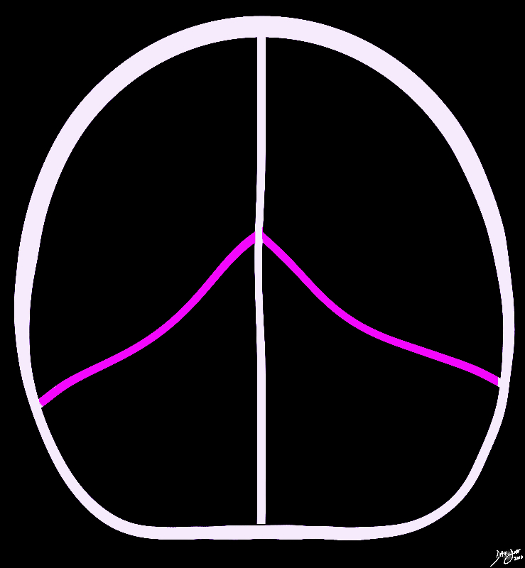
In this conceptual coronal drawing the distinction between the supratentorial and infratentorial space is made apparent by the bright pink tentorium that acts as a roof of the posterior fossa. The forebrain and the upper part of the midbrain lie above the tentorium, and the lower midbrain and hindbrain lie below. All the structures above the pink line are classified as supratentorial structures , and those below are infratentorial.
Courtesy Ashley Davidoff MD copyright 2010 all rights reserved 93914b07g02.8s


    [nodeType] => 1
    [parentNode] => (object value omitted)
    [childNodes] => (object value omitted)
    [firstChild] => (object value omitted)
    [lastChild] => (object value omitted)
    [previousSibling] => (object value omitted)
    [nextSibling] => (object value omitted)
    [attributes] => (object value omitted)
    [ownerDocument] => (object value omitted)
    [namespaceURI] =>
    [prefix] =>
    [localName] => table
    [baseURI] =>
    [textContent] =>


Space Position and the Tentorium in the Coronal Plane


In this conceptual coronal drawing the distinction between the supratentorial and infratentorial space is made apparent by the bright pink tentorium that acts as a roof of the posterior fossa. The forebrain and the upper part of the midbrain lie above the tentorium, and the lower midbrain and hindbrain lie below. All the structures above the pink line are classified as supratentorial structures , and those below are infratentorial.
Courtesy Ashley Davidoff MD copyright 2010 all rights reserved 93914b07g02.8s


)
DOMElement Object
(
    [schemaTypeInfo] =>
    [tagName] => td
    [firstElementChild] => (object value omitted)
    [lastElementChild] => (object value omitted)
    [childElementCount] => 2
    [previousElementSibling] =>
    [nextElementSibling] =>
    [nodeName] => td
    [nodeValue] =>
In this conceptual coronal drawing the distinction between the supratentorial and infratentorial space is made apparent by the bright pink tentorium that acts as a roof of the posterior fossa. The forebrain and the upper part of the midbrain lie above the tentorium, and the lower midbrain and hindbrain lie below. All the structures above the pink line are classified as supratentorial structures , and those below are infratentorial.
Courtesy Ashley Davidoff MD copyright 2010 all rights reserved 93914b07g02.8s

    [nodeType] => 1
    [parentNode] => (object value omitted)
    [childNodes] => (object value omitted)
    [firstChild] => (object value omitted)
    [lastChild] => (object value omitted)
    [previousSibling] => (object value omitted)
    [nextSibling] => (object value omitted)
    [attributes] => (object value omitted)
    [ownerDocument] => (object value omitted)
    [namespaceURI] =>
    [prefix] =>
    [localName] => td
    [baseURI] =>
    [textContent] =>
In this conceptual coronal drawing the distinction between the supratentorial and infratentorial space is made apparent by the bright pink tentorium that acts as a roof of the posterior fossa. The forebrain and the upper part of the midbrain lie above the tentorium, and the lower midbrain and hindbrain lie below. All the structures above the pink line are classified as supratentorial structures , and those below are infratentorial.
Courtesy Ashley Davidoff MD copyright 2010 all rights reserved 93914b07g02.8s

)
DOMElement Object
(
    [schemaTypeInfo] =>
    [tagName] => td
    [firstElementChild] => (object value omitted)
    [lastElementChild] => (object value omitted)
    [childElementCount] => 2
    [previousElementSibling] =>
    [nextElementSibling] =>
    [nodeName] => td
    [nodeValue] =>
Space Position and the Tentorium in the Coronal Plane
    [nodeType] => 1
    [parentNode] => (object value omitted)
    [childNodes] => (object value omitted)
    [firstChild] => (object value omitted)
    [lastChild] => (object value omitted)
    [previousSibling] => (object value omitted)
    [nextSibling] => (object value omitted)
    [attributes] => (object value omitted)
    [ownerDocument] => (object value omitted)
    [namespaceURI] =>
    [prefix] =>
    [localName] => td
    [baseURI] =>
    [textContent] =>
Space Position and the Tentorium in the Coronal Plane
)
DOMElement Object
(
    [schemaTypeInfo] =>
    [tagName] => table
    [firstElementChild] => (object value omitted)
    [lastElementChild] => (object value omitted)
    [childElementCount] => 1
    [previousElementSibling] => (object value omitted)
    [nextElementSibling] => (object value omitted)
    [nodeName] => table
    [nodeValue] =>


Concepts in Positional Relationships of the Brain in Axial Projection
Gray Matter on the Outside


The anchoring positional concept of the brain in the axial plane is reflected as two cerebral hemispheres with a series of structures reminiscent of backward and downward facing “C’s” symetrically positioned around the center. The most medial and central are the ventricular system (light blue) and the corpus callosum (purple) which lies superior and midline.   The orange circle is paramedian and consists of the thalamus and basal ganglia.  On the periphery of the brain is the gray matter, which umbrella all the other components of the brain.
Davidoff art Courtesy Ashley Davidoff copyright 2010 all rights reserved 93914.3ka12.8s


    [nodeType] => 1
    [parentNode] => (object value omitted)
    [childNodes] => (object value omitted)
    [firstChild] => (object value omitted)
    [lastChild] => (object value omitted)
    [previousSibling] => (object value omitted)
    [nextSibling] => (object value omitted)
    [attributes] => (object value omitted)
    [ownerDocument] => (object value omitted)
    [namespaceURI] =>
    [prefix] =>
    [localName] => table
    [baseURI] =>
    [textContent] =>


Concepts in Positional Relationships of the Brain in Axial Projection
Gray Matter on the Outside


The anchoring positional concept of the brain in the axial plane is reflected as two cerebral hemispheres with a series of structures reminiscent of backward and downward facing “C’s” symetrically positioned around the center. The most medial and central are the ventricular system (light blue) and the corpus callosum (purple) which lies superior and midline.   The orange circle is paramedian and consists of the thalamus and basal ganglia.  On the periphery of the brain is the gray matter, which umbrella all the other components of the brain.
Davidoff art Courtesy Ashley Davidoff copyright 2010 all rights reserved 93914.3ka12.8s


)
DOMElement Object
(
    [schemaTypeInfo] =>
    [tagName] => td
    [firstElementChild] => (object value omitted)
    [lastElementChild] => (object value omitted)
    [childElementCount] => 2
    [previousElementSibling] =>
    [nextElementSibling] =>
    [nodeName] => td
    [nodeValue] =>
The anchoring positional concept of the brain in the axial plane is reflected as two cerebral hemispheres with a series of structures reminiscent of backward and downward facing “C’s” symetrically positioned around the center. The most medial and central are the ventricular system (light blue) and the corpus callosum (purple) which lies superior and midline.   The orange circle is paramedian and consists of the thalamus and basal ganglia.  On the periphery of the brain is the gray matter, which umbrella all the other components of the brain.
Davidoff art Courtesy Ashley Davidoff copyright 2010 all rights reserved 93914.3ka12.8s

    [nodeType] => 1
    [parentNode] => (object value omitted)
    [childNodes] => (object value omitted)
    [firstChild] => (object value omitted)
    [lastChild] => (object value omitted)
    [previousSibling] => (object value omitted)
    [nextSibling] => (object value omitted)
    [attributes] => (object value omitted)
    [ownerDocument] => (object value omitted)
    [namespaceURI] =>
    [prefix] =>
    [localName] => td
    [baseURI] =>
    [textContent] =>
The anchoring positional concept of the brain in the axial plane is reflected as two cerebral hemispheres with a series of structures reminiscent of backward and downward facing “C’s” symetrically positioned around the center. The most medial and central are the ventricular system (light blue) and the corpus callosum (purple) which lies superior and midline.   The orange circle is paramedian and consists of the thalamus and basal ganglia.  On the periphery of the brain is the gray matter, which umbrella all the other components of the brain.
Davidoff art Courtesy Ashley Davidoff copyright 2010 all rights reserved 93914.3ka12.8s

)
DOMElement Object
(
    [schemaTypeInfo] =>
    [tagName] => td
    [firstElementChild] => (object value omitted)
    [lastElementChild] => (object value omitted)
    [childElementCount] => 3
    [previousElementSibling] =>
    [nextElementSibling] =>
    [nodeName] => td
    [nodeValue] =>

Concepts in Positional Relationships of the Brain in Axial Projection
Gray Matter on the Outside
    [nodeType] => 1
    [parentNode] => (object value omitted)
    [childNodes] => (object value omitted)
    [firstChild] => (object value omitted)
    [lastChild] => (object value omitted)
    [previousSibling] => (object value omitted)
    [nextSibling] => (object value omitted)
    [attributes] => (object value omitted)
    [ownerDocument] => (object value omitted)
    [namespaceURI] =>
    [prefix] =>
    [localName] => td
    [baseURI] =>
    [textContent] =>

Concepts in Positional Relationships of the Brain in Axial Projection
Gray Matter on the Outside
)
DOMElement Object
(
    [schemaTypeInfo] =>
    [tagName] => table
    [firstElementChild] => (object value omitted)
    [lastElementChild] => (object value omitted)
    [childElementCount] => 1
    [previousElementSibling] => (object value omitted)
    [nextElementSibling] => (object value omitted)
    [nodeName] => table
    [nodeValue] =>


Relative Position of Structures of the Forebrain


The forebrain has most of its components aligned in a series of inverted c- shaped rings starting from the outer membranes that culminate in the falx (pink), then extending inward smaller inner rings with each intimately connected to the others. The thalamus (dull orange) appears diagrammatically as the centre of these rings as seen from the sagittal view The outer ring is the falx (pink) followed by the sagittal sinus (blue) cerebral cortex (light green), cingulate gyrus (bright green) superiorly which becomes the parahippocampal gyrus inferiorly. The red ring represents the distribution of the main portion of the anterior cerebral artery. Next is the yellow ring which is the supracollosal gyrus (indusium griseum) superiorly and the hippocampus inferiorly. This is followed by the corpus callosum (purple) which enables the white ring of white matter to connect between hemispheres. The next ring is the thin bright pink ring which represents the fornix superiorly and the fimbria inferiorly. The innermost ring (light blue) represents the lateral horns of the ventricular system. The basal ganglia run just lateral to the lateral ventricles. The navy blue arrow headed structure is the septum pellucidum.
Courtesy Ashley Davidoff copyright 2010 all rights reserved 93907d13g03.8s


    [nodeType] => 1
    [parentNode] => (object value omitted)
    [childNodes] => (object value omitted)
    [firstChild] => (object value omitted)
    [lastChild] => (object value omitted)
    [previousSibling] => (object value omitted)
    [nextSibling] => (object value omitted)
    [attributes] => (object value omitted)
    [ownerDocument] => (object value omitted)
    [namespaceURI] =>
    [prefix] =>
    [localName] => table
    [baseURI] =>
    [textContent] =>


Relative Position of Structures of the Forebrain


The forebrain has most of its components aligned in a series of inverted c- shaped rings starting from the outer membranes that culminate in the falx (pink), then extending inward smaller inner rings with each intimately connected to the others. The thalamus (dull orange) appears diagrammatically as the centre of these rings as seen from the sagittal view The outer ring is the falx (pink) followed by the sagittal sinus (blue) cerebral cortex (light green), cingulate gyrus (bright green) superiorly which becomes the parahippocampal gyrus inferiorly. The red ring represents the distribution of the main portion of the anterior cerebral artery. Next is the yellow ring which is the supracollosal gyrus (indusium griseum) superiorly and the hippocampus inferiorly. This is followed by the corpus callosum (purple) which enables the white ring of white matter to connect between hemispheres. The next ring is the thin bright pink ring which represents the fornix superiorly and the fimbria inferiorly. The innermost ring (light blue) represents the lateral horns of the ventricular system. The basal ganglia run just lateral to the lateral ventricles. The navy blue arrow headed structure is the septum pellucidum.
Courtesy Ashley Davidoff copyright 2010 all rights reserved 93907d13g03.8s


)
DOMElement Object
(
    [schemaTypeInfo] =>
    [tagName] => td
    [firstElementChild] => (object value omitted)
    [lastElementChild] => (object value omitted)
    [childElementCount] => 2
    [previousElementSibling] =>
    [nextElementSibling] =>
    [nodeName] => td
    [nodeValue] =>
The forebrain has most of its components aligned in a series of inverted c- shaped rings starting from the outer membranes that culminate in the falx (pink), then extending inward smaller inner rings with each intimately connected to the others. The thalamus (dull orange) appears diagrammatically as the centre of these rings as seen from the sagittal view The outer ring is the falx (pink) followed by the sagittal sinus (blue) cerebral cortex (light green), cingulate gyrus (bright green) superiorly which becomes the parahippocampal gyrus inferiorly. The red ring represents the distribution of the main portion of the anterior cerebral artery. Next is the yellow ring which is the supracollosal gyrus (indusium griseum) superiorly and the hippocampus inferiorly. This is followed by the corpus callosum (purple) which enables the white ring of white matter to connect between hemispheres. The next ring is the thin bright pink ring which represents the fornix superiorly and the fimbria inferiorly. The innermost ring (light blue) represents the lateral horns of the ventricular system. The basal ganglia run just lateral to the lateral ventricles. The navy blue arrow headed structure is the septum pellucidum.
Courtesy Ashley Davidoff copyright 2010 all rights reserved 93907d13g03.8s

    [nodeType] => 1
    [parentNode] => (object value omitted)
    [childNodes] => (object value omitted)
    [firstChild] => (object value omitted)
    [lastChild] => (object value omitted)
    [previousSibling] => (object value omitted)
    [nextSibling] => (object value omitted)
    [attributes] => (object value omitted)
    [ownerDocument] => (object value omitted)
    [namespaceURI] =>
    [prefix] =>
    [localName] => td
    [baseURI] =>
    [textContent] =>
The forebrain has most of its components aligned in a series of inverted c- shaped rings starting from the outer membranes that culminate in the falx (pink), then extending inward smaller inner rings with each intimately connected to the others. The thalamus (dull orange) appears diagrammatically as the centre of these rings as seen from the sagittal view The outer ring is the falx (pink) followed by the sagittal sinus (blue) cerebral cortex (light green), cingulate gyrus (bright green) superiorly which becomes the parahippocampal gyrus inferiorly. The red ring represents the distribution of the main portion of the anterior cerebral artery. Next is the yellow ring which is the supracollosal gyrus (indusium griseum) superiorly and the hippocampus inferiorly. This is followed by the corpus callosum (purple) which enables the white ring of white matter to connect between hemispheres. The next ring is the thin bright pink ring which represents the fornix superiorly and the fimbria inferiorly. The innermost ring (light blue) represents the lateral horns of the ventricular system. The basal ganglia run just lateral to the lateral ventricles. The navy blue arrow headed structure is the septum pellucidum.
Courtesy Ashley Davidoff copyright 2010 all rights reserved 93907d13g03.8s

)
DOMElement Object
(
    [schemaTypeInfo] =>
    [tagName] => td
    [firstElementChild] => (object value omitted)
    [lastElementChild] => (object value omitted)
    [childElementCount] => 2
    [previousElementSibling] =>
    [nextElementSibling] =>
    [nodeName] => td
    [nodeValue] =>
Relative Position of Structures of the Forebrain
    [nodeType] => 1
    [parentNode] => (object value omitted)
    [childNodes] => (object value omitted)
    [firstChild] => (object value omitted)
    [lastChild] => (object value omitted)
    [previousSibling] => (object value omitted)
    [nextSibling] => (object value omitted)
    [attributes] => (object value omitted)
    [ownerDocument] => (object value omitted)
    [namespaceURI] =>
    [prefix] =>
    [localName] => td
    [baseURI] =>
    [textContent] =>
Relative Position of Structures of the Forebrain
)
DOMElement Object
(
    [schemaTypeInfo] =>
    [tagName] => table
    [firstElementChild] => (object value omitted)
    [lastElementChild] => (object value omitted)
    [childElementCount] => 1
    [previousElementSibling] => (object value omitted)
    [nextElementSibling] => (object value omitted)
    [nodeName] => table
    [nodeValue] =>


Between the Brain and The Skull


71422.800b01b brain meninges pia mater = pink dura mater = green subarachnnoid space bone somatosensory receptors rich in the dura inner outer pain periosteum MRI Courtesy Ashley DAvidoff Copyright 2008


    [nodeType] => 1
    [parentNode] => (object value omitted)
    [childNodes] => (object value omitted)
    [firstChild] => (object value omitted)
    [lastChild] => (object value omitted)
    [previousSibling] => (object value omitted)
    [nextSibling] => (object value omitted)
    [attributes] => (object value omitted)
    [ownerDocument] => (object value omitted)
    [namespaceURI] =>
    [prefix] =>
    [localName] => table
    [baseURI] =>
    [textContent] =>


Between the Brain and The Skull


71422.800b01b brain meninges pia mater = pink dura mater = green subarachnnoid space bone somatosensory receptors rich in the dura inner outer pain periosteum MRI Courtesy Ashley DAvidoff Copyright 2008


)
DOMElement Object
(
    [schemaTypeInfo] =>
    [tagName] => td
    [firstElementChild] =>
    [lastElementChild] =>
    [childElementCount] => 0
    [previousElementSibling] =>
    [nextElementSibling] =>
    [nodeName] => td
    [nodeValue] => 71422.800b01b brain meninges pia mater = pink dura mater = green subarachnnoid space bone somatosensory receptors rich in the dura inner outer pain periosteum MRI Courtesy Ashley DAvidoff Copyright 2008
    [nodeType] => 1
    [parentNode] => (object value omitted)
    [childNodes] => (object value omitted)
    [firstChild] => (object value omitted)
    [lastChild] => (object value omitted)
    [previousSibling] => (object value omitted)
    [nextSibling] => (object value omitted)
    [attributes] => (object value omitted)
    [ownerDocument] => (object value omitted)
    [namespaceURI] =>
    [prefix] =>
    [localName] => td
    [baseURI] =>
    [textContent] => 71422.800b01b brain meninges pia mater = pink dura mater = green subarachnnoid space bone somatosensory receptors rich in the dura inner outer pain periosteum MRI Courtesy Ashley DAvidoff Copyright 2008
)
DOMElement Object
(
    [schemaTypeInfo] =>
    [tagName] => td
    [firstElementChild] => (object value omitted)
    [lastElementChild] => (object value omitted)
    [childElementCount] => 2
    [previousElementSibling] =>
    [nextElementSibling] =>
    [nodeName] => td
    [nodeValue] =>
Between the Brain and The Skull
    [nodeType] => 1
    [parentNode] => (object value omitted)
    [childNodes] => (object value omitted)
    [firstChild] => (object value omitted)
    [lastChild] => (object value omitted)
    [previousSibling] => (object value omitted)
    [nextSibling] => (object value omitted)
    [attributes] => (object value omitted)
    [ownerDocument] => (object value omitted)
    [namespaceURI] =>
    [prefix] =>
    [localName] => td
    [baseURI] =>
    [textContent] =>
Between the Brain and The Skull
)
DOMElement Object
(
    [schemaTypeInfo] =>
    [tagName] => table
    [firstElementChild] => (object value omitted)
    [lastElementChild] => (object value omitted)
    [childElementCount] => 1
    [previousElementSibling] => (object value omitted)
    [nextElementSibling] => (object value omitted)
    [nodeName] => table
    [nodeValue] =>


The Cranial Vault in Coronal Projection


The posterior coronal view of the opened skull is a reconstruction from a normal CT scan. It reveals the 3 fossa of the skull; anterior (green) middle (red) and posterior (salmon pink) The frontal lobe is housed in the anterior cranial fossa while the temporal lobe and upper half of the midbrain  lie in the middle cranial fossa and the posterior fossa houses the lower portion of the midbrain and  hindbrain The whole brain is protected and lies snugly in the bony cranial vault.
Courtesy Ashley Davidoff MD Copyright 2010 All rights reserved 48720.2kc01.8s


    [nodeType] => 1
    [parentNode] => (object value omitted)
    [childNodes] => (object value omitted)
    [firstChild] => (object value omitted)
    [lastChild] => (object value omitted)
    [previousSibling] => (object value omitted)
    [nextSibling] => (object value omitted)
    [attributes] => (object value omitted)
    [ownerDocument] => (object value omitted)
    [namespaceURI] =>
    [prefix] =>
    [localName] => table
    [baseURI] =>
    [textContent] =>


The Cranial Vault in Coronal Projection


The posterior coronal view of the opened skull is a reconstruction from a normal CT scan. It reveals the 3 fossa of the skull; anterior (green) middle (red) and posterior (salmon pink) The frontal lobe is housed in the anterior cranial fossa while the temporal lobe and upper half of the midbrain  lie in the middle cranial fossa and the posterior fossa houses the lower portion of the midbrain and  hindbrain The whole brain is protected and lies snugly in the bony cranial vault.
Courtesy Ashley Davidoff MD Copyright 2010 All rights reserved 48720.2kc01.8s


)
DOMElement Object
(
    [schemaTypeInfo] =>
    [tagName] => td
    [firstElementChild] => (object value omitted)
    [lastElementChild] => (object value omitted)
    [childElementCount] => 2
    [previousElementSibling] =>
    [nextElementSibling] =>
    [nodeName] => td
    [nodeValue] =>
The posterior coronal view of the opened skull is a reconstruction from a normal CT scan. It reveals the 3 fossa of the skull; anterior (green) middle (red) and posterior (salmon pink) The frontal lobe is housed in the anterior cranial fossa while the temporal lobe and upper half of the midbrain  lie in the middle cranial fossa and the posterior fossa houses the lower portion of the midbrain and  hindbrain The whole brain is protected and lies snugly in the bony cranial vault.
Courtesy Ashley Davidoff MD Copyright 2010 All rights reserved 48720.2kc01.8s

    [nodeType] => 1
    [parentNode] => (object value omitted)
    [childNodes] => (object value omitted)
    [firstChild] => (object value omitted)
    [lastChild] => (object value omitted)
    [previousSibling] => (object value omitted)
    [nextSibling] => (object value omitted)
    [attributes] => (object value omitted)
    [ownerDocument] => (object value omitted)
    [namespaceURI] =>
    [prefix] =>
    [localName] => td
    [baseURI] =>
    [textContent] =>
The posterior coronal view of the opened skull is a reconstruction from a normal CT scan. It reveals the 3 fossa of the skull; anterior (green) middle (red) and posterior (salmon pink) The frontal lobe is housed in the anterior cranial fossa while the temporal lobe and upper half of the midbrain  lie in the middle cranial fossa and the posterior fossa houses the lower portion of the midbrain and  hindbrain The whole brain is protected and lies snugly in the bony cranial vault.
Courtesy Ashley Davidoff MD Copyright 2010 All rights reserved 48720.2kc01.8s

)
DOMElement Object
(
    [schemaTypeInfo] =>
    [tagName] => td
    [firstElementChild] => (object value omitted)
    [lastElementChild] => (object value omitted)
    [childElementCount] => 2
    [previousElementSibling] =>
    [nextElementSibling] =>
    [nodeName] => td
    [nodeValue] =>
The Cranial Vault in Coronal Projection
    [nodeType] => 1
    [parentNode] => (object value omitted)
    [childNodes] => (object value omitted)
    [firstChild] => (object value omitted)
    [lastChild] => (object value omitted)
    [previousSibling] => (object value omitted)
    [nextSibling] => (object value omitted)
    [attributes] => (object value omitted)
    [ownerDocument] => (object value omitted)
    [namespaceURI] =>
    [prefix] =>
    [localName] => td
    [baseURI] =>
    [textContent] =>
The Cranial Vault in Coronal Projection
)
DOMElement Object
(
    [schemaTypeInfo] =>
    [tagName] => table
    [firstElementChild] => (object value omitted)
    [lastElementChild] => (object value omitted)
    [childElementCount] => 1
    [previousElementSibling] => (object value omitted)
    [nextElementSibling] => (object value omitted)
    [nodeName] => table
    [nodeValue] =>


The Cranial Position of the Brain
Snug in the Calvarium


This sagittal MRI of the brain has ben artistically rendered, and reflects more detail of the anatomy of the forebrain, midbrain and hindbrain within the cranium.
Image Courtesy Philips medical Systems Davidoff art 92170b06b01.8s


    [nodeType] => 1
    [parentNode] => (object value omitted)
    [childNodes] => (object value omitted)
    [firstChild] => (object value omitted)
    [lastChild] => (object value omitted)
    [previousSibling] => (object value omitted)
    [nextSibling] => (object value omitted)
    [attributes] => (object value omitted)
    [ownerDocument] => (object value omitted)
    [namespaceURI] =>
    [prefix] =>
    [localName] => table
    [baseURI] =>
    [textContent] =>


The Cranial Position of the Brain
Snug in the Calvarium


This sagittal MRI of the brain has ben artistically rendered, and reflects more detail of the anatomy of the forebrain, midbrain and hindbrain within the cranium.
Image Courtesy Philips medical Systems Davidoff art 92170b06b01.8s


)
DOMElement Object
(
    [schemaTypeInfo] =>
    [tagName] => td
    [firstElementChild] => (object value omitted)
    [lastElementChild] => (object value omitted)
    [childElementCount] => 2
    [previousElementSibling] =>
    [nextElementSibling] =>
    [nodeName] => td
    [nodeValue] =>
This sagittal MRI of the brain has ben artistically rendered, and reflects more detail of the anatomy of the forebrain, midbrain and hindbrain within the cranium.
Image Courtesy Philips medical Systems Davidoff art 92170b06b01.8s

    [nodeType] => 1
    [parentNode] => (object value omitted)
    [childNodes] => (object value omitted)
    [firstChild] => (object value omitted)
    [lastChild] => (object value omitted)
    [previousSibling] => (object value omitted)
    [nextSibling] => (object value omitted)
    [attributes] => (object value omitted)
    [ownerDocument] => (object value omitted)
    [namespaceURI] =>
    [prefix] =>
    [localName] => td
    [baseURI] =>
    [textContent] =>
This sagittal MRI of the brain has ben artistically rendered, and reflects more detail of the anatomy of the forebrain, midbrain and hindbrain within the cranium.
Image Courtesy Philips medical Systems Davidoff art 92170b06b01.8s

)
DOMElement Object
(
    [schemaTypeInfo] =>
    [tagName] => td
    [firstElementChild] => (object value omitted)
    [lastElementChild] => (object value omitted)
    [childElementCount] => 3
    [previousElementSibling] =>
    [nextElementSibling] =>
    [nodeName] => td
    [nodeValue] =>

The Cranial Position of the Brain
Snug in the Calvarium
    [nodeType] => 1
    [parentNode] => (object value omitted)
    [childNodes] => (object value omitted)
    [firstChild] => (object value omitted)
    [lastChild] => (object value omitted)
    [previousSibling] => (object value omitted)
    [nextSibling] => (object value omitted)
    [attributes] => (object value omitted)
    [ownerDocument] => (object value omitted)
    [namespaceURI] =>
    [prefix] =>
    [localName] => td
    [baseURI] =>
    [textContent] =>

The Cranial Position of the Brain
Snug in the Calvarium
)
DOMElement Object
(
    [schemaTypeInfo] =>
    [tagName] => table
    [firstElementChild] => (object value omitted)
    [lastElementChild] => (object value omitted)
    [childElementCount] => 1
    [previousElementSibling] => (object value omitted)
    [nextElementSibling] => (object value omitted)
    [nodeName] => table
    [nodeValue] =>


The Brain – On Top of Things


The brain is the structurally and functionally the crowning glory of the human body. It sits on high in the loftiest position of the body, snugly within the cranial vault.
Artist Christa Wellman Created exclusively for The Common Vein Copyright 2010 All rights reserved 22392b09b.81


    [nodeType] => 1
    [parentNode] => (object value omitted)
    [childNodes] => (object value omitted)
    [firstChild] => (object value omitted)
    [lastChild] => (object value omitted)
    [previousSibling] => (object value omitted)
    [nextSibling] => (object value omitted)
    [attributes] => (object value omitted)
    [ownerDocument] => (object value omitted)
    [namespaceURI] =>
    [prefix] =>
    [localName] => table
    [baseURI] =>
    [textContent] =>


The Brain – On Top of Things


The brain is the structurally and functionally the crowning glory of the human body. It sits on high in the loftiest position of the body, snugly within the cranial vault.
Artist Christa Wellman Created exclusively for The Common Vein Copyright 2010 All rights reserved 22392b09b.81


)
DOMElement Object
(
    [schemaTypeInfo] =>
    [tagName] => td
    [firstElementChild] => (object value omitted)
    [lastElementChild] => (object value omitted)
    [childElementCount] => 2
    [previousElementSibling] =>
    [nextElementSibling] =>
    [nodeName] => td
    [nodeValue] =>
The brain is the structurally and functionally the crowning glory of the human body. It sits on high in the loftiest position of the body, snugly within the cranial vault.
Artist Christa Wellman Created exclusively for The Common Vein Copyright 2010 All rights reserved 22392b09b.81

    [nodeType] => 1
    [parentNode] => (object value omitted)
    [childNodes] => (object value omitted)
    [firstChild] => (object value omitted)
    [lastChild] => (object value omitted)
    [previousSibling] => (object value omitted)
    [nextSibling] => (object value omitted)
    [attributes] => (object value omitted)
    [ownerDocument] => (object value omitted)
    [namespaceURI] =>
    [prefix] =>
    [localName] => td
    [baseURI] =>
    [textContent] =>
The brain is the structurally and functionally the crowning glory of the human body. It sits on high in the loftiest position of the body, snugly within the cranial vault.
Artist Christa Wellman Created exclusively for The Common Vein Copyright 2010 All rights reserved 22392b09b.81

)
DOMElement Object
(
    [schemaTypeInfo] =>
    [tagName] => td
    [firstElementChild] => (object value omitted)
    [lastElementChild] => (object value omitted)
    [childElementCount] => 2
    [previousElementSibling] =>
    [nextElementSibling] =>
    [nodeName] => td
    [nodeValue] =>
The Brain – On Top of Things
    [nodeType] => 1
    [parentNode] => (object value omitted)
    [childNodes] => (object value omitted)
    [firstChild] => (object value omitted)
    [lastChild] => (object value omitted)
    [previousSibling] => (object value omitted)
    [nextSibling] => (object value omitted)
    [attributes] => (object value omitted)
    [ownerDocument] => (object value omitted)
    [namespaceURI] =>
    [prefix] =>
    [localName] => td
    [baseURI] =>
    [textContent] =>
The Brain – On Top of Things
)


