Parietal Gyri
The Common Vein Copyright 2009
Definition
Parietal gyri are the ridges of brain tissue seen on the surface of the parietal lobe that are separated by fissures and sulci consisting of mound of gray and white matted that have been thrown into folds enabling to increase the surface area of the brain in order to optimise space and hence optimise function.

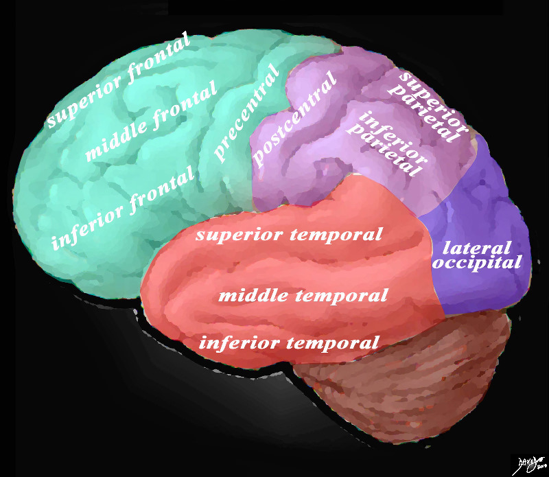
Overview of the Gyri of the Forebrain Lateral External View |
|
The lateral view of the brain shows the frontal lobe (green) parietal lobe (light mauve), the occipital lobe (purple) and the temporal lobe (red) In this view the frontal lobe gyri that are visible are; superior frontal, middle frontal, inferior frontal and precentral gyri. The parietal gyri include the post central, superior parietal and inferior parietal. The occipital gyrus that is visible is the lateral occipital. The temporal lobe gyri include the superior, middle and inferior temporal gyri.
Courtesy Ashley Davidoff MD Copyright 2010 83029d05g01.8s
|
Gyri of the Parietal Lobe
When viewed from the lateral aspect oft the brain three main gyri are seen. The post central gyrus is vertical and lies parallel with the precentral gyrus and central sulcus.
Posterior to the postcentral gyrus are the superior and the inferior parietal lobules.
The intraparietal sulcus runs posteriorly from the postcentral sulcus toward the occipital lobe almost horizontal and it separates the superior and inferior parietal lobules.
The inferior parietal lobule has two important subparts;
The supramarginal gyrus, and the angular gyrus.
Medial Surface of the Parietal Lobe
This surface of the parietal lobe contains a portion of the cingulate gyrus and the medial extension of the postcentral gyrus.
It is completed by an area called the precuneus.
This is bounded by the cingulate gyrus, the parietooccipital sulcus, and the marginal branch of the cingulate sulcus.
The extensions of the precentral and postcentral gyri onto the medial surface of the hemisphere are referred to together as the paracentral lobule.
This is partly in the frontal and parietal lobes.
DOMElement Object
(
    [schemaTypeInfo] =>
    [tagName] => table
    [firstElementChild] => (object value omitted)
    [lastElementChild] => (object value omitted)
    [childElementCount] => 1
    [previousElementSibling] => (object value omitted)
    [nextElementSibling] => (object value omitted)
    [nodeName] => table
    [nodeValue] =>


Overview of the Gyri of the Forebrain Lateral External View


The lateral view of the brain shows the frontal lobe (green) parietal lobe (light mauve), the occipital lobe (purple) and the temporal lobe (red) In this view the frontal lobe gyri that are visible are; superior frontal, middle frontal, inferior frontal and precentral gyri. The parietal gyri include the post central, superior parietal and inferior parietal. The occipital gyrus that is visible is the lateral occipital. The temporal lobe gyri include the superior, middle and inferior temporal gyri.
Courtesy Ashley Davidoff MD Copyright 2010 83029d05g01.8s


    [nodeType] => 1
    [parentNode] => (object value omitted)
    [childNodes] => (object value omitted)
    [firstChild] => (object value omitted)
    [lastChild] => (object value omitted)
    [previousSibling] => (object value omitted)
    [nextSibling] => (object value omitted)
    [attributes] => (object value omitted)
    [ownerDocument] => (object value omitted)
    [namespaceURI] =>
    [prefix] =>
    [localName] => table
    [baseURI] =>
    [textContent] =>


Overview of the Gyri of the Forebrain Lateral External View


The lateral view of the brain shows the frontal lobe (green) parietal lobe (light mauve), the occipital lobe (purple) and the temporal lobe (red) In this view the frontal lobe gyri that are visible are; superior frontal, middle frontal, inferior frontal and precentral gyri. The parietal gyri include the post central, superior parietal and inferior parietal. The occipital gyrus that is visible is the lateral occipital. The temporal lobe gyri include the superior, middle and inferior temporal gyri.
Courtesy Ashley Davidoff MD Copyright 2010 83029d05g01.8s


)
DOMElement Object
(
    [schemaTypeInfo] =>
    [tagName] => td
    [firstElementChild] => (object value omitted)
    [lastElementChild] => (object value omitted)
    [childElementCount] => 2
    [previousElementSibling] =>
    [nextElementSibling] =>
    [nodeName] => td
    [nodeValue] =>
The lateral view of the brain shows the frontal lobe (green) parietal lobe (light mauve), the occipital lobe (purple) and the temporal lobe (red) In this view the frontal lobe gyri that are visible are; superior frontal, middle frontal, inferior frontal and precentral gyri. The parietal gyri include the post central, superior parietal and inferior parietal. The occipital gyrus that is visible is the lateral occipital. The temporal lobe gyri include the superior, middle and inferior temporal gyri.
Courtesy Ashley Davidoff MD Copyright 2010 83029d05g01.8s

    [nodeType] => 1
    [parentNode] => (object value omitted)
    [childNodes] => (object value omitted)
    [firstChild] => (object value omitted)
    [lastChild] => (object value omitted)
    [previousSibling] => (object value omitted)
    [nextSibling] => (object value omitted)
    [attributes] => (object value omitted)
    [ownerDocument] => (object value omitted)
    [namespaceURI] =>
    [prefix] =>
    [localName] => td
    [baseURI] =>
    [textContent] =>
The lateral view of the brain shows the frontal lobe (green) parietal lobe (light mauve), the occipital lobe (purple) and the temporal lobe (red) In this view the frontal lobe gyri that are visible are; superior frontal, middle frontal, inferior frontal and precentral gyri. The parietal gyri include the post central, superior parietal and inferior parietal. The occipital gyrus that is visible is the lateral occipital. The temporal lobe gyri include the superior, middle and inferior temporal gyri.
Courtesy Ashley Davidoff MD Copyright 2010 83029d05g01.8s

)
DOMElement Object
(
    [schemaTypeInfo] =>
    [tagName] => td
    [firstElementChild] => (object value omitted)
    [lastElementChild] => (object value omitted)
    [childElementCount] => 2
    [previousElementSibling] =>
    [nextElementSibling] =>
    [nodeName] => td
    [nodeValue] =>
Overview of the Gyri of the Forebrain Lateral External View
    [nodeType] => 1
    [parentNode] => (object value omitted)
    [childNodes] => (object value omitted)
    [firstChild] => (object value omitted)
    [lastChild] => (object value omitted)
    [previousSibling] => (object value omitted)
    [nextSibling] => (object value omitted)
    [attributes] => (object value omitted)
    [ownerDocument] => (object value omitted)
    [namespaceURI] =>
    [prefix] =>
    [localName] => td
    [baseURI] =>
    [textContent] =>
Overview of the Gyri of the Forebrain Lateral External View
)

