Gradient Echo Sequence
GRE
Sumit Karia Md
Copyright 2010
Definition
Gradient echo sequences are those obtained by stimulating the object of study with a radiofrequency pulse at a flip angle below 90°. This will decrease the amount of magnetization in the transverse plane, and therefore a faster recovery in the z plane, which allows shorter TR?s and TE?s to be used, therefore decreasing scan time. Because of their speed, they are useful to minimize motion artifact that occurs during breathing, cardiac cycle, pulsations. Moving particles will have a bright signal. However, T2-weighted images will be more prone to artifacts. This occurs because the signal obtained is actually more T2*-weighted rather than T2-weighted – in other words, T2 is affected by static field inhomogeneities (and becomes called T2*). T2* is much shorter than T2.
 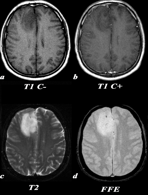
Oligodendroglioma – FFE Sequence for Calcification |
|
This 35 year old male presented with new onset of seizure.
T1 C- (a): On this T1 weighted image, there is a hypointense region in the right frontal lobe with blurring of the normally clear gray-white matter distinction.
T1 post C+ (b): No definite areas of enhancement were identified in this mass which is occasionally the case in oligodendrogliomas.
T2 (c): On T2 images, this lesion is more clearly defined as a region of increased signal. There is expansion of the involved gyri, as the adjacent sulci are not as clearly seen as they are in the same region on the contralateral side. Note the relative lack of surrounding edema around the lesion which would have the appearance of increased signal in the adjacent white matter sparing the gray matter.
FFE(d): This gradient echo MRI sequence is tailored for picking up substances which alter the local magnetic field, or paramagnetic substances, which will appear hypointense. This ?blooming? can be seen in areas of calcification, as in this case. Calcification is seen in a majority of oligodendrogliomas.
Image Courtesy Lawrence Chin MD 97673c.8
|
DOMElement Object
(
    [schemaTypeInfo] =>
    [tagName] => table
    [firstElementChild] => (object value omitted)
    [lastElementChild] => (object value omitted)
    [childElementCount] => 1
    [previousElementSibling] => (object value omitted)
    [nextElementSibling] =>
    [nodeName] => table
    [nodeValue] =>


Oligodendroglioma – FFE Sequence for Calcification


This 35 year old male presented with new onset of seizure.
T1 C- (a): On this T1 weighted image, there is a hypointense region in the right frontal lobe with blurring of the normally clear gray-white matter distinction.
T1 post C+ (b): No definite areas of enhancement were identified in this mass which is occasionally the case in oligodendrogliomas.
T2 (c): On T2 images, this lesion is more clearly defined as a region of increased signal. There is expansion of the involved gyri, as the adjacent sulci are not as clearly seen as they are in the same region on the contralateral side. Note the relative lack of surrounding edema around the lesion which would have the appearance of increased signal in the adjacent white matter sparing the gray matter.
FFE(d): This gradient echo MRI sequence is tailored for picking up substances which alter the local magnetic field, or paramagnetic substances, which will appear hypointense. This ?blooming? can be seen in areas of calcification, as in this case. Calcification is seen in a majority of oligodendrogliomas.
Image Courtesy Lawrence Chin MD 97673c.8


    [nodeType] => 1
    [parentNode] => (object value omitted)
    [childNodes] => (object value omitted)
    [firstChild] => (object value omitted)
    [lastChild] => (object value omitted)
    [previousSibling] => (object value omitted)
    [nextSibling] => (object value omitted)
    [attributes] => (object value omitted)
    [ownerDocument] => (object value omitted)
    [namespaceURI] =>
    [prefix] =>
    [localName] => table
    [baseURI] =>
    [textContent] =>


Oligodendroglioma – FFE Sequence for Calcification


This 35 year old male presented with new onset of seizure.
T1 C- (a): On this T1 weighted image, there is a hypointense region in the right frontal lobe with blurring of the normally clear gray-white matter distinction.
T1 post C+ (b): No definite areas of enhancement were identified in this mass which is occasionally the case in oligodendrogliomas.
T2 (c): On T2 images, this lesion is more clearly defined as a region of increased signal. There is expansion of the involved gyri, as the adjacent sulci are not as clearly seen as they are in the same region on the contralateral side. Note the relative lack of surrounding edema around the lesion which would have the appearance of increased signal in the adjacent white matter sparing the gray matter.
FFE(d): This gradient echo MRI sequence is tailored for picking up substances which alter the local magnetic field, or paramagnetic substances, which will appear hypointense. This ?blooming? can be seen in areas of calcification, as in this case. Calcification is seen in a majority of oligodendrogliomas.
Image Courtesy Lawrence Chin MD 97673c.8


)
DOMElement Object
(
    [schemaTypeInfo] =>
    [tagName] => td
    [firstElementChild] => (object value omitted)
    [lastElementChild] => (object value omitted)
    [childElementCount] => 6
    [previousElementSibling] =>
    [nextElementSibling] =>
    [nodeName] => td
    [nodeValue] =>
This 35 year old male presented with new onset of seizure.
T1 C- (a): On this T1 weighted image, there is a hypointense region in the right frontal lobe with blurring of the normally clear gray-white matter distinction.
T1 post C+ (b): No definite areas of enhancement were identified in this mass which is occasionally the case in oligodendrogliomas.
T2 (c): On T2 images, this lesion is more clearly defined as a region of increased signal. There is expansion of the involved gyri, as the adjacent sulci are not as clearly seen as they are in the same region on the contralateral side. Note the relative lack of surrounding edema around the lesion which would have the appearance of increased signal in the adjacent white matter sparing the gray matter.
FFE(d): This gradient echo MRI sequence is tailored for picking up substances which alter the local magnetic field, or paramagnetic substances, which will appear hypointense. This ?blooming? can be seen in areas of calcification, as in this case. Calcification is seen in a majority of oligodendrogliomas.
Image Courtesy Lawrence Chin MD 97673c.8

    [nodeType] => 1
    [parentNode] => (object value omitted)
    [childNodes] => (object value omitted)
    [firstChild] => (object value omitted)
    [lastChild] => (object value omitted)
    [previousSibling] => (object value omitted)
    [nextSibling] => (object value omitted)
    [attributes] => (object value omitted)
    [ownerDocument] => (object value omitted)
    [namespaceURI] =>
    [prefix] =>
    [localName] => td
    [baseURI] =>
    [textContent] =>
This 35 year old male presented with new onset of seizure.
T1 C- (a): On this T1 weighted image, there is a hypointense region in the right frontal lobe with blurring of the normally clear gray-white matter distinction.
T1 post C+ (b): No definite areas of enhancement were identified in this mass which is occasionally the case in oligodendrogliomas.
T2 (c): On T2 images, this lesion is more clearly defined as a region of increased signal. There is expansion of the involved gyri, as the adjacent sulci are not as clearly seen as they are in the same region on the contralateral side. Note the relative lack of surrounding edema around the lesion which would have the appearance of increased signal in the adjacent white matter sparing the gray matter.
FFE(d): This gradient echo MRI sequence is tailored for picking up substances which alter the local magnetic field, or paramagnetic substances, which will appear hypointense. This ?blooming? can be seen in areas of calcification, as in this case. Calcification is seen in a majority of oligodendrogliomas.
Image Courtesy Lawrence Chin MD 97673c.8

)
DOMElement Object
(
    [schemaTypeInfo] =>
    [tagName] => td
    [firstElementChild] => (object value omitted)
    [lastElementChild] => (object value omitted)
    [childElementCount] => 2
    [previousElementSibling] =>
    [nextElementSibling] =>
    [nodeName] => td
    [nodeValue] =>
Oligodendroglioma – FFE Sequence for Calcification
    [nodeType] => 1
    [parentNode] => (object value omitted)
    [childNodes] => (object value omitted)
    [firstChild] => (object value omitted)
    [lastChild] => (object value omitted)
    [previousSibling] => (object value omitted)
    [nextSibling] => (object value omitted)
    [attributes] => (object value omitted)
    [ownerDocument] => (object value omitted)
    [namespaceURI] =>
    [prefix] =>
    [localName] => td
    [baseURI] =>
    [textContent] =>
Oligodendroglioma – FFE Sequence for Calcification
)
https://beta.thecommonvein.net/wp-content/uploads/2023/05/97673c.8.jpg
http://www.thecommonvein.net/media/97673c.8_1.jpg
