Concepts
The Common Vein
Copyright 2009
Ashley Davidoff MD
Introduction
The brain is the most complex organ in the body, and the approach to understanding its structure must evolve from its most basic form that will stand as a foundation to the advancing complexity. As such one approach to understanding its structure, is to start with stick diagrams that draw simple intuitive lines of the vectors or axes of the brain, in an attempt to conceptualise the many parts of the brain and how they fit together.
Looking at the brain from its lateral aspect is very different from looking at it from above or from front. The sectional views in sagital axial and coronal projections add even more complexity as the vista of the structures changes every few mms. Even with this method it is difficult to understand brain structure because one has to conceptualise these vectors from many angles.
We will start by providing simple conceptual diagrams of the brain from the side, from above and from the front
The Sagittal View
|
The Basic Structure of the Brain from its Lateral Aspect The brain at its simplest |
|
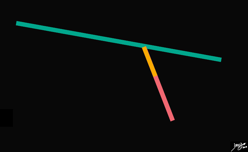
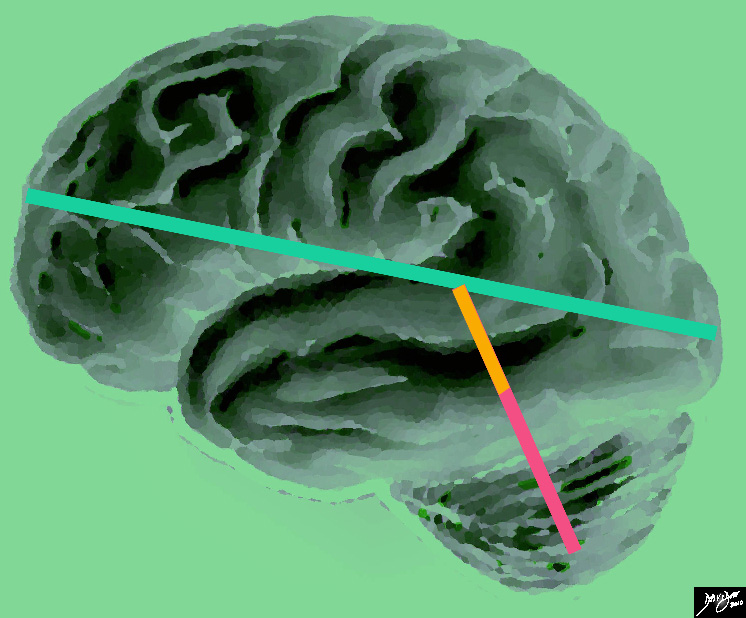
The stick diagram divides the brain into a relatively long almost horizontal component, and a relatively short almost vertical component. Anatomically the horizontal component is called the forebrain, and the vertical component will make up the midbrain (orange) and and hind brain (salmon) and extend ino the spinal cord Davidoff art Courtesy Ashley Davidoff MD copyright 2010 all rights reserved 93881b02.81s |
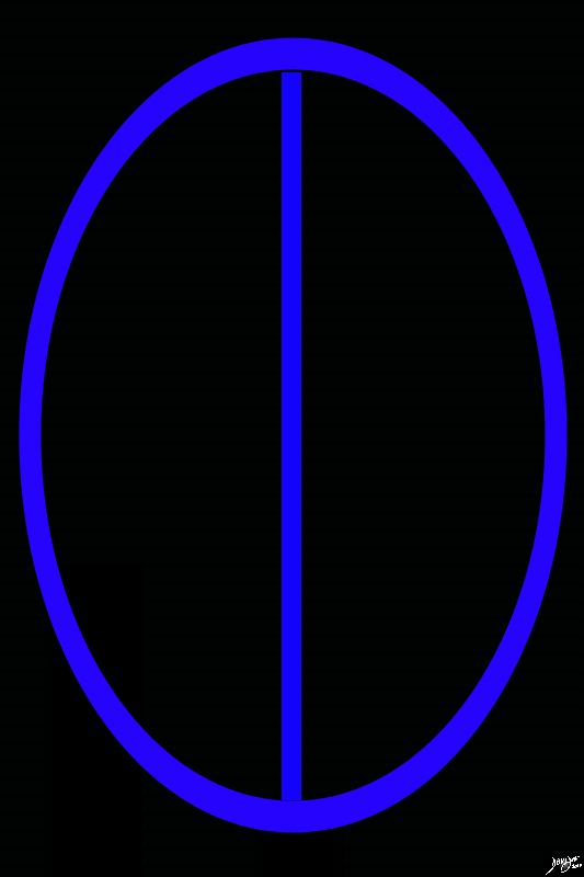
When the brain is approached from above the long axis is from front to back and the short axis is from side to side. The shape is almost an ovoid, but in truth the posterior aspect tends to be a little wider and the anterior aspect a tad narrower. The middle of the brain has a fissure called the interhemispheric fissure that divides the brain into two hemispheres.
Transverse or Axial View
Coronal View
DOMElement Object
(
    [schemaTypeInfo] =>
    [tagName] => table
    [firstElementChild] => (object value omitted)
    [lastElementChild] => (object value omitted)
    [childElementCount] => 1
    [previousElementSibling] => (object value omitted)
    [nextElementSibling] => (object value omitted)
    [nodeName] => table
    [nodeValue] =>


Frontal or Coronal View through the Middle of the Brain


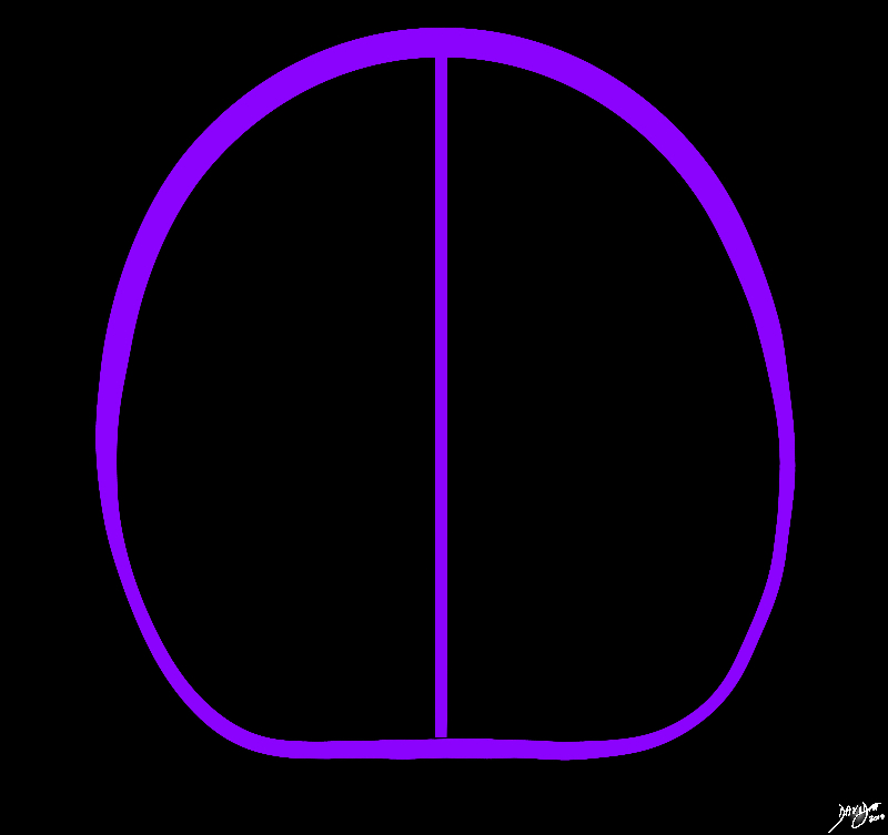
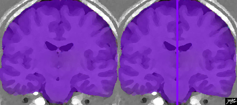
In the frontal or coronal view the brain has an almost rectangular shape, particulkalrly when viewed in a cut that traverses the temporal lobe. The midline structures of the brain, and predominantly the interhemispheric fissure divides the brain into two sides (purple divider). By convention, the cranial or superior part of the brain is above, the inferior part below with the right of the patient to your left and the left of the brain to your right. This rendered image of and MRI of the brain confirms the almost rectangular shape of the brain from this view and reveals the frontoparietal part of the brain in the anterior cranial fossa above, and the temporal lobes in the middle cranial fossa below. The internal anatomy will be discussed as we advance
Davidoff Art Courtesy Ashley Davidoff MD Copyright 2010 All rights reserved 92167c01.8s


    [nodeType] => 1
    [parentNode] => (object value omitted)
    [childNodes] => (object value omitted)
    [firstChild] => (object value omitted)
    [lastChild] => (object value omitted)
    [previousSibling] => (object value omitted)
    [nextSibling] => (object value omitted)
    [attributes] => (object value omitted)
    [ownerDocument] => (object value omitted)
    [namespaceURI] =>
    [prefix] =>
    [localName] => table
    [baseURI] =>
    [textContent] =>


Frontal or Coronal View through the Middle of the Brain


In the frontal or coronal view the brain has an almost rectangular shape, particulkalrly when viewed in a cut that traverses the temporal lobe. The midline structures of the brain, and predominantly the interhemispheric fissure divides the brain into two sides (purple divider). By convention, the cranial or superior part of the brain is above, the inferior part below with the right of the patient to your left and the left of the brain to your right. This rendered image of and MRI of the brain confirms the almost rectangular shape of the brain from this view and reveals the frontoparietal part of the brain in the anterior cranial fossa above, and the temporal lobes in the middle cranial fossa below. The internal anatomy will be discussed as we advance
Davidoff Art Courtesy Ashley Davidoff MD Copyright 2010 All rights reserved 92167c01.8s


)
DOMElement Object
(
    [schemaTypeInfo] =>
    [tagName] => td
    [firstElementChild] => (object value omitted)
    [lastElementChild] => (object value omitted)
    [childElementCount] => 2
    [previousElementSibling] =>
    [nextElementSibling] =>
    [nodeName] => td
    [nodeValue] =>
In the frontal or coronal view the brain has an almost rectangular shape, particulkalrly when viewed in a cut that traverses the temporal lobe. The midline structures of the brain, and predominantly the interhemispheric fissure divides the brain into two sides (purple divider). By convention, the cranial or superior part of the brain is above, the inferior part below with the right of the patient to your left and the left of the brain to your right. This rendered image of and MRI of the brain confirms the almost rectangular shape of the brain from this view and reveals the frontoparietal part of the brain in the anterior cranial fossa above, and the temporal lobes in the middle cranial fossa below. The internal anatomy will be discussed as we advance
Davidoff Art Courtesy Ashley Davidoff MD Copyright 2010 All rights reserved 92167c01.8s

    [nodeType] => 1
    [parentNode] => (object value omitted)
    [childNodes] => (object value omitted)
    [firstChild] => (object value omitted)
    [lastChild] => (object value omitted)
    [previousSibling] => (object value omitted)
    [nextSibling] => (object value omitted)
    [attributes] => (object value omitted)
    [ownerDocument] => (object value omitted)
    [namespaceURI] =>
    [prefix] =>
    [localName] => td
    [baseURI] =>
    [textContent] =>
In the frontal or coronal view the brain has an almost rectangular shape, particulkalrly when viewed in a cut that traverses the temporal lobe. The midline structures of the brain, and predominantly the interhemispheric fissure divides the brain into two sides (purple divider). By convention, the cranial or superior part of the brain is above, the inferior part below with the right of the patient to your left and the left of the brain to your right. This rendered image of and MRI of the brain confirms the almost rectangular shape of the brain from this view and reveals the frontoparietal part of the brain in the anterior cranial fossa above, and the temporal lobes in the middle cranial fossa below. The internal anatomy will be discussed as we advance
Davidoff Art Courtesy Ashley Davidoff MD Copyright 2010 All rights reserved 92167c01.8s

)
DOMElement Object
(
    [schemaTypeInfo] =>
    [tagName] => td
    [firstElementChild] => (object value omitted)
    [lastElementChild] => (object value omitted)
    [childElementCount] => 2
    [previousElementSibling] =>
    [nextElementSibling] =>
    [nodeName] => td
    [nodeValue] =>
Frontal or Coronal View through the Middle of the Brain

    [nodeType] => 1
    [parentNode] => (object value omitted)
    [childNodes] => (object value omitted)
    [firstChild] => (object value omitted)
    [lastChild] => (object value omitted)
    [previousSibling] => (object value omitted)
    [nextSibling] => (object value omitted)
    [attributes] => (object value omitted)
    [ownerDocument] => (object value omitted)
    [namespaceURI] =>
    [prefix] =>
    [localName] => td
    [baseURI] =>
    [textContent] =>
Frontal or Coronal View through the Middle of the Brain

)
DOMElement Object
(
    [schemaTypeInfo] =>
    [tagName] => table
    [firstElementChild] => (object value omitted)
    [lastElementChild] => (object value omitted)
    [childElementCount] => 1
    [previousElementSibling] => (object value omitted)
    [nextElementSibling] => (object value omitted)
    [nodeName] => table
    [nodeValue] =>


from the Front – Coronal Projection


In the frontal or coronal view the brain has an almost rectangular shapeMidline structures and predominantly the interhemispheric fissure divides the brain into two sides
Davidoff Art Copyright 2010 All rights reserved 93914b03.8s


    [nodeType] => 1
    [parentNode] => (object value omitted)
    [childNodes] => (object value omitted)
    [firstChild] => (object value omitted)
    [lastChild] => (object value omitted)
    [previousSibling] => (object value omitted)
    [nextSibling] => (object value omitted)
    [attributes] => (object value omitted)
    [ownerDocument] => (object value omitted)
    [namespaceURI] =>
    [prefix] =>
    [localName] => table
    [baseURI] =>
    [textContent] =>


from the Front – Coronal Projection


In the frontal or coronal view the brain has an almost rectangular shapeMidline structures and predominantly the interhemispheric fissure divides the brain into two sides
Davidoff Art Copyright 2010 All rights reserved 93914b03.8s


)
DOMElement Object
(
    [schemaTypeInfo] =>
    [tagName] => td
    [firstElementChild] => (object value omitted)
    [lastElementChild] => (object value omitted)
    [childElementCount] => 2
    [previousElementSibling] =>
    [nextElementSibling] =>
    [nodeName] => td
    [nodeValue] =>
In the frontal or coronal view the brain has an almost rectangular shapeMidline structures and predominantly the interhemispheric fissure divides the brain into two sides
Davidoff Art Copyright 2010 All rights reserved 93914b03.8s

    [nodeType] => 1
    [parentNode] => (object value omitted)
    [childNodes] => (object value omitted)
    [firstChild] => (object value omitted)
    [lastChild] => (object value omitted)
    [previousSibling] => (object value omitted)
    [nextSibling] => (object value omitted)
    [attributes] => (object value omitted)
    [ownerDocument] => (object value omitted)
    [namespaceURI] =>
    [prefix] =>
    [localName] => td
    [baseURI] =>
    [textContent] =>
In the frontal or coronal view the brain has an almost rectangular shapeMidline structures and predominantly the interhemispheric fissure divides the brain into two sides
Davidoff Art Copyright 2010 All rights reserved 93914b03.8s

)
DOMElement Object
(
    [schemaTypeInfo] =>
    [tagName] => td
    [firstElementChild] => (object value omitted)
    [lastElementChild] => (object value omitted)
    [childElementCount] => 2
    [previousElementSibling] =>
    [nextElementSibling] =>
    [nodeName] => td
    [nodeValue] =>
from the Front – Coronal Projection

    [nodeType] => 1
    [parentNode] => (object value omitted)
    [childNodes] => (object value omitted)
    [firstChild] => (object value omitted)
    [lastChild] => (object value omitted)
    [previousSibling] => (object value omitted)
    [nextSibling] => (object value omitted)
    [attributes] => (object value omitted)
    [ownerDocument] => (object value omitted)
    [namespaceURI] =>
    [prefix] =>
    [localName] => td
    [baseURI] =>
    [textContent] =>
from the Front – Coronal Projection

)
DOMElement Object
(
    [schemaTypeInfo] =>
    [tagName] => table
    [firstElementChild] => (object value omitted)
    [lastElementChild] => (object value omitted)
    [childElementCount] => 1
    [previousElementSibling] => (object value omitted)
    [nextElementSibling] => (object value omitted)
    [nodeName] => table
    [nodeValue] =>


Deeper in the Brain


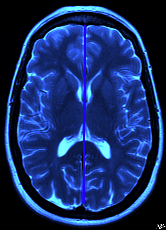
The classical axial position that enables reference points in transverse projection is at the level where the major portions of the ventricles are seen. In this rendered MROI t2 weighted image, the whiteness of the ventricles including the frontal horns, foramena of Munro, and occipital horns are visible . The symmetry of the the two hemispheres in structure is not matched by consistent symmetery in function
Courtesy Ashley Davidoff MD Copyright 2010 All rights reserved 94079.8a03.8s


    [nodeType] => 1
    [parentNode] => (object value omitted)
    [childNodes] => (object value omitted)
    [firstChild] => (object value omitted)
    [lastChild] => (object value omitted)
    [previousSibling] => (object value omitted)
    [nextSibling] => (object value omitted)
    [attributes] => (object value omitted)
    [ownerDocument] => (object value omitted)
    [namespaceURI] =>
    [prefix] =>
    [localName] => table
    [baseURI] =>
    [textContent] =>


Deeper in the Brain


The classical axial position that enables reference points in transverse projection is at the level where the major portions of the ventricles are seen. In this rendered MROI t2 weighted image, the whiteness of the ventricles including the frontal horns, foramena of Munro, and occipital horns are visible . The symmetry of the the two hemispheres in structure is not matched by consistent symmetery in function
Courtesy Ashley Davidoff MD Copyright 2010 All rights reserved 94079.8a03.8s


)
DOMElement Object
(
    [schemaTypeInfo] =>
    [tagName] => td
    [firstElementChild] => (object value omitted)
    [lastElementChild] => (object value omitted)
    [childElementCount] => 2
    [previousElementSibling] =>
    [nextElementSibling] =>
    [nodeName] => td
    [nodeValue] =>
The classical axial position that enables reference points in transverse projection is at the level where the major portions of the ventricles are seen. In this rendered MROI t2 weighted image, the whiteness of the ventricles including the frontal horns, foramena of Munro, and occipital horns are visible . The symmetry of the the two hemispheres in structure is not matched by consistent symmetery in function
Courtesy Ashley Davidoff MD Copyright 2010 All rights reserved 94079.8a03.8s

    [nodeType] => 1
    [parentNode] => (object value omitted)
    [childNodes] => (object value omitted)
    [firstChild] => (object value omitted)
    [lastChild] => (object value omitted)
    [previousSibling] => (object value omitted)
    [nextSibling] => (object value omitted)
    [attributes] => (object value omitted)
    [ownerDocument] => (object value omitted)
    [namespaceURI] =>
    [prefix] =>
    [localName] => td
    [baseURI] =>
    [textContent] =>
The classical axial position that enables reference points in transverse projection is at the level where the major portions of the ventricles are seen. In this rendered MROI t2 weighted image, the whiteness of the ventricles including the frontal horns, foramena of Munro, and occipital horns are visible . The symmetry of the the two hemispheres in structure is not matched by consistent symmetery in function
Courtesy Ashley Davidoff MD Copyright 2010 All rights reserved 94079.8a03.8s

)
DOMElement Object
(
    [schemaTypeInfo] =>
    [tagName] => td
    [firstElementChild] => (object value omitted)
    [lastElementChild] => (object value omitted)
    [childElementCount] => 2
    [previousElementSibling] =>
    [nextElementSibling] =>
    [nodeName] => td
    [nodeValue] =>
Deeper in the Brain

    [nodeType] => 1
    [parentNode] => (object value omitted)
    [childNodes] => (object value omitted)
    [firstChild] => (object value omitted)
    [lastChild] => (object value omitted)
    [previousSibling] => (object value omitted)
    [nextSibling] => (object value omitted)
    [attributes] => (object value omitted)
    [ownerDocument] => (object value omitted)
    [namespaceURI] =>
    [prefix] =>
    [localName] => td
    [baseURI] =>
    [textContent] =>
Deeper in the Brain

)
DOMElement Object
(
    [schemaTypeInfo] =>
    [tagName] => table
    [firstElementChild] => (object value omitted)
    [lastElementChild] => (object value omitted)
    [childElementCount] => 1
    [previousElementSibling] => (object value omitted)
    [nextElementSibling] => (object value omitted)
    [nodeName] => table
    [nodeValue] =>


The Axial Conceptual Vectors Overlying an External transverse View of the Brain


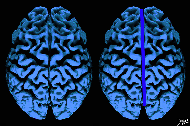
When the external surface is explored from above a fissure divides the oval of the brain into two hemisphere. The deep cleft is called the interhemishpheric fissure In additionthe serpiginous folds and crevices that characterize the outside surface of the brain start to become apparent as one explores the shape of the brain. It is obvious that it is more than a mere oval.
davidoff Art Copyright 2010 All rights reserved Davidoff Art Courtesy Ashley Davidoff MD 52981b05.8.44kb01c02.8s


    [nodeType] => 1
    [parentNode] => (object value omitted)
    [childNodes] => (object value omitted)
    [firstChild] => (object value omitted)
    [lastChild] => (object value omitted)
    [previousSibling] => (object value omitted)
    [nextSibling] => (object value omitted)
    [attributes] => (object value omitted)
    [ownerDocument] => (object value omitted)
    [namespaceURI] =>
    [prefix] =>
    [localName] => table
    [baseURI] =>
    [textContent] =>


The Axial Conceptual Vectors Overlying an External transverse View of the Brain


When the external surface is explored from above a fissure divides the oval of the brain into two hemisphere. The deep cleft is called the interhemishpheric fissure In additionthe serpiginous folds and crevices that characterize the outside surface of the brain start to become apparent as one explores the shape of the brain. It is obvious that it is more than a mere oval.
davidoff Art Copyright 2010 All rights reserved Davidoff Art Courtesy Ashley Davidoff MD 52981b05.8.44kb01c02.8s


)
DOMElement Object
(
    [schemaTypeInfo] =>
    [tagName] => td
    [firstElementChild] => (object value omitted)
    [lastElementChild] => (object value omitted)
    [childElementCount] => 2
    [previousElementSibling] =>
    [nextElementSibling] =>
    [nodeName] => td
    [nodeValue] =>
When the external surface is explored from above a fissure divides the oval of the brain into two hemisphere. The deep cleft is called the interhemishpheric fissure In additionthe serpiginous folds and crevices that characterize the outside surface of the brain start to become apparent as one explores the shape of the brain. It is obvious that it is more than a mere oval.
davidoff Art Copyright 2010 All rights reserved Davidoff Art Courtesy Ashley Davidoff MD 52981b05.8.44kb01c02.8s

    [nodeType] => 1
    [parentNode] => (object value omitted)
    [childNodes] => (object value omitted)
    [firstChild] => (object value omitted)
    [lastChild] => (object value omitted)
    [previousSibling] => (object value omitted)
    [nextSibling] => (object value omitted)
    [attributes] => (object value omitted)
    [ownerDocument] => (object value omitted)
    [namespaceURI] =>
    [prefix] =>
    [localName] => td
    [baseURI] =>
    [textContent] =>
When the external surface is explored from above a fissure divides the oval of the brain into two hemisphere. The deep cleft is called the interhemishpheric fissure In additionthe serpiginous folds and crevices that characterize the outside surface of the brain start to become apparent as one explores the shape of the brain. It is obvious that it is more than a mere oval.
davidoff Art Copyright 2010 All rights reserved Davidoff Art Courtesy Ashley Davidoff MD 52981b05.8.44kb01c02.8s

)
DOMElement Object
(
    [schemaTypeInfo] =>
    [tagName] => td
    [firstElementChild] => (object value omitted)
    [lastElementChild] => (object value omitted)
    [childElementCount] => 2
    [previousElementSibling] =>
    [nextElementSibling] =>
    [nodeName] => td
    [nodeValue] =>
The Axial Conceptual Vectors Overlying an External transverse View of the Brain
    [nodeType] => 1
    [parentNode] => (object value omitted)
    [childNodes] => (object value omitted)
    [firstChild] => (object value omitted)
    [lastChild] => (object value omitted)
    [previousSibling] => (object value omitted)
    [nextSibling] => (object value omitted)
    [attributes] => (object value omitted)
    [ownerDocument] => (object value omitted)
    [namespaceURI] =>
    [prefix] =>
    [localName] => td
    [baseURI] =>
    [textContent] =>
The Axial Conceptual Vectors Overlying an External transverse View of the Brain
)
DOMElement Object
(
    [schemaTypeInfo] =>
    [tagName] => table
    [firstElementChild] => (object value omitted)
    [lastElementChild] => (object value omitted)
    [childElementCount] => 1
    [previousElementSibling] => (object value omitted)
    [nextElementSibling] => (object value omitted)
    [nodeName] => table
    [nodeValue] =>


Axial Projection
Superior projection
The Two Cerebral Hemispheres Divided by the Interhemispheric Fissure


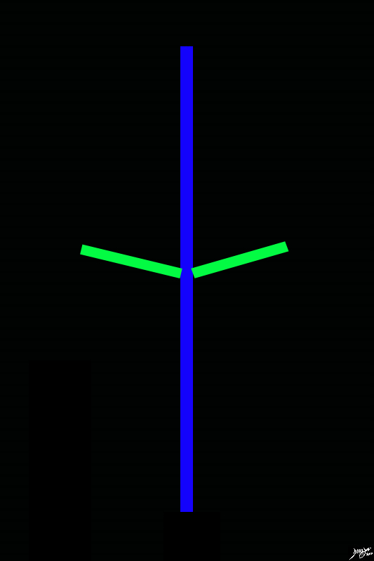
The diagram reflects the interhemispheric fissure as depicted in the axial plane dividing the brain into two halves. When looking at the top of the brain we are really looking at the forebrain, and the interhemispheric fissure divides the forebrain into two cerebral hemispheres.
 By convention anterior is on the top and posterior on the bottom, while the patients right is to our left and vice versa for the patients left brain stick diagram
Davidoff Art Copyright 2010 Courtesy Ash;ley Davidoff MD 93914.82s


    [nodeType] => 1
    [parentNode] => (object value omitted)
    [childNodes] => (object value omitted)
    [firstChild] => (object value omitted)
    [lastChild] => (object value omitted)
    [previousSibling] => (object value omitted)
    [nextSibling] => (object value omitted)
    [attributes] => (object value omitted)
    [ownerDocument] => (object value omitted)
    [namespaceURI] =>
    [prefix] =>
    [localName] => table
    [baseURI] =>
    [textContent] =>


Axial Projection
Superior projection
The Two Cerebral Hemispheres Divided by the Interhemispheric Fissure


The diagram reflects the interhemispheric fissure as depicted in the axial plane dividing the brain into two halves. When looking at the top of the brain we are really looking at the forebrain, and the interhemispheric fissure divides the forebrain into two cerebral hemispheres.
 By convention anterior is on the top and posterior on the bottom, while the patients right is to our left and vice versa for the patients left brain stick diagram
Davidoff Art Copyright 2010 Courtesy Ash;ley Davidoff MD 93914.82s


)
DOMElement Object
(
    [schemaTypeInfo] =>
    [tagName] => td
    [firstElementChild] => (object value omitted)
    [lastElementChild] => (object value omitted)
    [childElementCount] => 3
    [previousElementSibling] =>
    [nextElementSibling] =>
    [nodeName] => td
    [nodeValue] =>
The diagram reflects the interhemispheric fissure as depicted in the axial plane dividing the brain into two halves. When looking at the top of the brain we are really looking at the forebrain, and the interhemispheric fissure divides the forebrain into two cerebral hemispheres.
 By convention anterior is on the top and posterior on the bottom, while the patients right is to our left and vice versa for the patients left brain stick diagram
Davidoff Art Copyright 2010 Courtesy Ash;ley Davidoff MD 93914.82s

    [nodeType] => 1
    [parentNode] => (object value omitted)
    [childNodes] => (object value omitted)
    [firstChild] => (object value omitted)
    [lastChild] => (object value omitted)
    [previousSibling] => (object value omitted)
    [nextSibling] => (object value omitted)
    [attributes] => (object value omitted)
    [ownerDocument] => (object value omitted)
    [namespaceURI] =>
    [prefix] =>
    [localName] => td
    [baseURI] =>
    [textContent] =>
The diagram reflects the interhemispheric fissure as depicted in the axial plane dividing the brain into two halves. When looking at the top of the brain we are really looking at the forebrain, and the interhemispheric fissure divides the forebrain into two cerebral hemispheres.
 By convention anterior is on the top and posterior on the bottom, while the patients right is to our left and vice versa for the patients left brain stick diagram
Davidoff Art Copyright 2010 Courtesy Ash;ley Davidoff MD 93914.82s

)
DOMElement Object
(
    [schemaTypeInfo] =>
    [tagName] => td
    [firstElementChild] => (object value omitted)
    [lastElementChild] => (object value omitted)
    [childElementCount] => 4
    [previousElementSibling] =>
    [nextElementSibling] =>
    [nodeName] => td
    [nodeValue] =>
Axial Projection
Superior projection
The Two Cerebral Hemispheres Divided by the Interhemispheric Fissure

    [nodeType] => 1
    [parentNode] => (object value omitted)
    [childNodes] => (object value omitted)
    [firstChild] => (object value omitted)
    [lastChild] => (object value omitted)
    [previousSibling] => (object value omitted)
    [nextSibling] => (object value omitted)
    [attributes] => (object value omitted)
    [ownerDocument] => (object value omitted)
    [namespaceURI] =>
    [prefix] =>
    [localName] => td
    [baseURI] =>
    [textContent] =>
Axial Projection
Superior projection
The Two Cerebral Hemispheres Divided by the Interhemispheric Fissure

)
DOMElement Object
(
    [schemaTypeInfo] =>
    [tagName] => table
    [firstElementChild] => (object value omitted)
    [lastElementChild] => (object value omitted)
    [childElementCount] => 1
    [previousElementSibling] => (object value omitted)
    [nextElementSibling] => (object value omitted)
    [nodeName] => table
    [nodeValue] =>


The Sagital Conceptual Vectors Overlying an External Lateral View of the Brain


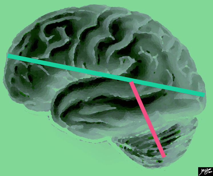
The vectors of the brain from a side view are superimposed on a specimen of the brain, showing a long anteroposterior vector (light green) and a shorter more vertical vector (salmon pink)
brain normal green basic principles concepts vectors Davidoff Art courtesy Ashley Davidoff copyright 2010 all rights reserved 83029e04b01.8s


    [nodeType] => 1
    [parentNode] => (object value omitted)
    [childNodes] => (object value omitted)
    [firstChild] => (object value omitted)
    [lastChild] => (object value omitted)
    [previousSibling] => (object value omitted)
    [nextSibling] => (object value omitted)
    [attributes] => (object value omitted)
    [ownerDocument] => (object value omitted)
    [namespaceURI] =>
    [prefix] =>
    [localName] => table
    [baseURI] =>
    [textContent] =>


The Sagital Conceptual Vectors Overlying an External Lateral View of the Brain


The vectors of the brain from a side view are superimposed on a specimen of the brain, showing a long anteroposterior vector (light green) and a shorter more vertical vector (salmon pink)
brain normal green basic principles concepts vectors Davidoff Art courtesy Ashley Davidoff copyright 2010 all rights reserved 83029e04b01.8s


)
DOMElement Object
(
    [schemaTypeInfo] =>
    [tagName] => td
    [firstElementChild] => (object value omitted)
    [lastElementChild] => (object value omitted)
    [childElementCount] => 2
    [previousElementSibling] =>
    [nextElementSibling] =>
    [nodeName] => td
    [nodeValue] =>
The vectors of the brain from a side view are superimposed on a specimen of the brain, showing a long anteroposterior vector (light green) and a shorter more vertical vector (salmon pink)
brain normal green basic principles concepts vectors Davidoff Art courtesy Ashley Davidoff copyright 2010 all rights reserved 83029e04b01.8s

    [nodeType] => 1
    [parentNode] => (object value omitted)
    [childNodes] => (object value omitted)
    [firstChild] => (object value omitted)
    [lastChild] => (object value omitted)
    [previousSibling] => (object value omitted)
    [nextSibling] => (object value omitted)
    [attributes] => (object value omitted)
    [ownerDocument] => (object value omitted)
    [namespaceURI] =>
    [prefix] =>
    [localName] => td
    [baseURI] =>
    [textContent] =>
The vectors of the brain from a side view are superimposed on a specimen of the brain, showing a long anteroposterior vector (light green) and a shorter more vertical vector (salmon pink)
brain normal green basic principles concepts vectors Davidoff Art courtesy Ashley Davidoff copyright 2010 all rights reserved 83029e04b01.8s

)
DOMElement Object
(
    [schemaTypeInfo] =>
    [tagName] => td
    [firstElementChild] => (object value omitted)
    [lastElementChild] => (object value omitted)
    [childElementCount] => 2
    [previousElementSibling] =>
    [nextElementSibling] =>
    [nodeName] => td
    [nodeValue] =>
The Sagital Conceptual Vectors Overlying an External Lateral View of the Brain

    [nodeType] => 1
    [parentNode] => (object value omitted)
    [childNodes] => (object value omitted)
    [firstChild] => (object value omitted)
    [lastChild] => (object value omitted)
    [previousSibling] => (object value omitted)
    [nextSibling] => (object value omitted)
    [attributes] => (object value omitted)
    [ownerDocument] => (object value omitted)
    [namespaceURI] =>
    [prefix] =>
    [localName] => td
    [baseURI] =>
    [textContent] =>
The Sagital Conceptual Vectors Overlying an External Lateral View of the Brain

)
DOMElement Object
(
    [schemaTypeInfo] =>
    [tagName] => table
    [firstElementChild] => (object value omitted)
    [lastElementChild] => (object value omitted)
    [childElementCount] => 1
    [previousElementSibling] => (object value omitted)
    [nextElementSibling] => (object value omitted)
    [nodeName] => table
    [nodeValue] =>


The Basic Structure of the Brain from its Lateral Aspect
The brain at its simplest


The stick diagram divides the brain into a relatively long almost horizontal component, and a relatively short almost vertical component. Anatomically the horizontal component is called the forebrain, and the vertical component will make up the midbrain (orange) and  and hind brain (salmon)  and extend ino the spinal cord
Davidoff art Courtesy Ashley Davidoff MD copyright 2010 all rights reserved 93881b02.81s


    [nodeType] => 1
    [parentNode] => (object value omitted)
    [childNodes] => (object value omitted)
    [firstChild] => (object value omitted)
    [lastChild] => (object value omitted)
    [previousSibling] => (object value omitted)
    [nextSibling] => (object value omitted)
    [attributes] => (object value omitted)
    [ownerDocument] => (object value omitted)
    [namespaceURI] =>
    [prefix] =>
    [localName] => table
    [baseURI] =>
    [textContent] =>


The Basic Structure of the Brain from its Lateral Aspect
The brain at its simplest


The stick diagram divides the brain into a relatively long almost horizontal component, and a relatively short almost vertical component. Anatomically the horizontal component is called the forebrain, and the vertical component will make up the midbrain (orange) and  and hind brain (salmon)  and extend ino the spinal cord
Davidoff art Courtesy Ashley Davidoff MD copyright 2010 all rights reserved 93881b02.81s


)
DOMElement Object
(
    [schemaTypeInfo] =>
    [tagName] => td
    [firstElementChild] => (object value omitted)
    [lastElementChild] => (object value omitted)
    [childElementCount] => 2
    [previousElementSibling] =>
    [nextElementSibling] =>
    [nodeName] => td
    [nodeValue] =>
The stick diagram divides the brain into a relatively long almost horizontal component, and a relatively short almost vertical component. Anatomically the horizontal component is called the forebrain, and the vertical component will make up the midbrain (orange) and  and hind brain (salmon)  and extend ino the spinal cord
Davidoff art Courtesy Ashley Davidoff MD copyright 2010 all rights reserved 93881b02.81s

    [nodeType] => 1
    [parentNode] => (object value omitted)
    [childNodes] => (object value omitted)
    [firstChild] => (object value omitted)
    [lastChild] => (object value omitted)
    [previousSibling] => (object value omitted)
    [nextSibling] => (object value omitted)
    [attributes] => (object value omitted)
    [ownerDocument] => (object value omitted)
    [namespaceURI] =>
    [prefix] =>
    [localName] => td
    [baseURI] =>
    [textContent] =>
The stick diagram divides the brain into a relatively long almost horizontal component, and a relatively short almost vertical component. Anatomically the horizontal component is called the forebrain, and the vertical component will make up the midbrain (orange) and  and hind brain (salmon)  and extend ino the spinal cord
Davidoff art Courtesy Ashley Davidoff MD copyright 2010 all rights reserved 93881b02.81s

)
DOMElement Object
(
    [schemaTypeInfo] =>
    [tagName] => td
    [firstElementChild] => (object value omitted)
    [lastElementChild] => (object value omitted)
    [childElementCount] => 3
    [previousElementSibling] =>
    [nextElementSibling] =>
    [nodeName] => td
    [nodeValue] =>

The Basic Structure of the Brain from its Lateral Aspect
The brain at its simplest

    [nodeType] => 1
    [parentNode] => (object value omitted)
    [childNodes] => (object value omitted)
    [firstChild] => (object value omitted)
    [lastChild] => (object value omitted)
    [previousSibling] => (object value omitted)
    [nextSibling] => (object value omitted)
    [attributes] => (object value omitted)
    [ownerDocument] => (object value omitted)
    [namespaceURI] =>
    [prefix] =>
    [localName] => td
    [baseURI] =>
    [textContent] =>

The Basic Structure of the Brain from its Lateral Aspect
The brain at its simplest

)

