Ventricles of the Brain ? Concepts
Ashley Davidoff MD
The Common Vein Copyright 2010
Introduction ? Sagittal Projection
The ventricular system can be conceptualized as paired structures running horizontally and superiorly connecting with a vertical component.
Transverse or Axial View
Coronal View
DOMElement Object
(
    [schemaTypeInfo] =>
    [tagName] => table
    [firstElementChild] => (object value omitted)
    [lastElementChild] => (object value omitted)
    [childElementCount] => 1
    [previousElementSibling] => (object value omitted)
    [nextElementSibling] =>
    [nodeName] => table
    [nodeValue] =>


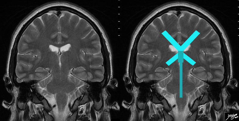
Coronal T2 Weighted Image


The ?X? of the horizontal limb represents the ventricular system that subtends the frontal horns (upper V) and the temporal horns( lower invetred V). These will not be seen on the same cut since the frontal horns are anterior and the temporal horns are more posterior. The most superior and widened portion of the vertical limb (darker green) represents the third ventricle Inferior to the third ventricle along the vertical limb is the aqueduct of Sylvius and 4th ventricle
Courtesy Ashley Davidoff MD copyright 2010 all rights reserved MRI from 99721 series 94458e10.81s


    [nodeType] => 1
    [parentNode] => (object value omitted)
    [childNodes] => (object value omitted)
    [firstChild] => (object value omitted)
    [lastChild] => (object value omitted)
    [previousSibling] => (object value omitted)
    [nextSibling] => (object value omitted)
    [attributes] => (object value omitted)
    [ownerDocument] => (object value omitted)
    [namespaceURI] =>
    [prefix] =>
    [localName] => table
    [baseURI] =>
    [textContent] =>


Coronal T2 Weighted Image


The ?X? of the horizontal limb represents the ventricular system that subtends the frontal horns (upper V) and the temporal horns( lower invetred V). These will not be seen on the same cut since the frontal horns are anterior and the temporal horns are more posterior. The most superior and widened portion of the vertical limb (darker green) represents the third ventricle Inferior to the third ventricle along the vertical limb is the aqueduct of Sylvius and 4th ventricle
Courtesy Ashley Davidoff MD copyright 2010 all rights reserved MRI from 99721 series 94458e10.81s


)
DOMElement Object
(
    [schemaTypeInfo] =>
    [tagName] => td
    [firstElementChild] => (object value omitted)
    [lastElementChild] => (object value omitted)
    [childElementCount] => 2
    [previousElementSibling] =>
    [nextElementSibling] =>
    [nodeName] => td
    [nodeValue] =>
The ?X? of the horizontal limb represents the ventricular system that subtends the frontal horns (upper V) and the temporal horns( lower invetred V). These will not be seen on the same cut since the frontal horns are anterior and the temporal horns are more posterior. The most superior and widened portion of the vertical limb (darker green) represents the third ventricle Inferior to the third ventricle along the vertical limb is the aqueduct of Sylvius and 4th ventricle
Courtesy Ashley Davidoff MD copyright 2010 all rights reserved MRI from 99721 series 94458e10.81s

    [nodeType] => 1
    [parentNode] => (object value omitted)
    [childNodes] => (object value omitted)
    [firstChild] => (object value omitted)
    [lastChild] => (object value omitted)
    [previousSibling] => (object value omitted)
    [nextSibling] => (object value omitted)
    [attributes] => (object value omitted)
    [ownerDocument] => (object value omitted)
    [namespaceURI] =>
    [prefix] =>
    [localName] => td
    [baseURI] =>
    [textContent] =>
The ?X? of the horizontal limb represents the ventricular system that subtends the frontal horns (upper V) and the temporal horns( lower invetred V). These will not be seen on the same cut since the frontal horns are anterior and the temporal horns are more posterior. The most superior and widened portion of the vertical limb (darker green) represents the third ventricle Inferior to the third ventricle along the vertical limb is the aqueduct of Sylvius and 4th ventricle
Courtesy Ashley Davidoff MD copyright 2010 all rights reserved MRI from 99721 series 94458e10.81s

)
DOMElement Object
(
    [schemaTypeInfo] =>
    [tagName] => td
    [firstElementChild] => (object value omitted)
    [lastElementChild] => (object value omitted)
    [childElementCount] => 2
    [previousElementSibling] =>
    [nextElementSibling] =>
    [nodeName] => td
    [nodeValue] =>
Coronal T2 Weighted Image

    [nodeType] => 1
    [parentNode] => (object value omitted)
    [childNodes] => (object value omitted)
    [firstChild] => (object value omitted)
    [lastChild] => (object value omitted)
    [previousSibling] => (object value omitted)
    [nextSibling] => (object value omitted)
    [attributes] => (object value omitted)
    [ownerDocument] => (object value omitted)
    [namespaceURI] =>
    [prefix] =>
    [localName] => td
    [baseURI] =>
    [textContent] =>
Coronal T2 Weighted Image

)
DOMElement Object
(
    [schemaTypeInfo] =>
    [tagName] => table
    [firstElementChild] => (object value omitted)
    [lastElementChild] => (object value omitted)
    [childElementCount] => 1
    [previousElementSibling] => (object value omitted)
    [nextElementSibling] => (object value omitted)
    [nodeName] => table
    [nodeValue] =>


The Coronal View of the Ventricular System


The distribution of the ventricular system is demonstrated in this diagram with the horizontall oriented components in teal, and the vertically oriented components in light green. They are both centered around the midlinewith all the components of the vertcial system being exactly in midline and all the components of the horizontal system connected and confluent in the midline. The horizontal structures include the lateral ventricles, frontal horns, atria,and temporal horns. The vertical midline structures include the third ventricle, aqueduct of Sylvius, and 4th ventricle.
Courtesy Ashley Davidoff MD copyright 2010 all rights reserved 93914b04d06b.81s


    [nodeType] => 1
    [parentNode] => (object value omitted)
    [childNodes] => (object value omitted)
    [firstChild] => (object value omitted)
    [lastChild] => (object value omitted)
    [previousSibling] => (object value omitted)
    [nextSibling] => (object value omitted)
    [attributes] => (object value omitted)
    [ownerDocument] => (object value omitted)
    [namespaceURI] =>
    [prefix] =>
    [localName] => table
    [baseURI] =>
    [textContent] =>


The Coronal View of the Ventricular System


The distribution of the ventricular system is demonstrated in this diagram with the horizontall oriented components in teal, and the vertically oriented components in light green. They are both centered around the midlinewith all the components of the vertcial system being exactly in midline and all the components of the horizontal system connected and confluent in the midline. The horizontal structures include the lateral ventricles, frontal horns, atria,and temporal horns. The vertical midline structures include the third ventricle, aqueduct of Sylvius, and 4th ventricle.
Courtesy Ashley Davidoff MD copyright 2010 all rights reserved 93914b04d06b.81s


)
DOMElement Object
(
    [schemaTypeInfo] =>
    [tagName] => td
    [firstElementChild] => (object value omitted)
    [lastElementChild] => (object value omitted)
    [childElementCount] => 2
    [previousElementSibling] =>
    [nextElementSibling] =>
    [nodeName] => td
    [nodeValue] =>
The distribution of the ventricular system is demonstrated in this diagram with the horizontall oriented components in teal, and the vertically oriented components in light green. They are both centered around the midlinewith all the components of the vertcial system being exactly in midline and all the components of the horizontal system connected and confluent in the midline. The horizontal structures include the lateral ventricles, frontal horns, atria,and temporal horns. The vertical midline structures include the third ventricle, aqueduct of Sylvius, and 4th ventricle.
Courtesy Ashley Davidoff MD copyright 2010 all rights reserved 93914b04d06b.81s

    [nodeType] => 1
    [parentNode] => (object value omitted)
    [childNodes] => (object value omitted)
    [firstChild] => (object value omitted)
    [lastChild] => (object value omitted)
    [previousSibling] => (object value omitted)
    [nextSibling] => (object value omitted)
    [attributes] => (object value omitted)
    [ownerDocument] => (object value omitted)
    [namespaceURI] =>
    [prefix] =>
    [localName] => td
    [baseURI] =>
    [textContent] =>
The distribution of the ventricular system is demonstrated in this diagram with the horizontall oriented components in teal, and the vertically oriented components in light green. They are both centered around the midlinewith all the components of the vertcial system being exactly in midline and all the components of the horizontal system connected and confluent in the midline. The horizontal structures include the lateral ventricles, frontal horns, atria,and temporal horns. The vertical midline structures include the third ventricle, aqueduct of Sylvius, and 4th ventricle.
Courtesy Ashley Davidoff MD copyright 2010 all rights reserved 93914b04d06b.81s

)
DOMElement Object
(
    [schemaTypeInfo] =>
    [tagName] => td
    [firstElementChild] => (object value omitted)
    [lastElementChild] => (object value omitted)
    [childElementCount] => 2
    [previousElementSibling] =>
    [nextElementSibling] =>
    [nodeName] => td
    [nodeValue] => The Coronal View of the Ventricular System
    [nodeType] => 1
    [parentNode] => (object value omitted)
    [childNodes] => (object value omitted)
    [firstChild] => (object value omitted)
    [lastChild] => (object value omitted)
    [previousSibling] => (object value omitted)
    [nextSibling] => (object value omitted)
    [attributes] => (object value omitted)
    [ownerDocument] => (object value omitted)
    [namespaceURI] =>
    [prefix] =>
    [localName] => td
    [baseURI] =>
    [textContent] => The Coronal View of the Ventricular System
)
DOMElement Object
(
    [schemaTypeInfo] =>
    [tagName] => table
    [firstElementChild] => (object value omitted)
    [lastElementChild] => (object value omitted)
    [childElementCount] => 1
    [previousElementSibling] => (object value omitted)
    [nextElementSibling] => (object value omitted)
    [nodeName] => table
    [nodeValue] =>


Axial T2 weighted Image


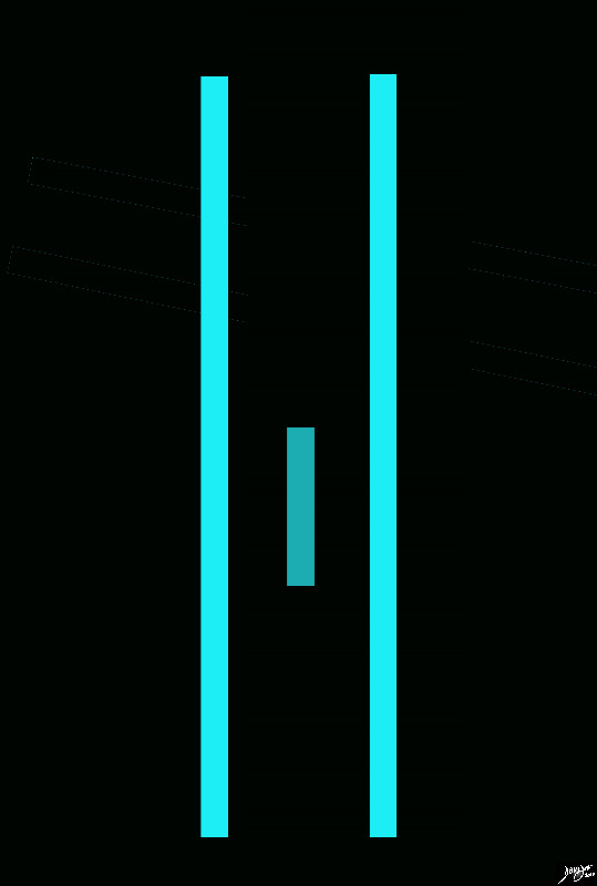
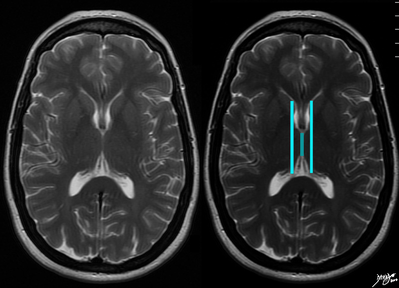
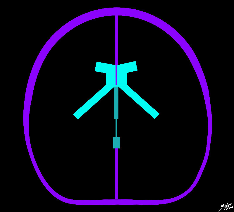
The horizontal limbsreviewed in the sagittal section are reflected as two parallelvectors that include parrt of the frontal horns and occipital components, while the teal green limb in the middle represents the conglomerate vertical limb that includes the third ventricle, the aqueduct of Sylvius and the fourth ventricle.
Courtesy Ashley Davidoff MD copyright 2010 all rights reserved 94458e08a.8s


    [nodeType] => 1
    [parentNode] => (object value omitted)
    [childNodes] => (object value omitted)
    [firstChild] => (object value omitted)
    [lastChild] => (object value omitted)
    [previousSibling] => (object value omitted)
    [nextSibling] => (object value omitted)
    [attributes] => (object value omitted)
    [ownerDocument] => (object value omitted)
    [namespaceURI] =>
    [prefix] =>
    [localName] => table
    [baseURI] =>
    [textContent] =>


Axial T2 weighted Image


The horizontal limbsreviewed in the sagittal section are reflected as two parallelvectors that include parrt of the frontal horns and occipital components, while the teal green limb in the middle represents the conglomerate vertical limb that includes the third ventricle, the aqueduct of Sylvius and the fourth ventricle.
Courtesy Ashley Davidoff MD copyright 2010 all rights reserved 94458e08a.8s


)
DOMElement Object
(
    [schemaTypeInfo] =>
    [tagName] => td
    [firstElementChild] => (object value omitted)
    [lastElementChild] => (object value omitted)
    [childElementCount] => 2
    [previousElementSibling] =>
    [nextElementSibling] =>
    [nodeName] => td
    [nodeValue] =>
The horizontal limbsreviewed in the sagittal section are reflected as two parallelvectors that include parrt of the frontal horns and occipital components, while the teal green limb in the middle represents the conglomerate vertical limb that includes the third ventricle, the aqueduct of Sylvius and the fourth ventricle.
Courtesy Ashley Davidoff MD copyright 2010 all rights reserved 94458e08a.8s

    [nodeType] => 1
    [parentNode] => (object value omitted)
    [childNodes] => (object value omitted)
    [firstChild] => (object value omitted)
    [lastChild] => (object value omitted)
    [previousSibling] => (object value omitted)
    [nextSibling] => (object value omitted)
    [attributes] => (object value omitted)
    [ownerDocument] => (object value omitted)
    [namespaceURI] =>
    [prefix] =>
    [localName] => td
    [baseURI] =>
    [textContent] =>
The horizontal limbsreviewed in the sagittal section are reflected as two parallelvectors that include parrt of the frontal horns and occipital components, while the teal green limb in the middle represents the conglomerate vertical limb that includes the third ventricle, the aqueduct of Sylvius and the fourth ventricle.
Courtesy Ashley Davidoff MD copyright 2010 all rights reserved 94458e08a.8s

)
DOMElement Object
(
    [schemaTypeInfo] =>
    [tagName] => td
    [firstElementChild] => (object value omitted)
    [lastElementChild] => (object value omitted)
    [childElementCount] => 2
    [previousElementSibling] =>
    [nextElementSibling] =>
    [nodeName] => td
    [nodeValue] => Axial T2 weighted Image
    [nodeType] => 1
    [parentNode] => (object value omitted)
    [childNodes] => (object value omitted)
    [firstChild] => (object value omitted)
    [lastChild] => (object value omitted)
    [previousSibling] => (object value omitted)
    [nextSibling] => (object value omitted)
    [attributes] => (object value omitted)
    [ownerDocument] => (object value omitted)
    [namespaceURI] =>
    [prefix] =>
    [localName] => td
    [baseURI] =>
    [textContent] => Axial T2 weighted Image
)
DOMElement Object
(
    [schemaTypeInfo] =>
    [tagName] => table
    [firstElementChild] => (object value omitted)
    [lastElementChild] => (object value omitted)
    [childElementCount] => 1
    [previousElementSibling] => (object value omitted)
    [nextElementSibling] => (object value omitted)
    [nodeName] => table
    [nodeValue] =>


The Transverse Plane


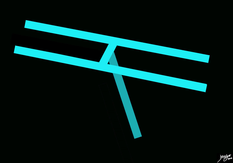
The axis of the brain in the sagittal view consists of an anteroposterior or relatively horozontal vector and a relatively vertical, craniocaudal vector. The ventricular system folows these vectors and consists of a paired horizontal system and a single vertical system
Courtesy Ashley Davidoff MD copyright 2010 all rights reserved  94458d02.8s


    [nodeType] => 1
    [parentNode] => (object value omitted)
    [childNodes] => (object value omitted)
    [firstChild] => (object value omitted)
    [lastChild] => (object value omitted)
    [previousSibling] => (object value omitted)
    [nextSibling] => (object value omitted)
    [attributes] => (object value omitted)
    [ownerDocument] => (object value omitted)
    [namespaceURI] =>
    [prefix] =>
    [localName] => table
    [baseURI] =>
    [textContent] =>


The Transverse Plane


The axis of the brain in the sagittal view consists of an anteroposterior or relatively horozontal vector and a relatively vertical, craniocaudal vector. The ventricular system folows these vectors and consists of a paired horizontal system and a single vertical system
Courtesy Ashley Davidoff MD copyright 2010 all rights reserved  94458d02.8s


)
DOMElement Object
(
    [schemaTypeInfo] =>
    [tagName] => td
    [firstElementChild] => (object value omitted)
    [lastElementChild] => (object value omitted)
    [childElementCount] => 2
    [previousElementSibling] =>
    [nextElementSibling] =>
    [nodeName] => td
    [nodeValue] =>
The axis of the brain in the sagittal view consists of an anteroposterior or relatively horozontal vector and a relatively vertical, craniocaudal vector. The ventricular system folows these vectors and consists of a paired horizontal system and a single vertical system
Courtesy Ashley Davidoff MD copyright 2010 all rights reserved  94458d02.8s

    [nodeType] => 1
    [parentNode] => (object value omitted)
    [childNodes] => (object value omitted)
    [firstChild] => (object value omitted)
    [lastChild] => (object value omitted)
    [previousSibling] => (object value omitted)
    [nextSibling] => (object value omitted)
    [attributes] => (object value omitted)
    [ownerDocument] => (object value omitted)
    [namespaceURI] =>
    [prefix] =>
    [localName] => td
    [baseURI] =>
    [textContent] =>
The axis of the brain in the sagittal view consists of an anteroposterior or relatively horozontal vector and a relatively vertical, craniocaudal vector. The ventricular system folows these vectors and consists of a paired horizontal system and a single vertical system
Courtesy Ashley Davidoff MD copyright 2010 all rights reserved  94458d02.8s

)
DOMElement Object
(
    [schemaTypeInfo] =>
    [tagName] => td
    [firstElementChild] => (object value omitted)
    [lastElementChild] => (object value omitted)
    [childElementCount] => 2
    [previousElementSibling] =>
    [nextElementSibling] =>
    [nodeName] => td
    [nodeValue] => The Transverse Plane
    [nodeType] => 1
    [parentNode] => (object value omitted)
    [childNodes] => (object value omitted)
    [firstChild] => (object value omitted)
    [lastChild] => (object value omitted)
    [previousSibling] => (object value omitted)
    [nextSibling] => (object value omitted)
    [attributes] => (object value omitted)
    [ownerDocument] => (object value omitted)
    [namespaceURI] =>
    [prefix] =>
    [localName] => td
    [baseURI] =>
    [textContent] => The Transverse Plane
)
DOMElement Object
(
    [schemaTypeInfo] =>
    [tagName] => table
    [firstElementChild] => (object value omitted)
    [lastElementChild] => (object value omitted)
    [childElementCount] => 1
    [previousElementSibling] => (object value omitted)
    [nextElementSibling] => (object value omitted)
    [nodeName] => table
    [nodeValue] =>


The vectors of the ventricular System Overlaid on the Brain


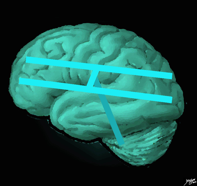
The ventricular system is overlaid on a drawing of the brain. Each limb of the horizantal component is situated in a cerebral hemisphere The vertical limb includes the third and fourth ventricles
Courtesy Ashley Davidoff MD copyright 2010 all rights reserved  94458e07.8s


    [nodeType] => 1
    [parentNode] => (object value omitted)
    [childNodes] => (object value omitted)
    [firstChild] => (object value omitted)
    [lastChild] => (object value omitted)
    [previousSibling] => (object value omitted)
    [nextSibling] => (object value omitted)
    [attributes] => (object value omitted)
    [ownerDocument] => (object value omitted)
    [namespaceURI] =>
    [prefix] =>
    [localName] => table
    [baseURI] =>
    [textContent] =>


The vectors of the ventricular System Overlaid on the Brain


The ventricular system is overlaid on a drawing of the brain. Each limb of the horizantal component is situated in a cerebral hemisphere The vertical limb includes the third and fourth ventricles
Courtesy Ashley Davidoff MD copyright 2010 all rights reserved  94458e07.8s


)
DOMElement Object
(
    [schemaTypeInfo] =>
    [tagName] => td
    [firstElementChild] => (object value omitted)
    [lastElementChild] => (object value omitted)
    [childElementCount] => 2
    [previousElementSibling] =>
    [nextElementSibling] =>
    [nodeName] => td
    [nodeValue] =>
The ventricular system is overlaid on a drawing of the brain. Each limb of the horizantal component is situated in a cerebral hemisphere The vertical limb includes the third and fourth ventricles
Courtesy Ashley Davidoff MD copyright 2010 all rights reserved  94458e07.8s

    [nodeType] => 1
    [parentNode] => (object value omitted)
    [childNodes] => (object value omitted)
    [firstChild] => (object value omitted)
    [lastChild] => (object value omitted)
    [previousSibling] => (object value omitted)
    [nextSibling] => (object value omitted)
    [attributes] => (object value omitted)
    [ownerDocument] => (object value omitted)
    [namespaceURI] =>
    [prefix] =>
    [localName] => td
    [baseURI] =>
    [textContent] =>
The ventricular system is overlaid on a drawing of the brain. Each limb of the horizantal component is situated in a cerebral hemisphere The vertical limb includes the third and fourth ventricles
Courtesy Ashley Davidoff MD copyright 2010 all rights reserved  94458e07.8s

)
DOMElement Object
(
    [schemaTypeInfo] =>
    [tagName] => td
    [firstElementChild] => (object value omitted)
    [lastElementChild] => (object value omitted)
    [childElementCount] => 2
    [previousElementSibling] =>
    [nextElementSibling] =>
    [nodeName] => td
    [nodeValue] => The vectors of the ventricular System Overlaid on the Brain
    [nodeType] => 1
    [parentNode] => (object value omitted)
    [childNodes] => (object value omitted)
    [firstChild] => (object value omitted)
    [lastChild] => (object value omitted)
    [previousSibling] => (object value omitted)
    [nextSibling] => (object value omitted)
    [attributes] => (object value omitted)
    [ownerDocument] => (object value omitted)
    [namespaceURI] =>
    [prefix] =>
    [localName] => td
    [baseURI] =>
    [textContent] => The vectors of the ventricular System Overlaid on the Brain
)
DOMElement Object
(
    [schemaTypeInfo] =>
    [tagName] => table
    [firstElementChild] => (object value omitted)
    [lastElementChild] => (object value omitted)
    [childElementCount] => 1
    [previousElementSibling] => (object value omitted)
    [nextElementSibling] => (object value omitted)
    [nodeName] => table
    [nodeValue] =>


The Sagital Vectors


The axis of the brain in the sagittal view consists of an anteroposterior or relatively horozontal vector and a relatively vertical, craniocaudal vector. The ventricular system folows these vectors and consists of a paired horizontal system and a single vertical system
Courtesy Ashley Davidoff MD copyright 2010 all rights reserved 94458b02b01.8s


    [nodeType] => 1
    [parentNode] => (object value omitted)
    [childNodes] => (object value omitted)
    [firstChild] => (object value omitted)
    [lastChild] => (object value omitted)
    [previousSibling] => (object value omitted)
    [nextSibling] => (object value omitted)
    [attributes] => (object value omitted)
    [ownerDocument] => (object value omitted)
    [namespaceURI] =>
    [prefix] =>
    [localName] => table
    [baseURI] =>
    [textContent] =>


The Sagital Vectors


The axis of the brain in the sagittal view consists of an anteroposterior or relatively horozontal vector and a relatively vertical, craniocaudal vector. The ventricular system folows these vectors and consists of a paired horizontal system and a single vertical system
Courtesy Ashley Davidoff MD copyright 2010 all rights reserved 94458b02b01.8s


)
DOMElement Object
(
    [schemaTypeInfo] =>
    [tagName] => td
    [firstElementChild] => (object value omitted)
    [lastElementChild] => (object value omitted)
    [childElementCount] => 2
    [previousElementSibling] =>
    [nextElementSibling] =>
    [nodeName] => td
    [nodeValue] =>
The axis of the brain in the sagittal view consists of an anteroposterior or relatively horozontal vector and a relatively vertical, craniocaudal vector. The ventricular system folows these vectors and consists of a paired horizontal system and a single vertical system
Courtesy Ashley Davidoff MD copyright 2010 all rights reserved 94458b02b01.8s

    [nodeType] => 1
    [parentNode] => (object value omitted)
    [childNodes] => (object value omitted)
    [firstChild] => (object value omitted)
    [lastChild] => (object value omitted)
    [previousSibling] => (object value omitted)
    [nextSibling] => (object value omitted)
    [attributes] => (object value omitted)
    [ownerDocument] => (object value omitted)
    [namespaceURI] =>
    [prefix] =>
    [localName] => td
    [baseURI] =>
    [textContent] =>
The axis of the brain in the sagittal view consists of an anteroposterior or relatively horozontal vector and a relatively vertical, craniocaudal vector. The ventricular system folows these vectors and consists of a paired horizontal system and a single vertical system
Courtesy Ashley Davidoff MD copyright 2010 all rights reserved 94458b02b01.8s

)
DOMElement Object
(
    [schemaTypeInfo] =>
    [tagName] => td
    [firstElementChild] => (object value omitted)
    [lastElementChild] => (object value omitted)
    [childElementCount] => 2
    [previousElementSibling] =>
    [nextElementSibling] =>
    [nodeName] => td
    [nodeValue] => The Sagital Vectors
    [nodeType] => 1
    [parentNode] => (object value omitted)
    [childNodes] => (object value omitted)
    [firstChild] => (object value omitted)
    [lastChild] => (object value omitted)
    [previousSibling] => (object value omitted)
    [nextSibling] => (object value omitted)
    [attributes] => (object value omitted)
    [ownerDocument] => (object value omitted)
    [namespaceURI] =>
    [prefix] =>
    [localName] => td
    [baseURI] =>
    [textContent] => The Sagital Vectors
)

