Choroid Plexus Papilloma /Carcinoma
Elisa Flower MD Asim Mian MD
The Common Vein Copyright 2010
Definition
Choroid plexus papillomas and carcinomas are neoplasms that arise from the choroid plexus. They are most commonly seen in children, usually by the age of 5. They are typically located in the trigone or atria of the lateral ventricles. When found in the adults, they generally arise from the fourth ventricle.
It is thought that Simian virus 40 may be a predisposing factor. There is also an association between choroid plexus tumors with Li-fraumeni and Aicardi syndrome.
Histologically these tumors are characterized by papillary growths that have the appearance of normal choroid plexus. Both choroid plexus papillomas and carcinomas may result in CSF dissemination.
Clinically these patients present with hydrocephalus by two different mechanisms. Not only can these massesy obstruct the normal flow of CSF, they also cause an overproduction of CSF.
Diagnosis is suggested by classic imaging findings in the proper patient demographic however it is confirmed by pathology.
Both CT and MRI are useful in diagnosis, however MRI is better for tissue characterization and evaluation of the entire neuroaxis for CNS dissemination. On CT, these lesions are often a hyperdense lobulated intraventricular mass. Calcification and hemorrhage can also frequently be seen. On MRI, these lesions are T1 hypointense and heterogeneous on T2 due to areas of hemorrhage, calcifications, and vascular structures. Classically these masses enhance avidly. Choroid plexus papillomas and carcinomas can be quite difficult to distinguish on imaging with the only feature more suggestive of carcinoma is frank invasion of the adjacent parenchyma but even that can be seen in choroid plexus papilloma.
Treatment of choice is total surgical resection.
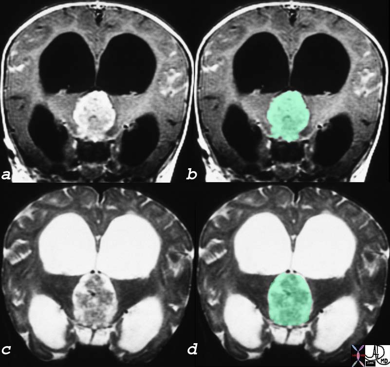
Papilloma of the Choroid Plexus Causing Hydrocephalus and Headache
|
| The coronal MRI shows a gadolinium enhanced T1 weighted image (a,b) with black CSF in the dilated ventricles and a T2 weighted image with bright CSF (c,d). A large mass in the third ventricle (overlaid ingreen) dominates the study and is complicated by severe hydrocephalus. The mass was a choroid plexus papilloma of the third ventricle. In patients with brain tumor one of the causes of headache is hydrocephalus. This is an extreme case of hydrocephalus.
23223c01 head brain choroid plexus third ventricle hydrocephalus fx mass neoplasm papilloma MRI T1 T2 coronal with contrast C Courtesy James Donnelly MD |
DOMElement Object
(
    [schemaTypeInfo] =>
    [tagName] => table
    [firstElementChild] => (object value omitted)
    [lastElementChild] => (object value omitted)
    [childElementCount] => 1
    [previousElementSibling] => (object value omitted)
    [nextElementSibling] =>
    [nodeName] => table
    [nodeValue] =>


Papilloma of the Choroid Plexus Causing Hydrocephalus and Headache


The coronal MRI shows a gadolinium enhanced T1 weighted image (a,b) with black CSF in the dilated ventricles and a T2 weighted image with bright CSF (c,d).   A large mass in the third ventricle (overlaid ingreen) dominates the study and is complicated by severe hydrocephalus.  The mass was a choroid plexus papilloma of the third ventricle.  In patients with brain tumor one of the causes of headache is hydrocephalus.  This is an extreme case of hydrocephalus.
23223c01 head brain choroid plexus third ventricle hydrocephalus fx mass neoplasm papilloma MRI T1 T2 coronal with contrast C Courtesy James Donnelly MD


    [nodeType] => 1
    [parentNode] => (object value omitted)
    [childNodes] => (object value omitted)
    [firstChild] => (object value omitted)
    [lastChild] => (object value omitted)
    [previousSibling] => (object value omitted)
    [nextSibling] => (object value omitted)
    [attributes] => (object value omitted)
    [ownerDocument] => (object value omitted)
    [namespaceURI] =>
    [prefix] =>
    [localName] => table
    [baseURI] =>
    [textContent] =>


Papilloma of the Choroid Plexus Causing Hydrocephalus and Headache


The coronal MRI shows a gadolinium enhanced T1 weighted image (a,b) with black CSF in the dilated ventricles and a T2 weighted image with bright CSF (c,d).   A large mass in the third ventricle (overlaid ingreen) dominates the study and is complicated by severe hydrocephalus.  The mass was a choroid plexus papilloma of the third ventricle.  In patients with brain tumor one of the causes of headache is hydrocephalus.  This is an extreme case of hydrocephalus.
23223c01 head brain choroid plexus third ventricle hydrocephalus fx mass neoplasm papilloma MRI T1 T2 coronal with contrast C Courtesy James Donnelly MD


)
DOMElement Object
(
    [schemaTypeInfo] =>
    [tagName] => td
    [firstElementChild] => (object value omitted)
    [lastElementChild] => (object value omitted)
    [childElementCount] => 3
    [previousElementSibling] =>
    [nextElementSibling] =>
    [nodeName] => td
    [nodeValue] => The coronal MRI shows a gadolinium enhanced T1 weighted image (a,b) with black CSF in the dilated ventricles and a T2 weighted image with bright CSF (c,d).   A large mass in the third ventricle (overlaid ingreen) dominates the study and is complicated by severe hydrocephalus.  The mass was a choroid plexus papilloma of the third ventricle.  In patients with brain tumor one of the causes of headache is hydrocephalus.  This is an extreme case of hydrocephalus.
23223c01 head brain choroid plexus third ventricle hydrocephalus fx mass neoplasm papilloma MRI T1 T2 coronal with contrast C Courtesy James Donnelly MD
    [nodeType] => 1
    [parentNode] => (object value omitted)
    [childNodes] => (object value omitted)
    [firstChild] => (object value omitted)
    [lastChild] => (object value omitted)
    [previousSibling] => (object value omitted)
    [nextSibling] => (object value omitted)
    [attributes] => (object value omitted)
    [ownerDocument] => (object value omitted)
    [namespaceURI] =>
    [prefix] =>
    [localName] => td
    [baseURI] =>
    [textContent] => The coronal MRI shows a gadolinium enhanced T1 weighted image (a,b) with black CSF in the dilated ventricles and a T2 weighted image with bright CSF (c,d).   A large mass in the third ventricle (overlaid ingreen) dominates the study and is complicated by severe hydrocephalus.  The mass was a choroid plexus papilloma of the third ventricle.  In patients with brain tumor one of the causes of headache is hydrocephalus.  This is an extreme case of hydrocephalus.
23223c01 head brain choroid plexus third ventricle hydrocephalus fx mass neoplasm papilloma MRI T1 T2 coronal with contrast C Courtesy James Donnelly MD
)
DOMElement Object
(
    [schemaTypeInfo] =>
    [tagName] => td
    [firstElementChild] => (object value omitted)
    [lastElementChild] => (object value omitted)
    [childElementCount] => 2
    [previousElementSibling] =>
    [nextElementSibling] =>
    [nodeName] => td
    [nodeValue] =>
Papilloma of the Choroid Plexus Causing Hydrocephalus and Headache

    [nodeType] => 1
    [parentNode] => (object value omitted)
    [childNodes] => (object value omitted)
    [firstChild] => (object value omitted)
    [lastChild] => (object value omitted)
    [previousSibling] => (object value omitted)
    [nextSibling] => (object value omitted)
    [attributes] => (object value omitted)
    [ownerDocument] => (object value omitted)
    [namespaceURI] =>
    [prefix] =>
    [localName] => td
    [baseURI] =>
    [textContent] =>
Papilloma of the Choroid Plexus Causing Hydrocephalus and Headache

)
