Ashley Davidoff MD
The Common Vein Copyright 2010
Definition
The cerebellum (Latin for little brain) is a part of the hindbrain that lies with the pons and medulla below the tentorium.
It is structurally part of the rhombencephalon and more specifically part of the metencephalon with the pons.
Although it is smaller than the cerebral hemispheres it does in fact have more cells because of its tightly packed external folds called folia which increases its surface area, but also because of the inordinately large number of tightly packed granule cells in the granular layer of the gray matter.
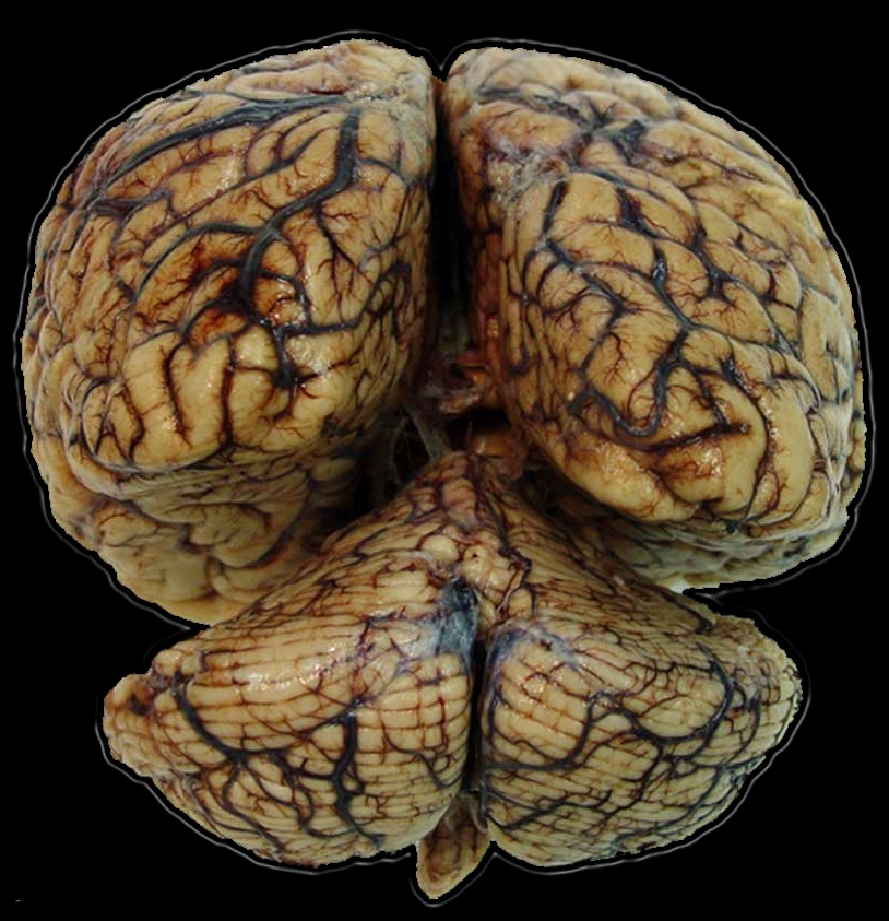
It has an unusual shape and looks almost like an inverted top. It lies in the posterior fossa below the occipital and temporal lobes. Unlike the irregular contours of the cerebral gyri and sulci, the folds of the cerebellum are neatly ordered and tightly bound, being almost the same height and running parallel with each other in horizontal plane. The cerebellum has similar graymatter-white matter arangenment as the cerebral hemispheres
It is divided into two hemispheres and a narrow midline zone called the vermis. Another method of division is by dividing it into three lobes: the flocculonodular, anterior, and posterior lobes.
Functionally, the cerebellum it is most important in motor function but also is active in sensory perception, and cognitive function. Learned motor responses, and intricate patterns of motor function such as dance and playing an instrument or spport are stored in the cerebellum.
Diseases of the cerebellum result in abnormalities of equilibrium, muscle tone, postural control, and co-ordination of voluntary movements.
Diagnostic for cerebellum dysfunction includes fMRI.
|
The Cerebellum in Context The Cerebellum (red) Part of the Hindbrain(Rhombencephalon) Member of the Metencephalon |
|
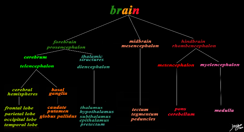
The basic and simplest classification of the brain into forebrain midbrain and hindbrain is shown in this diagram and advanced to a more complex tree using the embryological and evolutionary terminologies. The cerebellum is part of the hindbrain The forebrain consists of the cerebrum also called the prosencephalon, which contains the more advanced form of the brain and the thalamic structures which contain more basic structures. The cerebrum (telencephalon) itself consists of two cerebral hemispheres and paired basal ganglial structures. Each cerebral hemisphere will have gray and white matter distributed in the frontal parietal temporal and occipital lobe, with the basal ganglia being part of the gray matter deep in the cerebral hemispheres. The most important thalamic structures arising from the diencephalons include the thalamus itself and the hypothalamus. The midbrain (mesencepaholon) consists of the tectum tegmentum and cerebral peduncles. The hindbrain has two major branch points based on the evolutionary development. The pons and cerebellum(part of the metencephalon) are grouped and the medulla (part of the myelencephalon is the second branch. Courtesy Ashley Davidoff MD Copyright 2010 All rights reserved 97686.8s |
|
Cerebellum as seen from Below |
|
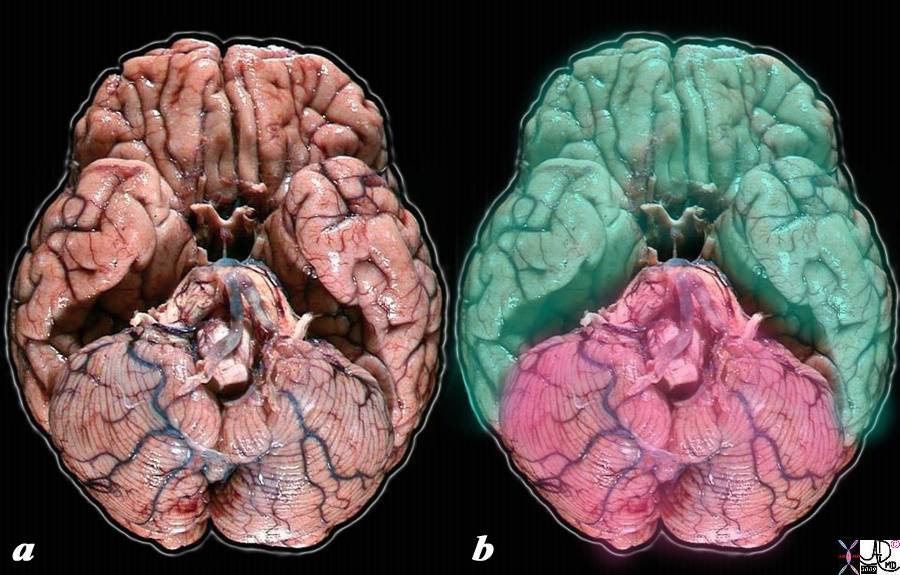
When the anatomic specimen is viewed from below, the forebrain (torquoise green) and the hindbrain (salmon pink) dominated by the cerebellum are best seen. The midbrain which is a relatively small structure is not visualized. code brain anatomy surface view sulcus sulci gyrus gyri cerebellum forebrain hindbrain normal structure anatomy brain Source unknown Modified by Davidoff MD 53605c02.8s |
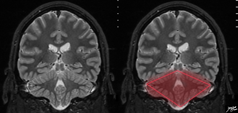
Coronal View |
|
The T2 weighted MRI of the brain in coronal projection shows an almost rhomboid shaped structure Courtesy Ashley DAvidoff MD copyright 2010 all rights reserved 89742.3kcb01b.8s |
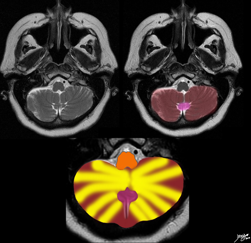
The cerebellum and The Winking Tabby Cat |
|
The MRI of the posterior fossa and cerebellum was rendered and reminded the author of a winking tabby cat Copyright 2010 all rights reserved 49037c01.8s |
Sensory Input
The sensory input consists of three large bundles of white matter that enter tthe cerebellum called the cerebellar peduncles
Circulatory Diseases
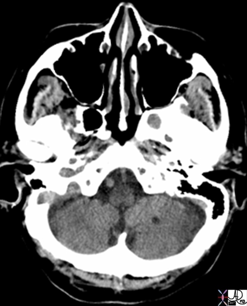
Chronic Infarct Left Cerebellar Hemisphere |
|
The axial CT scan shows a low density lesion in the left cerebellar hemisphere consistent with chronic infarcts. Courtesy Ashley Davidoff MD 71563 |
References
Schuenke M, Schulte E, Schumacher Udo Thieme Atlas of Anatomy: head and neuroanatomy Thieme germany 2007
DOMElement Object
(
    [schemaTypeInfo] =>
    [tagName] => table
    [firstElementChild] => (object value omitted)
    [lastElementChild] => (object value omitted)
    [childElementCount] => 1
    [previousElementSibling] => (object value omitted)
    [nextElementSibling] => (object value omitted)
    [nodeName] => table
    [nodeValue] =>


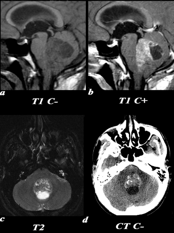
Medulloblastoma – Most Common Pediatric Malignant Brain Tumor


This 26 year old male presented with a three week history of progressive headache, nausea and vomiting. T1 pre (a) and post (b,): The exact origin of this mass can be difficult to ascertain given its large size. It clearly grows into the fourth ventricle, widening the lower portion of the Sylvian aqueduct. Note the smooth interface with the posterior aspect of the pons with associated mass effect verses the more irregular margin posteriorly where it arises from the cerebellum. T2 (c): The solid component of the medulloblastoma matches gray matter signal. Cystic or necrotic areas are demonstrated by higher T2 signal areas. CT(d): The unenhanced CT scan demonstrates a predominately hyperdense mass with punctuate calcifications in the region of the fourth ventricle . The low density area posteriorly is a cystic or necrotic component of the mass while the fourth ventricle is completely effaced.
Image Courtesy Elisa Flower MD and Asim Mian MD 97668c01.81


    [nodeType] => 1
    [parentNode] => (object value omitted)
    [childNodes] => (object value omitted)
    [firstChild] => (object value omitted)
    [lastChild] => (object value omitted)
    [previousSibling] => (object value omitted)
    [nextSibling] => (object value omitted)
    [attributes] => (object value omitted)
    [ownerDocument] => (object value omitted)
    [namespaceURI] =>
    [prefix] =>
    [localName] => table
    [baseURI] =>
    [textContent] =>


Medulloblastoma – Most Common Pediatric Malignant Brain Tumor


This 26 year old male presented with a three week history of progressive headache, nausea and vomiting. T1 pre (a) and post (b,): The exact origin of this mass can be difficult to ascertain given its large size. It clearly grows into the fourth ventricle, widening the lower portion of the Sylvian aqueduct. Note the smooth interface with the posterior aspect of the pons with associated mass effect verses the more irregular margin posteriorly where it arises from the cerebellum. T2 (c): The solid component of the medulloblastoma matches gray matter signal. Cystic or necrotic areas are demonstrated by higher T2 signal areas. CT(d): The unenhanced CT scan demonstrates a predominately hyperdense mass with punctuate calcifications in the region of the fourth ventricle . The low density area posteriorly is a cystic or necrotic component of the mass while the fourth ventricle is completely effaced.
Image Courtesy Elisa Flower MD and Asim Mian MD 97668c01.81


)
DOMElement Object
(
    [schemaTypeInfo] =>
    [tagName] => td
    [firstElementChild] => (object value omitted)
    [lastElementChild] => (object value omitted)
    [childElementCount] => 2
    [previousElementSibling] =>
    [nextElementSibling] =>
    [nodeName] => td
    [nodeValue] =>
This 26 year old male presented with a three week history of progressive headache, nausea and vomiting. T1 pre (a) and post (b,): The exact origin of this mass can be difficult to ascertain given its large size. It clearly grows into the fourth ventricle, widening the lower portion of the Sylvian aqueduct. Note the smooth interface with the posterior aspect of the pons with associated mass effect verses the more irregular margin posteriorly where it arises from the cerebellum. T2 (c): The solid component of the medulloblastoma matches gray matter signal. Cystic or necrotic areas are demonstrated by higher T2 signal areas. CT(d): The unenhanced CT scan demonstrates a predominately hyperdense mass with punctuate calcifications in the region of the fourth ventricle . The low density area posteriorly is a cystic or necrotic component of the mass while the fourth ventricle is completely effaced.
Image Courtesy Elisa Flower MD and Asim Mian MD 97668c01.81

    [nodeType] => 1
    [parentNode] => (object value omitted)
    [childNodes] => (object value omitted)
    [firstChild] => (object value omitted)
    [lastChild] => (object value omitted)
    [previousSibling] => (object value omitted)
    [nextSibling] => (object value omitted)
    [attributes] => (object value omitted)
    [ownerDocument] => (object value omitted)
    [namespaceURI] =>
    [prefix] =>
    [localName] => td
    [baseURI] =>
    [textContent] =>
This 26 year old male presented with a three week history of progressive headache, nausea and vomiting. T1 pre (a) and post (b,): The exact origin of this mass can be difficult to ascertain given its large size. It clearly grows into the fourth ventricle, widening the lower portion of the Sylvian aqueduct. Note the smooth interface with the posterior aspect of the pons with associated mass effect verses the more irregular margin posteriorly where it arises from the cerebellum. T2 (c): The solid component of the medulloblastoma matches gray matter signal. Cystic or necrotic areas are demonstrated by higher T2 signal areas. CT(d): The unenhanced CT scan demonstrates a predominately hyperdense mass with punctuate calcifications in the region of the fourth ventricle . The low density area posteriorly is a cystic or necrotic component of the mass while the fourth ventricle is completely effaced.
Image Courtesy Elisa Flower MD and Asim Mian MD 97668c01.81

)
DOMElement Object
(
    [schemaTypeInfo] =>
    [tagName] => td
    [firstElementChild] => (object value omitted)
    [lastElementChild] => (object value omitted)
    [childElementCount] => 2
    [previousElementSibling] =>
    [nextElementSibling] =>
    [nodeName] => td
    [nodeValue] =>
Medulloblastoma – Most Common Pediatric Malignant Brain Tumor
    [nodeType] => 1
    [parentNode] => (object value omitted)
    [childNodes] => (object value omitted)
    [firstChild] => (object value omitted)
    [lastChild] => (object value omitted)
    [previousSibling] => (object value omitted)
    [nextSibling] => (object value omitted)
    [attributes] => (object value omitted)
    [ownerDocument] => (object value omitted)
    [namespaceURI] =>
    [prefix] =>
    [localName] => td
    [baseURI] =>
    [textContent] =>
Medulloblastoma – Most Common Pediatric Malignant Brain Tumor
)
DOMElement Object
(
    [schemaTypeInfo] =>
    [tagName] => table
    [firstElementChild] => (object value omitted)
    [lastElementChild] => (object value omitted)
    [childElementCount] => 1
    [previousElementSibling] => (object value omitted)
    [nextElementSibling] => (object value omitted)
    [nodeName] => table
    [nodeValue] =>


Chronic Infarct Left Cerebellar Hemisphere


The axial CT scan shows a low density lesion in the left cerebellar hemisphere consistent with chronic infarcts.
Courtesy Ashley Davidoff MD 71563


    [nodeType] => 1
    [parentNode] => (object value omitted)
    [childNodes] => (object value omitted)
    [firstChild] => (object value omitted)
    [lastChild] => (object value omitted)
    [previousSibling] => (object value omitted)
    [nextSibling] => (object value omitted)
    [attributes] => (object value omitted)
    [ownerDocument] => (object value omitted)
    [namespaceURI] =>
    [prefix] =>
    [localName] => table
    [baseURI] =>
    [textContent] =>


Chronic Infarct Left Cerebellar Hemisphere


The axial CT scan shows a low density lesion in the left cerebellar hemisphere consistent with chronic infarcts.
Courtesy Ashley Davidoff MD 71563


)
DOMElement Object
(
    [schemaTypeInfo] =>
    [tagName] => td
    [firstElementChild] => (object value omitted)
    [lastElementChild] => (object value omitted)
    [childElementCount] => 2
    [previousElementSibling] =>
    [nextElementSibling] =>
    [nodeName] => td
    [nodeValue] =>
The axial CT scan shows a low density lesion in the left cerebellar hemisphere consistent with chronic infarcts.
Courtesy Ashley Davidoff MD 71563

    [nodeType] => 1
    [parentNode] => (object value omitted)
    [childNodes] => (object value omitted)
    [firstChild] => (object value omitted)
    [lastChild] => (object value omitted)
    [previousSibling] => (object value omitted)
    [nextSibling] => (object value omitted)
    [attributes] => (object value omitted)
    [ownerDocument] => (object value omitted)
    [namespaceURI] =>
    [prefix] =>
    [localName] => td
    [baseURI] =>
    [textContent] =>
The axial CT scan shows a low density lesion in the left cerebellar hemisphere consistent with chronic infarcts.
Courtesy Ashley Davidoff MD 71563

)
DOMElement Object
(
    [schemaTypeInfo] =>
    [tagName] => td
    [firstElementChild] => (object value omitted)
    [lastElementChild] => (object value omitted)
    [childElementCount] => 2
    [previousElementSibling] =>
    [nextElementSibling] =>
    [nodeName] => td
    [nodeValue] =>
Chronic Infarct Left Cerebellar Hemisphere
    [nodeType] => 1
    [parentNode] => (object value omitted)
    [childNodes] => (object value omitted)
    [firstChild] => (object value omitted)
    [lastChild] => (object value omitted)
    [previousSibling] => (object value omitted)
    [nextSibling] => (object value omitted)
    [attributes] => (object value omitted)
    [ownerDocument] => (object value omitted)
    [namespaceURI] =>
    [prefix] =>
    [localName] => td
    [baseURI] =>
    [textContent] =>
Chronic Infarct Left Cerebellar Hemisphere
)
DOMElement Object
(
    [schemaTypeInfo] =>
    [tagName] => table
    [firstElementChild] => (object value omitted)
    [lastElementChild] => (object value omitted)
    [childElementCount] => 1
    [previousElementSibling] => (object value omitted)
    [nextElementSibling] => (object value omitted)
    [nodeName] => table
    [nodeValue] =>


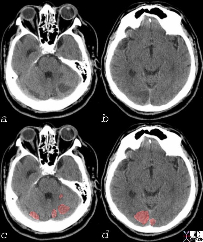
Acute Multicentric Non Hemorrhagic Infarcts Consistent with Embolic Disease


Multicentric Infarcts in the Cerebellum and Occipital Lobes Multicentric low density regions in both posterior cerebellar hemispheres and the occipital lobes bilaterally seen in (a and b) correspondingly overlaid in light red in c and d, are consistent with non hemorrhagic infarcts. Embolic disease is most likely
Courtesy Ashley DAvidoff MD 74923c01


    [nodeType] => 1
    [parentNode] => (object value omitted)
    [childNodes] => (object value omitted)
    [firstChild] => (object value omitted)
    [lastChild] => (object value omitted)
    [previousSibling] => (object value omitted)
    [nextSibling] => (object value omitted)
    [attributes] => (object value omitted)
    [ownerDocument] => (object value omitted)
    [namespaceURI] =>
    [prefix] =>
    [localName] => table
    [baseURI] =>
    [textContent] =>


Acute Multicentric Non Hemorrhagic Infarcts Consistent with Embolic Disease


Multicentric Infarcts in the Cerebellum and Occipital Lobes Multicentric low density regions in both posterior cerebellar hemispheres and the occipital lobes bilaterally seen in (a and b) correspondingly overlaid in light red in c and d, are consistent with non hemorrhagic infarcts. Embolic disease is most likely
Courtesy Ashley DAvidoff MD 74923c01


)
DOMElement Object
(
    [schemaTypeInfo] =>
    [tagName] => td
    [firstElementChild] => (object value omitted)
    [lastElementChild] => (object value omitted)
    [childElementCount] => 2
    [previousElementSibling] =>
    [nextElementSibling] =>
    [nodeName] => td
    [nodeValue] =>
Multicentric Infarcts in the Cerebellum and Occipital Lobes Multicentric low density regions in both posterior cerebellar hemispheres and the occipital lobes bilaterally seen in (a and b) correspondingly overlaid in light red in c and d, are consistent with non hemorrhagic infarcts. Embolic disease is most likely
Courtesy Ashley DAvidoff MD 74923c01

    [nodeType] => 1
    [parentNode] => (object value omitted)
    [childNodes] => (object value omitted)
    [firstChild] => (object value omitted)
    [lastChild] => (object value omitted)
    [previousSibling] => (object value omitted)
    [nextSibling] => (object value omitted)
    [attributes] => (object value omitted)
    [ownerDocument] => (object value omitted)
    [namespaceURI] =>
    [prefix] =>
    [localName] => td
    [baseURI] =>
    [textContent] =>
Multicentric Infarcts in the Cerebellum and Occipital Lobes Multicentric low density regions in both posterior cerebellar hemispheres and the occipital lobes bilaterally seen in (a and b) correspondingly overlaid in light red in c and d, are consistent with non hemorrhagic infarcts. Embolic disease is most likely
Courtesy Ashley DAvidoff MD 74923c01

)
DOMElement Object
(
    [schemaTypeInfo] =>
    [tagName] => td
    [firstElementChild] => (object value omitted)
    [lastElementChild] => (object value omitted)
    [childElementCount] => 2
    [previousElementSibling] =>
    [nextElementSibling] =>
    [nodeName] => td
    [nodeValue] =>
Acute Multicentric Non Hemorrhagic Infarcts Consistent with Embolic Disease
    [nodeType] => 1
    [parentNode] => (object value omitted)
    [childNodes] => (object value omitted)
    [firstChild] => (object value omitted)
    [lastChild] => (object value omitted)
    [previousSibling] => (object value omitted)
    [nextSibling] => (object value omitted)
    [attributes] => (object value omitted)
    [ownerDocument] => (object value omitted)
    [namespaceURI] =>
    [prefix] =>
    [localName] => td
    [baseURI] =>
    [textContent] =>
Acute Multicentric Non Hemorrhagic Infarcts Consistent with Embolic Disease
)
DOMElement Object
(
    [schemaTypeInfo] =>
    [tagName] => table
    [firstElementChild] => (object value omitted)
    [lastElementChild] => (object value omitted)
    [childElementCount] => 1
    [previousElementSibling] => (object value omitted)
    [nextElementSibling] => (object value omitted)
    [nodeName] => table
    [nodeValue] =>


Acute Frontoparietal Infarction
Normal Cerebellum


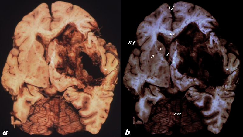
The brain has a characteristic appearance characterised by its creamy color and characteristic shape and fissured surface. This pathological specimen shows an acute hemorrhagic infarct in the left frontoparietal region with necrosis of the brain tissue and mass effect on the lateral ventricle and midline shift.
The horizontal folds, darker color, and overall shape of the cerebellum are characteristic.
The specimen also serves to reveal the normal right side with its creamy color and darker brownish gray matter that is accentuated by increasing the contrast on the right image (b). Other structures that are seen include the corpus callosum that lies above the ventricles (cc), the caudate nucleus (c) that lies inferior to the lateral ventricle, the line of white matter between the caudate and putamen called the internal capsule, the 3rd ventricle (3) medial to these structures , and the thalamus (th) inferior to them. The interhemispheric fissure (if) is seen superiorly and the Sylvian fissure (Sf) is seen laterally.
Courtesy Ashley Davidoff MD Copyright 2010 10407c01


    [nodeType] => 1
    [parentNode] => (object value omitted)
    [childNodes] => (object value omitted)
    [firstChild] => (object value omitted)
    [lastChild] => (object value omitted)
    [previousSibling] => (object value omitted)
    [nextSibling] => (object value omitted)
    [attributes] => (object value omitted)
    [ownerDocument] => (object value omitted)
    [namespaceURI] =>
    [prefix] =>
    [localName] => table
    [baseURI] =>
    [textContent] =>


Acute Frontoparietal Infarction
Normal Cerebellum


The brain has a characteristic appearance characterised by its creamy color and characteristic shape and fissured surface. This pathological specimen shows an acute hemorrhagic infarct in the left frontoparietal region with necrosis of the brain tissue and mass effect on the lateral ventricle and midline shift.
The horizontal folds, darker color, and overall shape of the cerebellum are characteristic.
The specimen also serves to reveal the normal right side with its creamy color and darker brownish gray matter that is accentuated by increasing the contrast on the right image (b). Other structures that are seen include the corpus callosum that lies above the ventricles (cc), the caudate nucleus (c) that lies inferior to the lateral ventricle, the line of white matter between the caudate and putamen called the internal capsule, the 3rd ventricle (3) medial to these structures , and the thalamus (th) inferior to them. The interhemispheric fissure (if) is seen superiorly and the Sylvian fissure (Sf) is seen laterally.
Courtesy Ashley Davidoff MD Copyright 2010 10407c01


)
DOMElement Object
(
    [schemaTypeInfo] =>
    [tagName] => td
    [firstElementChild] => (object value omitted)
    [lastElementChild] => (object value omitted)
    [childElementCount] => 4
    [previousElementSibling] =>
    [nextElementSibling] =>
    [nodeName] => td
    [nodeValue] =>
The brain has a characteristic appearance characterised by its creamy color and characteristic shape and fissured surface. This pathological specimen shows an acute hemorrhagic infarct in the left frontoparietal region with necrosis of the brain tissue and mass effect on the lateral ventricle and midline shift.
The horizontal folds, darker color, and overall shape of the cerebellum are characteristic.
The specimen also serves to reveal the normal right side with its creamy color and darker brownish gray matter that is accentuated by increasing the contrast on the right image (b). Other structures that are seen include the corpus callosum that lies above the ventricles (cc), the caudate nucleus (c) that lies inferior to the lateral ventricle, the line of white matter between the caudate and putamen called the internal capsule, the 3rd ventricle (3) medial to these structures , and the thalamus (th) inferior to them. The interhemispheric fissure (if) is seen superiorly and the Sylvian fissure (Sf) is seen laterally.
Courtesy Ashley Davidoff MD Copyright 2010 10407c01

    [nodeType] => 1
    [parentNode] => (object value omitted)
    [childNodes] => (object value omitted)
    [firstChild] => (object value omitted)
    [lastChild] => (object value omitted)
    [previousSibling] => (object value omitted)
    [nextSibling] => (object value omitted)
    [attributes] => (object value omitted)
    [ownerDocument] => (object value omitted)
    [namespaceURI] =>
    [prefix] =>
    [localName] => td
    [baseURI] =>
    [textContent] =>
The brain has a characteristic appearance characterised by its creamy color and characteristic shape and fissured surface. This pathological specimen shows an acute hemorrhagic infarct in the left frontoparietal region with necrosis of the brain tissue and mass effect on the lateral ventricle and midline shift.
The horizontal folds, darker color, and overall shape of the cerebellum are characteristic.
The specimen also serves to reveal the normal right side with its creamy color and darker brownish gray matter that is accentuated by increasing the contrast on the right image (b). Other structures that are seen include the corpus callosum that lies above the ventricles (cc), the caudate nucleus (c) that lies inferior to the lateral ventricle, the line of white matter between the caudate and putamen called the internal capsule, the 3rd ventricle (3) medial to these structures , and the thalamus (th) inferior to them. The interhemispheric fissure (if) is seen superiorly and the Sylvian fissure (Sf) is seen laterally.
Courtesy Ashley Davidoff MD Copyright 2010 10407c01

)
DOMElement Object
(
    [schemaTypeInfo] =>
    [tagName] => td
    [firstElementChild] => (object value omitted)
    [lastElementChild] => (object value omitted)
    [childElementCount] => 3
    [previousElementSibling] =>
    [nextElementSibling] =>
    [nodeName] => td
    [nodeValue] =>
Acute Frontoparietal Infarction
Normal Cerebellum
    [nodeType] => 1
    [parentNode] => (object value omitted)
    [childNodes] => (object value omitted)
    [firstChild] => (object value omitted)
    [lastChild] => (object value omitted)
    [previousSibling] => (object value omitted)
    [nextSibling] => (object value omitted)
    [attributes] => (object value omitted)
    [ownerDocument] => (object value omitted)
    [namespaceURI] =>
    [prefix] =>
    [localName] => td
    [baseURI] =>
    [textContent] =>
Acute Frontoparietal Infarction
Normal Cerebellum
)
DOMElement Object
(
    [schemaTypeInfo] =>
    [tagName] => table
    [firstElementChild] => (object value omitted)
    [lastElementChild] => (object value omitted)
    [childElementCount] => 1
    [previousElementSibling] => (object value omitted)
    [nextElementSibling] => (object value omitted)
    [nodeName] => table
    [nodeValue] =>


The cerebellum and The Winking Tabby Cat


The MRI of the posterior fossa and cerebellum was rendered and reminded the author of a winking tabby cat
Copyright 2010 all rights reserved 49037c01.8s


    [nodeType] => 1
    [parentNode] => (object value omitted)
    [childNodes] => (object value omitted)
    [firstChild] => (object value omitted)
    [lastChild] => (object value omitted)
    [previousSibling] => (object value omitted)
    [nextSibling] => (object value omitted)
    [attributes] => (object value omitted)
    [ownerDocument] => (object value omitted)
    [namespaceURI] =>
    [prefix] =>
    [localName] => table
    [baseURI] =>
    [textContent] =>


The cerebellum and The Winking Tabby Cat


The MRI of the posterior fossa and cerebellum was rendered and reminded the author of a winking tabby cat
Copyright 2010 all rights reserved 49037c01.8s


)
DOMElement Object
(
    [schemaTypeInfo] =>
    [tagName] => td
    [firstElementChild] => (object value omitted)
    [lastElementChild] => (object value omitted)
    [childElementCount] => 2
    [previousElementSibling] =>
    [nextElementSibling] =>
    [nodeName] => td
    [nodeValue] =>
The MRI of the posterior fossa and cerebellum was rendered and reminded the author of a winking tabby cat
Copyright 2010 all rights reserved 49037c01.8s

    [nodeType] => 1
    [parentNode] => (object value omitted)
    [childNodes] => (object value omitted)
    [firstChild] => (object value omitted)
    [lastChild] => (object value omitted)
    [previousSibling] => (object value omitted)
    [nextSibling] => (object value omitted)
    [attributes] => (object value omitted)
    [ownerDocument] => (object value omitted)
    [namespaceURI] =>
    [prefix] =>
    [localName] => td
    [baseURI] =>
    [textContent] =>
The MRI of the posterior fossa and cerebellum was rendered and reminded the author of a winking tabby cat
Copyright 2010 all rights reserved 49037c01.8s

)
DOMElement Object
(
    [schemaTypeInfo] =>
    [tagName] => td
    [firstElementChild] => (object value omitted)
    [lastElementChild] => (object value omitted)
    [childElementCount] => 2
    [previousElementSibling] =>
    [nextElementSibling] =>
    [nodeName] => td
    [nodeValue] =>
The cerebellum and The Winking Tabby Cat
    [nodeType] => 1
    [parentNode] => (object value omitted)
    [childNodes] => (object value omitted)
    [firstChild] => (object value omitted)
    [lastChild] => (object value omitted)
    [previousSibling] => (object value omitted)
    [nextSibling] => (object value omitted)
    [attributes] => (object value omitted)
    [ownerDocument] => (object value omitted)
    [namespaceURI] =>
    [prefix] =>
    [localName] => td
    [baseURI] =>
    [textContent] =>
The cerebellum and The Winking Tabby Cat
)
DOMElement Object
(
    [schemaTypeInfo] =>
    [tagName] => table
    [firstElementChild] => (object value omitted)
    [lastElementChild] => (object value omitted)
    [childElementCount] => 1
    [previousElementSibling] => (object value omitted)
    [nextElementSibling] => (object value omitted)
    [nodeName] => table
    [nodeValue] =>


The Cerebellum in Relation to the Pons Medulla, Tentorium and Midbrain


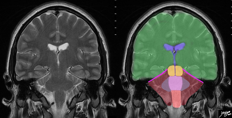
In this T2 weighted coronal MRI image the distinction between the supratentorial and infratentorial structures is made apparent by the bright pink tentorium that acts as a roof of the posterior fossa. The forebrain (green) midbrain (orange) and hindbrain (pink salmon and maroon) and the cerebellum (maroon), with other parts of the hindbrain filled in including the pons (light pink) middle cerebellar peduncles (mauve) and medulla (salmon) All the structures above the pink line are supratentorial, and those below are infratentorial. Part of the midbrain is supratentorial and part is infratentorial. The ventricular system is outlined in blue
Courtesy Ashley Davidoff MD copyright 2010 all rights reserved 89721c06b.8sg01


    [nodeType] => 1
    [parentNode] => (object value omitted)
    [childNodes] => (object value omitted)
    [firstChild] => (object value omitted)
    [lastChild] => (object value omitted)
    [previousSibling] => (object value omitted)
    [nextSibling] => (object value omitted)
    [attributes] => (object value omitted)
    [ownerDocument] => (object value omitted)
    [namespaceURI] =>
    [prefix] =>
    [localName] => table
    [baseURI] =>
    [textContent] =>


The Cerebellum in Relation to the Pons Medulla, Tentorium and Midbrain


In this T2 weighted coronal MRI image the distinction between the supratentorial and infratentorial structures is made apparent by the bright pink tentorium that acts as a roof of the posterior fossa. The forebrain (green) midbrain (orange) and hindbrain (pink salmon and maroon) and the cerebellum (maroon), with other parts of the hindbrain filled in including the pons (light pink) middle cerebellar peduncles (mauve) and medulla (salmon) All the structures above the pink line are supratentorial, and those below are infratentorial. Part of the midbrain is supratentorial and part is infratentorial. The ventricular system is outlined in blue
Courtesy Ashley Davidoff MD copyright 2010 all rights reserved 89721c06b.8sg01


)
DOMElement Object
(
    [schemaTypeInfo] =>
    [tagName] => td
    [firstElementChild] => (object value omitted)
    [lastElementChild] => (object value omitted)
    [childElementCount] => 2
    [previousElementSibling] =>
    [nextElementSibling] =>
    [nodeName] => td
    [nodeValue] =>
In this T2 weighted coronal MRI image the distinction between the supratentorial and infratentorial structures is made apparent by the bright pink tentorium that acts as a roof of the posterior fossa. The forebrain (green) midbrain (orange) and hindbrain (pink salmon and maroon) and the cerebellum (maroon), with other parts of the hindbrain filled in including the pons (light pink) middle cerebellar peduncles (mauve) and medulla (salmon) All the structures above the pink line are supratentorial, and those below are infratentorial. Part of the midbrain is supratentorial and part is infratentorial. The ventricular system is outlined in blue
Courtesy Ashley Davidoff MD copyright 2010 all rights reserved 89721c06b.8sg01

    [nodeType] => 1
    [parentNode] => (object value omitted)
    [childNodes] => (object value omitted)
    [firstChild] => (object value omitted)
    [lastChild] => (object value omitted)
    [previousSibling] => (object value omitted)
    [nextSibling] => (object value omitted)
    [attributes] => (object value omitted)
    [ownerDocument] => (object value omitted)
    [namespaceURI] =>
    [prefix] =>
    [localName] => td
    [baseURI] =>
    [textContent] =>
In this T2 weighted coronal MRI image the distinction between the supratentorial and infratentorial structures is made apparent by the bright pink tentorium that acts as a roof of the posterior fossa. The forebrain (green) midbrain (orange) and hindbrain (pink salmon and maroon) and the cerebellum (maroon), with other parts of the hindbrain filled in including the pons (light pink) middle cerebellar peduncles (mauve) and medulla (salmon) All the structures above the pink line are supratentorial, and those below are infratentorial. Part of the midbrain is supratentorial and part is infratentorial. The ventricular system is outlined in blue
Courtesy Ashley Davidoff MD copyright 2010 all rights reserved 89721c06b.8sg01

)
DOMElement Object
(
    [schemaTypeInfo] =>
    [tagName] => td
    [firstElementChild] => (object value omitted)
    [lastElementChild] => (object value omitted)
    [childElementCount] => 2
    [previousElementSibling] =>
    [nextElementSibling] =>
    [nodeName] => td
    [nodeValue] =>
The Cerebellum in Relation to the Pons Medulla, Tentorium and Midbrain
    [nodeType] => 1
    [parentNode] => (object value omitted)
    [childNodes] => (object value omitted)
    [firstChild] => (object value omitted)
    [lastChild] => (object value omitted)
    [previousSibling] => (object value omitted)
    [nextSibling] => (object value omitted)
    [attributes] => (object value omitted)
    [ownerDocument] => (object value omitted)
    [namespaceURI] =>
    [prefix] =>
    [localName] => td
    [baseURI] =>
    [textContent] =>
The Cerebellum in Relation to the Pons Medulla, Tentorium and Midbrain
)
DOMElement Object
(
    [schemaTypeInfo] =>
    [tagName] => table
    [firstElementChild] => (object value omitted)
    [lastElementChild] => (object value omitted)
    [childElementCount] => 1
    [previousElementSibling] => (object value omitted)
    [nextElementSibling] => (object value omitted)
    [nodeName] => table
    [nodeValue] =>


The Cerebellar hemispherses, Vermis, and Tonsils


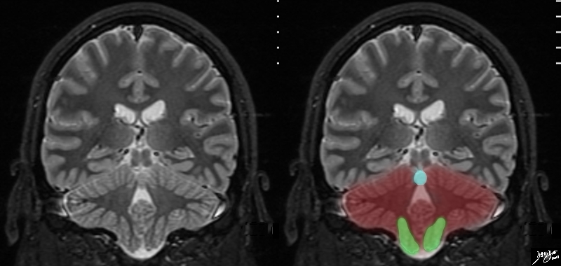
The T2 weighted MRI of the brain in coronal projection shows the cerebellum with the vermis superiorly (light blue, and the cerebellar tonsils situated medially and inferiorly
Courtesy Ashley Davidoff MD copyright 2010 all rights reserved 89742.3kcb02.8s


    [nodeType] => 1
    [parentNode] => (object value omitted)
    [childNodes] => (object value omitted)
    [firstChild] => (object value omitted)
    [lastChild] => (object value omitted)
    [previousSibling] => (object value omitted)
    [nextSibling] => (object value omitted)
    [attributes] => (object value omitted)
    [ownerDocument] => (object value omitted)
    [namespaceURI] =>
    [prefix] =>
    [localName] => table
    [baseURI] =>
    [textContent] =>


The Cerebellar hemispherses, Vermis, and Tonsils


The T2 weighted MRI of the brain in coronal projection shows the cerebellum with the vermis superiorly (light blue, and the cerebellar tonsils situated medially and inferiorly
Courtesy Ashley Davidoff MD copyright 2010 all rights reserved 89742.3kcb02.8s


)
DOMElement Object
(
    [schemaTypeInfo] =>
    [tagName] => td
    [firstElementChild] => (object value omitted)
    [lastElementChild] => (object value omitted)
    [childElementCount] => 2
    [previousElementSibling] =>
    [nextElementSibling] =>
    [nodeName] => td
    [nodeValue] =>
The T2 weighted MRI of the brain in coronal projection shows the cerebellum with the vermis superiorly (light blue, and the cerebellar tonsils situated medially and inferiorly
Courtesy Ashley Davidoff MD copyright 2010 all rights reserved 89742.3kcb02.8s

    [nodeType] => 1
    [parentNode] => (object value omitted)
    [childNodes] => (object value omitted)
    [firstChild] => (object value omitted)
    [lastChild] => (object value omitted)
    [previousSibling] => (object value omitted)
    [nextSibling] => (object value omitted)
    [attributes] => (object value omitted)
    [ownerDocument] => (object value omitted)
    [namespaceURI] =>
    [prefix] =>
    [localName] => td
    [baseURI] =>
    [textContent] =>
The T2 weighted MRI of the brain in coronal projection shows the cerebellum with the vermis superiorly (light blue, and the cerebellar tonsils situated medially and inferiorly
Courtesy Ashley Davidoff MD copyright 2010 all rights reserved 89742.3kcb02.8s

)
DOMElement Object
(
    [schemaTypeInfo] =>
    [tagName] => td
    [firstElementChild] => (object value omitted)
    [lastElementChild] => (object value omitted)
    [childElementCount] => 2
    [previousElementSibling] =>
    [nextElementSibling] =>
    [nodeName] => td
    [nodeValue] =>
The Cerebellar hemispherses, Vermis, and Tonsils
    [nodeType] => 1
    [parentNode] => (object value omitted)
    [childNodes] => (object value omitted)
    [firstChild] => (object value omitted)
    [lastChild] => (object value omitted)
    [previousSibling] => (object value omitted)
    [nextSibling] => (object value omitted)
    [attributes] => (object value omitted)
    [ownerDocument] => (object value omitted)
    [namespaceURI] =>
    [prefix] =>
    [localName] => td
    [baseURI] =>
    [textContent] =>
The Cerebellar hemispherses, Vermis, and Tonsils
)
DOMElement Object
(
    [schemaTypeInfo] =>
    [tagName] => table
    [firstElementChild] => (object value omitted)
    [lastElementChild] => (object value omitted)
    [childElementCount] => 1
    [previousElementSibling] => (object value omitted)
    [nextElementSibling] => (object value omitted)
    [nodeName] => table
    [nodeValue] =>


Coronal View


The T2 weighted MRI of the brain in coronal projection shows an almost rhomboid shaped structure
Courtesy Ashley DAvidoff MD copyright 2010 all rights reserved  89742.3kcb01b.8s


    [nodeType] => 1
    [parentNode] => (object value omitted)
    [childNodes] => (object value omitted)
    [firstChild] => (object value omitted)
    [lastChild] => (object value omitted)
    [previousSibling] => (object value omitted)
    [nextSibling] => (object value omitted)
    [attributes] => (object value omitted)
    [ownerDocument] => (object value omitted)
    [namespaceURI] =>
    [prefix] =>
    [localName] => table
    [baseURI] =>
    [textContent] =>


Coronal View


The T2 weighted MRI of the brain in coronal projection shows an almost rhomboid shaped structure
Courtesy Ashley DAvidoff MD copyright 2010 all rights reserved  89742.3kcb01b.8s


)
DOMElement Object
(
    [schemaTypeInfo] =>
    [tagName] => td
    [firstElementChild] => (object value omitted)
    [lastElementChild] => (object value omitted)
    [childElementCount] => 2
    [previousElementSibling] =>
    [nextElementSibling] =>
    [nodeName] => td
    [nodeValue] =>
The T2 weighted MRI of the brain in coronal projection shows an almost rhomboid shaped structure
Courtesy Ashley DAvidoff MD copyright 2010 all rights reserved  89742.3kcb01b.8s

    [nodeType] => 1
    [parentNode] => (object value omitted)
    [childNodes] => (object value omitted)
    [firstChild] => (object value omitted)
    [lastChild] => (object value omitted)
    [previousSibling] => (object value omitted)
    [nextSibling] => (object value omitted)
    [attributes] => (object value omitted)
    [ownerDocument] => (object value omitted)
    [namespaceURI] =>
    [prefix] =>
    [localName] => td
    [baseURI] =>
    [textContent] =>
The T2 weighted MRI of the brain in coronal projection shows an almost rhomboid shaped structure
Courtesy Ashley DAvidoff MD copyright 2010 all rights reserved  89742.3kcb01b.8s

)
DOMElement Object
(
    [schemaTypeInfo] =>
    [tagName] => td
    [firstElementChild] => (object value omitted)
    [lastElementChild] => (object value omitted)
    [childElementCount] => 2
    [previousElementSibling] =>
    [nextElementSibling] =>
    [nodeName] => td
    [nodeValue] =>
Coronal View
    [nodeType] => 1
    [parentNode] => (object value omitted)
    [childNodes] => (object value omitted)
    [firstChild] => (object value omitted)
    [lastChild] => (object value omitted)
    [previousSibling] => (object value omitted)
    [nextSibling] => (object value omitted)
    [attributes] => (object value omitted)
    [ownerDocument] => (object value omitted)
    [namespaceURI] =>
    [prefix] =>
    [localName] => td
    [baseURI] =>
    [textContent] =>
Coronal View
)
DOMElement Object
(
    [schemaTypeInfo] =>
    [tagName] => table
    [firstElementChild] => (object value omitted)
    [lastElementChild] => (object value omitted)
    [childElementCount] => 1
    [previousElementSibling] => (object value omitted)
    [nextElementSibling] => (object value omitted)
    [nodeName] => table
    [nodeValue] =>


MRI of the cerebellum in the Sagittal Plane


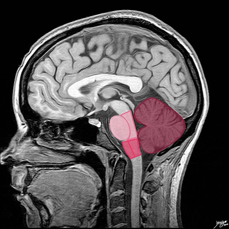
The shapes in fact are not quite ovoid. The pons (lightest pink) has shapes reminiscent of an oversize egg lying in a small bed The anterior eggshaped portion is called the ventral or anterior pons and the posterior portion called the tegmentum which is a continuation of the tegmentum of the midbrain. The medulla oblongata is almost rectangular but has a subtle forward bulging to its anterior surface. The anterior portion is called the ventral or anterior medulla and the posterior portion is called the tegmentum, similar in name and a continuation of the tegmentum of the midbrain and pons.  The cerebellum is situated posteriorly, is the largest of the three major components and it consists of two hemispheres and a central vermis better appreciated in the axial plane.
Courtesy Ashley Davidoff MD Copyright 2010 all rights reserved 92141.3kd03b04b.8s


    [nodeType] => 1
    [parentNode] => (object value omitted)
    [childNodes] => (object value omitted)
    [firstChild] => (object value omitted)
    [lastChild] => (object value omitted)
    [previousSibling] => (object value omitted)
    [nextSibling] => (object value omitted)
    [attributes] => (object value omitted)
    [ownerDocument] => (object value omitted)
    [namespaceURI] =>
    [prefix] =>
    [localName] => table
    [baseURI] =>
    [textContent] =>


MRI of the cerebellum in the Sagittal Plane


The shapes in fact are not quite ovoid. The pons (lightest pink) has shapes reminiscent of an oversize egg lying in a small bed The anterior eggshaped portion is called the ventral or anterior pons and the posterior portion called the tegmentum which is a continuation of the tegmentum of the midbrain. The medulla oblongata is almost rectangular but has a subtle forward bulging to its anterior surface. The anterior portion is called the ventral or anterior medulla and the posterior portion is called the tegmentum, similar in name and a continuation of the tegmentum of the midbrain and pons.  The cerebellum is situated posteriorly, is the largest of the three major components and it consists of two hemispheres and a central vermis better appreciated in the axial plane.
Courtesy Ashley Davidoff MD Copyright 2010 all rights reserved 92141.3kd03b04b.8s


)
DOMElement Object
(
    [schemaTypeInfo] =>
    [tagName] => td
    [firstElementChild] => (object value omitted)
    [lastElementChild] => (object value omitted)
    [childElementCount] => 2
    [previousElementSibling] =>
    [nextElementSibling] =>
    [nodeName] => td
    [nodeValue] =>
The shapes in fact are not quite ovoid. The pons (lightest pink) has shapes reminiscent of an oversize egg lying in a small bed The anterior eggshaped portion is called the ventral or anterior pons and the posterior portion called the tegmentum which is a continuation of the tegmentum of the midbrain. The medulla oblongata is almost rectangular but has a subtle forward bulging to its anterior surface. The anterior portion is called the ventral or anterior medulla and the posterior portion is called the tegmentum, similar in name and a continuation of the tegmentum of the midbrain and pons.  The cerebellum is situated posteriorly, is the largest of the three major components and it consists of two hemispheres and a central vermis better appreciated in the axial plane.
Courtesy Ashley Davidoff MD Copyright 2010 all rights reserved 92141.3kd03b04b.8s

    [nodeType] => 1
    [parentNode] => (object value omitted)
    [childNodes] => (object value omitted)
    [firstChild] => (object value omitted)
    [lastChild] => (object value omitted)
    [previousSibling] => (object value omitted)
    [nextSibling] => (object value omitted)
    [attributes] => (object value omitted)
    [ownerDocument] => (object value omitted)
    [namespaceURI] =>
    [prefix] =>
    [localName] => td
    [baseURI] =>
    [textContent] =>
The shapes in fact are not quite ovoid. The pons (lightest pink) has shapes reminiscent of an oversize egg lying in a small bed The anterior eggshaped portion is called the ventral or anterior pons and the posterior portion called the tegmentum which is a continuation of the tegmentum of the midbrain. The medulla oblongata is almost rectangular but has a subtle forward bulging to its anterior surface. The anterior portion is called the ventral or anterior medulla and the posterior portion is called the tegmentum, similar in name and a continuation of the tegmentum of the midbrain and pons.  The cerebellum is situated posteriorly, is the largest of the three major components and it consists of two hemispheres and a central vermis better appreciated in the axial plane.
Courtesy Ashley Davidoff MD Copyright 2010 all rights reserved 92141.3kd03b04b.8s

)
DOMElement Object
(
    [schemaTypeInfo] =>
    [tagName] => td
    [firstElementChild] => (object value omitted)
    [lastElementChild] => (object value omitted)
    [childElementCount] => 2
    [previousElementSibling] =>
    [nextElementSibling] =>
    [nodeName] => td
    [nodeValue] =>
MRI of the cerebellum in the Sagittal Plane
    [nodeType] => 1
    [parentNode] => (object value omitted)
    [childNodes] => (object value omitted)
    [firstChild] => (object value omitted)
    [lastChild] => (object value omitted)
    [previousSibling] => (object value omitted)
    [nextSibling] => (object value omitted)
    [attributes] => (object value omitted)
    [ownerDocument] => (object value omitted)
    [namespaceURI] =>
    [prefix] =>
    [localName] => td
    [baseURI] =>
    [textContent] =>
MRI of the cerebellum in the Sagittal Plane
)
DOMElement Object
(
    [schemaTypeInfo] =>
    [tagName] => table
    [firstElementChild] => (object value omitted)
    [lastElementChild] => (object value omitted)
    [childElementCount] => 1
    [previousElementSibling] => (object value omitted)
    [nextElementSibling] => (object value omitted)
    [nodeName] => table
    [nodeValue] =>


Cerebellum as seen from Below


When the anatomic specimen is viewed from below, the forebrain (torquoise green) and the hindbrain (salmon pink) dominated by the cerebellum are best seen. The midbrain which is a relatively small structure is not visualized. code brain anatomy surface view sulcus sulci gyrus gyri cerebellum forebrain hindbrain normal structure anatomy brain
Source unknown Modified by Davidoff MD 53605c02.8s


    [nodeType] => 1
    [parentNode] => (object value omitted)
    [childNodes] => (object value omitted)
    [firstChild] => (object value omitted)
    [lastChild] => (object value omitted)
    [previousSibling] => (object value omitted)
    [nextSibling] => (object value omitted)
    [attributes] => (object value omitted)
    [ownerDocument] => (object value omitted)
    [namespaceURI] =>
    [prefix] =>
    [localName] => table
    [baseURI] =>
    [textContent] =>


Cerebellum as seen from Below


When the anatomic specimen is viewed from below, the forebrain (torquoise green) and the hindbrain (salmon pink) dominated by the cerebellum are best seen. The midbrain which is a relatively small structure is not visualized. code brain anatomy surface view sulcus sulci gyrus gyri cerebellum forebrain hindbrain normal structure anatomy brain
Source unknown Modified by Davidoff MD 53605c02.8s


)
DOMElement Object
(
    [schemaTypeInfo] =>
    [tagName] => td
    [firstElementChild] => (object value omitted)
    [lastElementChild] => (object value omitted)
    [childElementCount] => 2
    [previousElementSibling] =>
    [nextElementSibling] =>
    [nodeName] => td
    [nodeValue] =>
When the anatomic specimen is viewed from below, the forebrain (torquoise green) and the hindbrain (salmon pink) dominated by the cerebellum are best seen. The midbrain which is a relatively small structure is not visualized. code brain anatomy surface view sulcus sulci gyrus gyri cerebellum forebrain hindbrain normal structure anatomy brain
Source unknown Modified by Davidoff MD 53605c02.8s

    [nodeType] => 1
    [parentNode] => (object value omitted)
    [childNodes] => (object value omitted)
    [firstChild] => (object value omitted)
    [lastChild] => (object value omitted)
    [previousSibling] => (object value omitted)
    [nextSibling] => (object value omitted)
    [attributes] => (object value omitted)
    [ownerDocument] => (object value omitted)
    [namespaceURI] =>
    [prefix] =>
    [localName] => td
    [baseURI] =>
    [textContent] =>
When the anatomic specimen is viewed from below, the forebrain (torquoise green) and the hindbrain (salmon pink) dominated by the cerebellum are best seen. The midbrain which is a relatively small structure is not visualized. code brain anatomy surface view sulcus sulci gyrus gyri cerebellum forebrain hindbrain normal structure anatomy brain
Source unknown Modified by Davidoff MD 53605c02.8s

)
DOMElement Object
(
    [schemaTypeInfo] =>
    [tagName] => td
    [firstElementChild] => (object value omitted)
    [lastElementChild] => (object value omitted)
    [childElementCount] => 2
    [previousElementSibling] =>
    [nextElementSibling] =>
    [nodeName] => td
    [nodeValue] =>

Cerebellum as seen from Below
    [nodeType] => 1
    [parentNode] => (object value omitted)
    [childNodes] => (object value omitted)
    [firstChild] => (object value omitted)
    [lastChild] => (object value omitted)
    [previousSibling] => (object value omitted)
    [nextSibling] => (object value omitted)
    [attributes] => (object value omitted)
    [ownerDocument] => (object value omitted)
    [namespaceURI] =>
    [prefix] =>
    [localName] => td
    [baseURI] =>
    [textContent] =>

Cerebellum as seen from Below
)
DOMElement Object
(
    [schemaTypeInfo] =>
    [tagName] => table
    [firstElementChild] => (object value omitted)
    [lastElementChild] => (object value omitted)
    [childElementCount] => 1
    [previousElementSibling] => (object value omitted)
    [nextElementSibling] => (object value omitted)
    [nodeName] => table
    [nodeValue] =>


Gross Anatomy of the Cerebellum


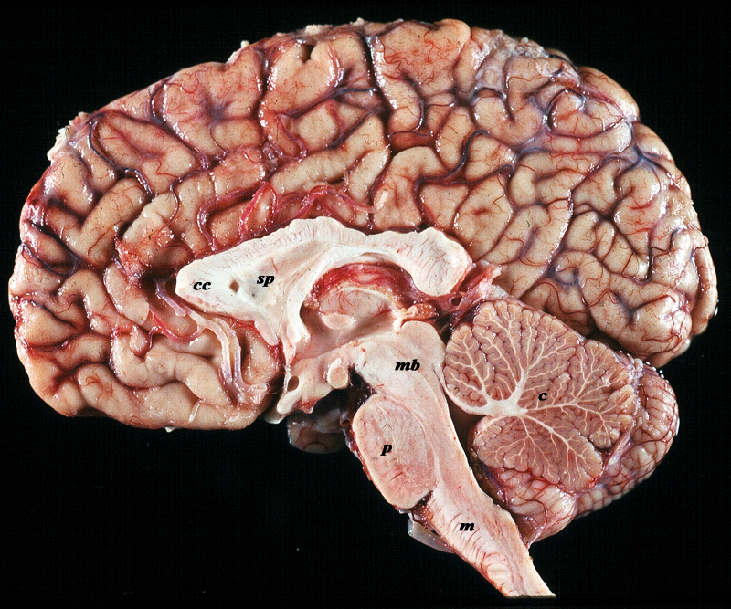
The midsagittal section view of brain reveals the distinctive shape position and character of the midline structures of the brain. The distinction between the character of the cerebral cortex which has a creamy color and the white matter exemplified by the corpus callosum (c) and septum pellucidum (sp) which are white, and the midbrain (mb) pons (p) and medulla (m) which are off white as opposed to the color of the cerebellum (c) which is light salmon pink is well demonstrated.
The distinction between the folds of the cerebrum (gyri) and , and cerebellar folds (folia) are also exemplified in this image.  The gyri of the cerebral hemispheres (gyri) are broad, irregular in shape, and course in different directions. The folds of the cerebellum on this view look like the branches of a tree.
The cerebellum is smaller (Latin for little brain),  and lies below the cerebral hemispheres.The relative sizes of the forebrain, midbrain and hindbrain and their components are well appreciated in this section.
Image Courtesy of Thomas W.Smith, MD; Department of Pathology, University of Massachusetts Medical School. 97805b02


    [nodeType] => 1
    [parentNode] => (object value omitted)
    [childNodes] => (object value omitted)
    [firstChild] => (object value omitted)
    [lastChild] => (object value omitted)
    [previousSibling] => (object value omitted)
    [nextSibling] => (object value omitted)
    [attributes] => (object value omitted)
    [ownerDocument] => (object value omitted)
    [namespaceURI] =>
    [prefix] =>
    [localName] => table
    [baseURI] =>
    [textContent] =>


Gross Anatomy of the Cerebellum


The midsagittal section view of brain reveals the distinctive shape position and character of the midline structures of the brain. The distinction between the character of the cerebral cortex which has a creamy color and the white matter exemplified by the corpus callosum (c) and septum pellucidum (sp) which are white, and the midbrain (mb) pons (p) and medulla (m) which are off white as opposed to the color of the cerebellum (c) which is light salmon pink is well demonstrated.
The distinction between the folds of the cerebrum (gyri) and , and cerebellar folds (folia) are also exemplified in this image.  The gyri of the cerebral hemispheres (gyri) are broad, irregular in shape, and course in different directions. The folds of the cerebellum on this view look like the branches of a tree.
The cerebellum is smaller (Latin for little brain),  and lies below the cerebral hemispheres.The relative sizes of the forebrain, midbrain and hindbrain and their components are well appreciated in this section.
Image Courtesy of Thomas W.Smith, MD; Department of Pathology, University of Massachusetts Medical School. 97805b02


)
DOMElement Object
(
    [schemaTypeInfo] =>
    [tagName] => td
    [firstElementChild] => (object value omitted)
    [lastElementChild] => (object value omitted)
    [childElementCount] => 4
    [previousElementSibling] =>
    [nextElementSibling] =>
    [nodeName] => td
    [nodeValue] =>
The midsagittal section view of brain reveals the distinctive shape position and character of the midline structures of the brain. The distinction between the character of the cerebral cortex which has a creamy color and the white matter exemplified by the corpus callosum (c) and septum pellucidum (sp) which are white, and the midbrain (mb) pons (p) and medulla (m) which are off white as opposed to the color of the cerebellum (c) which is light salmon pink is well demonstrated.
The distinction between the folds of the cerebrum (gyri) and , and cerebellar folds (folia) are also exemplified in this image.  The gyri of the cerebral hemispheres (gyri) are broad, irregular in shape, and course in different directions. The folds of the cerebellum on this view look like the branches of a tree.
The cerebellum is smaller (Latin for little brain),  and lies below the cerebral hemispheres.The relative sizes of the forebrain, midbrain and hindbrain and their components are well appreciated in this section.
Image Courtesy of Thomas W.Smith, MD; Department of Pathology, University of Massachusetts Medical School. 97805b02

    [nodeType] => 1
    [parentNode] => (object value omitted)
    [childNodes] => (object value omitted)
    [firstChild] => (object value omitted)
    [lastChild] => (object value omitted)
    [previousSibling] => (object value omitted)
    [nextSibling] => (object value omitted)
    [attributes] => (object value omitted)
    [ownerDocument] => (object value omitted)
    [namespaceURI] =>
    [prefix] =>
    [localName] => td
    [baseURI] =>
    [textContent] =>
The midsagittal section view of brain reveals the distinctive shape position and character of the midline structures of the brain. The distinction between the character of the cerebral cortex which has a creamy color and the white matter exemplified by the corpus callosum (c) and septum pellucidum (sp) which are white, and the midbrain (mb) pons (p) and medulla (m) which are off white as opposed to the color of the cerebellum (c) which is light salmon pink is well demonstrated.
The distinction between the folds of the cerebrum (gyri) and , and cerebellar folds (folia) are also exemplified in this image.  The gyri of the cerebral hemispheres (gyri) are broad, irregular in shape, and course in different directions. The folds of the cerebellum on this view look like the branches of a tree.
The cerebellum is smaller (Latin for little brain),  and lies below the cerebral hemispheres.The relative sizes of the forebrain, midbrain and hindbrain and their components are well appreciated in this section.
Image Courtesy of Thomas W.Smith, MD; Department of Pathology, University of Massachusetts Medical School. 97805b02

)
DOMElement Object
(
    [schemaTypeInfo] =>
    [tagName] => td
    [firstElementChild] => (object value omitted)
    [lastElementChild] => (object value omitted)
    [childElementCount] => 2
    [previousElementSibling] =>
    [nextElementSibling] =>
    [nodeName] => td
    [nodeValue] =>
Gross Anatomy of the Cerebellum
    [nodeType] => 1
    [parentNode] => (object value omitted)
    [childNodes] => (object value omitted)
    [firstChild] => (object value omitted)
    [lastChild] => (object value omitted)
    [previousSibling] => (object value omitted)
    [nextSibling] => (object value omitted)
    [attributes] => (object value omitted)
    [ownerDocument] => (object value omitted)
    [namespaceURI] =>
    [prefix] =>
    [localName] => td
    [baseURI] =>
    [textContent] =>
Gross Anatomy of the Cerebellum
)
DOMElement Object
(
    [schemaTypeInfo] =>
    [tagName] => table
    [firstElementChild] => (object value omitted)
    [lastElementChild] => (object value omitted)
    [childElementCount] => 1
    [previousElementSibling] => (object value omitted)
    [nextElementSibling] => (object value omitted)
    [nodeName] => table
    [nodeValue] =>


External Possterior View
The Cerebellum Below and the cerebral hemisphere Above
Difference in Morphology


The posterior external view of brain reveals the distinction between the character of the cerebral hemispheres and the cerebellum, exemplified in this instance by the morphology of the folds, and the difference in size shape and position . The cerebellar folds called folia have a banded appearance and are regularly spaced, tightly packed in neat almost equal rows, and distributed horizontally. The folds of the cerebral hemispheres (gyri) are broader, irregular in shape, and course in different directions. The cerebellum is smaller (Latin for little brain), has the shape of an inverted top, or the roof of a Russian Orthodox church) and lies below the cerebral hemispheres. The veins of both the cerebral hemispheres and the cerebellum are seen coursing over the surface,
Image Courtesy of Thomas W.Smith, MD; Department of Pathology, University of Massachusetts Medical School. 97625b03


    [nodeType] => 1
    [parentNode] => (object value omitted)
    [childNodes] => (object value omitted)
    [firstChild] => (object value omitted)
    [lastChild] => (object value omitted)
    [previousSibling] => (object value omitted)
    [nextSibling] => (object value omitted)
    [attributes] => (object value omitted)
    [ownerDocument] => (object value omitted)
    [namespaceURI] =>
    [prefix] =>
    [localName] => table
    [baseURI] =>
    [textContent] =>


External Possterior View
The Cerebellum Below and the cerebral hemisphere Above
Difference in Morphology


The posterior external view of brain reveals the distinction between the character of the cerebral hemispheres and the cerebellum, exemplified in this instance by the morphology of the folds, and the difference in size shape and position . The cerebellar folds called folia have a banded appearance and are regularly spaced, tightly packed in neat almost equal rows, and distributed horizontally. The folds of the cerebral hemispheres (gyri) are broader, irregular in shape, and course in different directions. The cerebellum is smaller (Latin for little brain), has the shape of an inverted top, or the roof of a Russian Orthodox church) and lies below the cerebral hemispheres. The veins of both the cerebral hemispheres and the cerebellum are seen coursing over the surface,
Image Courtesy of Thomas W.Smith, MD; Department of Pathology, University of Massachusetts Medical School. 97625b03


)
DOMElement Object
(
    [schemaTypeInfo] =>
    [tagName] => td
    [firstElementChild] => (object value omitted)
    [lastElementChild] => (object value omitted)
    [childElementCount] => 2
    [previousElementSibling] =>
    [nextElementSibling] =>
    [nodeName] => td
    [nodeValue] =>
The posterior external view of brain reveals the distinction between the character of the cerebral hemispheres and the cerebellum, exemplified in this instance by the morphology of the folds, and the difference in size shape and position . The cerebellar folds called folia have a banded appearance and are regularly spaced, tightly packed in neat almost equal rows, and distributed horizontally. The folds of the cerebral hemispheres (gyri) are broader, irregular in shape, and course in different directions. The cerebellum is smaller (Latin for little brain), has the shape of an inverted top, or the roof of a Russian Orthodox church) and lies below the cerebral hemispheres. The veins of both the cerebral hemispheres and the cerebellum are seen coursing over the surface,
Image Courtesy of Thomas W.Smith, MD; Department of Pathology, University of Massachusetts Medical School. 97625b03

    [nodeType] => 1
    [parentNode] => (object value omitted)
    [childNodes] => (object value omitted)
    [firstChild] => (object value omitted)
    [lastChild] => (object value omitted)
    [previousSibling] => (object value omitted)
    [nextSibling] => (object value omitted)
    [attributes] => (object value omitted)
    [ownerDocument] => (object value omitted)
    [namespaceURI] =>
    [prefix] =>
    [localName] => td
    [baseURI] =>
    [textContent] =>
The posterior external view of brain reveals the distinction between the character of the cerebral hemispheres and the cerebellum, exemplified in this instance by the morphology of the folds, and the difference in size shape and position . The cerebellar folds called folia have a banded appearance and are regularly spaced, tightly packed in neat almost equal rows, and distributed horizontally. The folds of the cerebral hemispheres (gyri) are broader, irregular in shape, and course in different directions. The cerebellum is smaller (Latin for little brain), has the shape of an inverted top, or the roof of a Russian Orthodox church) and lies below the cerebral hemispheres. The veins of both the cerebral hemispheres and the cerebellum are seen coursing over the surface,
Image Courtesy of Thomas W.Smith, MD; Department of Pathology, University of Massachusetts Medical School. 97625b03

)
DOMElement Object
(
    [schemaTypeInfo] =>
    [tagName] => td
    [firstElementChild] => (object value omitted)
    [lastElementChild] => (object value omitted)
    [childElementCount] => 4
    [previousElementSibling] =>
    [nextElementSibling] =>
    [nodeName] => td
    [nodeValue] =>
External Possterior View
The Cerebellum Below and the cerebral hemisphere Above
Difference in Morphology
    [nodeType] => 1
    [parentNode] => (object value omitted)
    [childNodes] => (object value omitted)
    [firstChild] => (object value omitted)
    [lastChild] => (object value omitted)
    [previousSibling] => (object value omitted)
    [nextSibling] => (object value omitted)
    [attributes] => (object value omitted)
    [ownerDocument] => (object value omitted)
    [namespaceURI] =>
    [prefix] =>
    [localName] => td
    [baseURI] =>
    [textContent] =>
External Possterior View
The Cerebellum Below and the cerebral hemisphere Above
Difference in Morphology
)
DOMElement Object
(
    [schemaTypeInfo] =>
    [tagName] => table
    [firstElementChild] => (object value omitted)
    [lastElementChild] => (object value omitted)
    [childElementCount] => 1
    [previousElementSibling] => (object value omitted)
    [nextElementSibling] => (object value omitted)
    [nodeName] => table
    [nodeValue] =>


External Lateral View of the Brain


The brain has a characteristic appearance manifest by its creamy color, characteristic shape and fissured surface. To the feel it is said to feel like soft butter. The anatomic specimen reveals an external view of the brain. The characteristic cream color of the brain can be appreciated on this specimen. This reflects the color of the gray matter and external portion of the cortex The surface of the hemispheres is known as cortex. It presents as a system of irregular prominences (called convolutions or gyri) that circumscribe depressions (called fissures or sulci) which are somewhat profound and full of twists and turns. The sulci and gyri of the forebrain are distinctly different from the pattern of the cerebellum. The folia of the cerebellum have a banded appearance and are regularly spaced, tightly packed in neat almost equal rows, and distributed horizontally.  The relative size, shape and position of the forebrain compared to the hindbrain is also well demonstrated in this view.
Image Courtesy of Thomas W.Smith, MD; Department of Pathology, University of Massachusetts Medical School. 97804


    [nodeType] => 1
    [parentNode] => (object value omitted)
    [childNodes] => (object value omitted)
    [firstChild] => (object value omitted)
    [lastChild] => (object value omitted)
    [previousSibling] => (object value omitted)
    [nextSibling] => (object value omitted)
    [attributes] => (object value omitted)
    [ownerDocument] => (object value omitted)
    [namespaceURI] =>
    [prefix] =>
    [localName] => table
    [baseURI] =>
    [textContent] =>


External Lateral View of the Brain


The brain has a characteristic appearance manifest by its creamy color, characteristic shape and fissured surface. To the feel it is said to feel like soft butter. The anatomic specimen reveals an external view of the brain. The characteristic cream color of the brain can be appreciated on this specimen. This reflects the color of the gray matter and external portion of the cortex The surface of the hemispheres is known as cortex. It presents as a system of irregular prominences (called convolutions or gyri) that circumscribe depressions (called fissures or sulci) which are somewhat profound and full of twists and turns. The sulci and gyri of the forebrain are distinctly different from the pattern of the cerebellum. The folia of the cerebellum have a banded appearance and are regularly spaced, tightly packed in neat almost equal rows, and distributed horizontally.  The relative size, shape and position of the forebrain compared to the hindbrain is also well demonstrated in this view.
Image Courtesy of Thomas W.Smith, MD; Department of Pathology, University of Massachusetts Medical School. 97804


)
DOMElement Object
(
    [schemaTypeInfo] =>
    [tagName] => td
    [firstElementChild] => (object value omitted)
    [lastElementChild] => (object value omitted)
    [childElementCount] => 2
    [previousElementSibling] =>
    [nextElementSibling] =>
    [nodeName] => td
    [nodeValue] =>
The brain has a characteristic appearance manifest by its creamy color, characteristic shape and fissured surface. To the feel it is said to feel like soft butter. The anatomic specimen reveals an external view of the brain. The characteristic cream color of the brain can be appreciated on this specimen. This reflects the color of the gray matter and external portion of the cortex The surface of the hemispheres is known as cortex. It presents as a system of irregular prominences (called convolutions or gyri) that circumscribe depressions (called fissures or sulci) which are somewhat profound and full of twists and turns. The sulci and gyri of the forebrain are distinctly different from the pattern of the cerebellum. The folia of the cerebellum have a banded appearance and are regularly spaced, tightly packed in neat almost equal rows, and distributed horizontally.  The relative size, shape and position of the forebrain compared to the hindbrain is also well demonstrated in this view.
Image Courtesy of Thomas W.Smith, MD; Department of Pathology, University of Massachusetts Medical School. 97804

    [nodeType] => 1
    [parentNode] => (object value omitted)
    [childNodes] => (object value omitted)
    [firstChild] => (object value omitted)
    [lastChild] => (object value omitted)
    [previousSibling] => (object value omitted)
    [nextSibling] => (object value omitted)
    [attributes] => (object value omitted)
    [ownerDocument] => (object value omitted)
    [namespaceURI] =>
    [prefix] =>
    [localName] => td
    [baseURI] =>
    [textContent] =>
The brain has a characteristic appearance manifest by its creamy color, characteristic shape and fissured surface. To the feel it is said to feel like soft butter. The anatomic specimen reveals an external view of the brain. The characteristic cream color of the brain can be appreciated on this specimen. This reflects the color of the gray matter and external portion of the cortex The surface of the hemispheres is known as cortex. It presents as a system of irregular prominences (called convolutions or gyri) that circumscribe depressions (called fissures or sulci) which are somewhat profound and full of twists and turns. The sulci and gyri of the forebrain are distinctly different from the pattern of the cerebellum. The folia of the cerebellum have a banded appearance and are regularly spaced, tightly packed in neat almost equal rows, and distributed horizontally.  The relative size, shape and position of the forebrain compared to the hindbrain is also well demonstrated in this view.
Image Courtesy of Thomas W.Smith, MD; Department of Pathology, University of Massachusetts Medical School. 97804

)
DOMElement Object
(
    [schemaTypeInfo] =>
    [tagName] => td
    [firstElementChild] => (object value omitted)
    [lastElementChild] => (object value omitted)
    [childElementCount] => 2
    [previousElementSibling] =>
    [nextElementSibling] =>
    [nodeName] => td
    [nodeValue] =>
External Lateral View of the Brain
    [nodeType] => 1
    [parentNode] => (object value omitted)
    [childNodes] => (object value omitted)
    [firstChild] => (object value omitted)
    [lastChild] => (object value omitted)
    [previousSibling] => (object value omitted)
    [nextSibling] => (object value omitted)
    [attributes] => (object value omitted)
    [ownerDocument] => (object value omitted)
    [namespaceURI] =>
    [prefix] =>
    [localName] => td
    [baseURI] =>
    [textContent] =>
External Lateral View of the Brain
)
DOMElement Object
(
    [schemaTypeInfo] =>
    [tagName] => table
    [firstElementChild] => (object value omitted)
    [lastElementChild] => (object value omitted)
    [childElementCount] => 1
    [previousElementSibling] => (object value omitted)
    [nextElementSibling] => (object value omitted)
    [nodeName] => table
    [nodeValue] =>


The Cerebellum in Context
The Cerebellum (red)
Part of the Hindbrain(Rhombencephalon)
Member of the Metencephalon


The basic and simplest classification of the brain into forebrain midbrain and hindbrain is shown in this diagram and advanced to a more complex tree using the embryological and evolutionary terminologies.
The cerebellum is part of the hindbrain
The forebrain consists of the cerebrum also called the prosencephalon, which contains the more advanced form of the brain and the thalamic structures which contain more basic structures. The cerebrum (telencephalon) itself consists of two cerebral hemispheres and paired basal ganglial structures. Each cerebral hemisphere will have gray and white matter distributed in the frontal parietal temporal and occipital lobe, with the basal ganglia being part of the gray matter deep in the cerebral hemispheres. The most important thalamic structures arising from the diencephalons include the thalamus itself and the hypothalamus. The midbrain (mesencepaholon) consists of the tectum tegmentum and cerebral peduncles. The hindbrain has two major branch points based on the evolutionary development. The pons and cerebellum(part of the metencephalon) are grouped and the medulla (part of the myelencephalon is the second branch.
Courtesy Ashley Davidoff MD Copyright 2010 All rights reserved 97686.8s


    [nodeType] => 1
    [parentNode] => (object value omitted)
    [childNodes] => (object value omitted)
    [firstChild] => (object value omitted)
    [lastChild] => (object value omitted)
    [previousSibling] => (object value omitted)
    [nextSibling] => (object value omitted)
    [attributes] => (object value omitted)
    [ownerDocument] => (object value omitted)
    [namespaceURI] =>
    [prefix] =>
    [localName] => table
    [baseURI] =>
    [textContent] =>


The Cerebellum in Context
The Cerebellum (red)
Part of the Hindbrain(Rhombencephalon)
Member of the Metencephalon


The basic and simplest classification of the brain into forebrain midbrain and hindbrain is shown in this diagram and advanced to a more complex tree using the embryological and evolutionary terminologies.
The cerebellum is part of the hindbrain
The forebrain consists of the cerebrum also called the prosencephalon, which contains the more advanced form of the brain and the thalamic structures which contain more basic structures. The cerebrum (telencephalon) itself consists of two cerebral hemispheres and paired basal ganglial structures. Each cerebral hemisphere will have gray and white matter distributed in the frontal parietal temporal and occipital lobe, with the basal ganglia being part of the gray matter deep in the cerebral hemispheres. The most important thalamic structures arising from the diencephalons include the thalamus itself and the hypothalamus. The midbrain (mesencepaholon) consists of the tectum tegmentum and cerebral peduncles. The hindbrain has two major branch points based on the evolutionary development. The pons and cerebellum(part of the metencephalon) are grouped and the medulla (part of the myelencephalon is the second branch.
Courtesy Ashley Davidoff MD Copyright 2010 All rights reserved 97686.8s


)
DOMElement Object
(
    [schemaTypeInfo] =>
    [tagName] => td
    [firstElementChild] => (object value omitted)
    [lastElementChild] => (object value omitted)
    [childElementCount] => 4
    [previousElementSibling] =>
    [nextElementSibling] =>
    [nodeName] => td
    [nodeValue] =>
The basic and simplest classification of the brain into forebrain midbrain and hindbrain is shown in this diagram and advanced to a more complex tree using the embryological and evolutionary terminologies.
The cerebellum is part of the hindbrain
The forebrain consists of the cerebrum also called the prosencephalon, which contains the more advanced form of the brain and the thalamic structures which contain more basic structures. The cerebrum (telencephalon) itself consists of two cerebral hemispheres and paired basal ganglial structures. Each cerebral hemisphere will have gray and white matter distributed in the frontal parietal temporal and occipital lobe, with the basal ganglia being part of the gray matter deep in the cerebral hemispheres. The most important thalamic structures arising from the diencephalons include the thalamus itself and the hypothalamus. The midbrain (mesencepaholon) consists of the tectum tegmentum and cerebral peduncles. The hindbrain has two major branch points based on the evolutionary development. The pons and cerebellum(part of the metencephalon) are grouped and the medulla (part of the myelencephalon is the second branch.
Courtesy Ashley Davidoff MD Copyright 2010 All rights reserved 97686.8s

    [nodeType] => 1
    [parentNode] => (object value omitted)
    [childNodes] => (object value omitted)
    [firstChild] => (object value omitted)
    [lastChild] => (object value omitted)
    [previousSibling] => (object value omitted)
    [nextSibling] => (object value omitted)
    [attributes] => (object value omitted)
    [ownerDocument] => (object value omitted)
    [namespaceURI] =>
    [prefix] =>
    [localName] => td
    [baseURI] =>
    [textContent] =>
The basic and simplest classification of the brain into forebrain midbrain and hindbrain is shown in this diagram and advanced to a more complex tree using the embryological and evolutionary terminologies.
The cerebellum is part of the hindbrain
The forebrain consists of the cerebrum also called the prosencephalon, which contains the more advanced form of the brain and the thalamic structures which contain more basic structures. The cerebrum (telencephalon) itself consists of two cerebral hemispheres and paired basal ganglial structures. Each cerebral hemisphere will have gray and white matter distributed in the frontal parietal temporal and occipital lobe, with the basal ganglia being part of the gray matter deep in the cerebral hemispheres. The most important thalamic structures arising from the diencephalons include the thalamus itself and the hypothalamus. The midbrain (mesencepaholon) consists of the tectum tegmentum and cerebral peduncles. The hindbrain has two major branch points based on the evolutionary development. The pons and cerebellum(part of the metencephalon) are grouped and the medulla (part of the myelencephalon is the second branch.
Courtesy Ashley Davidoff MD Copyright 2010 All rights reserved 97686.8s

)
DOMElement Object
(
    [schemaTypeInfo] =>
    [tagName] => td
    [firstElementChild] => (object value omitted)
    [lastElementChild] => (object value omitted)
    [childElementCount] => 5
    [previousElementSibling] =>
    [nextElementSibling] =>
    [nodeName] => td
    [nodeValue] =>

The Cerebellum in Context
The Cerebellum (red)
Part of the Hindbrain(Rhombencephalon)
Member of the Metencephalon
    [nodeType] => 1
    [parentNode] => (object value omitted)
    [childNodes] => (object value omitted)
    [firstChild] => (object value omitted)
    [lastChild] => (object value omitted)
    [previousSibling] => (object value omitted)
    [nextSibling] => (object value omitted)
    [attributes] => (object value omitted)
    [ownerDocument] => (object value omitted)
    [namespaceURI] =>
    [prefix] =>
    [localName] => td
    [baseURI] =>
    [textContent] =>

The Cerebellum in Context
The Cerebellum (red)
Part of the Hindbrain(Rhombencephalon)
Member of the Metencephalon
)

