Central Sulcus or Central Fissure or Fissure of Rolando
Ashley Davidoff MD
The Common Vein Copyright 2010
Definition
The central sulcus is groove or cleft between that divides the frontal lobe from the parietal lobe and also divides the motor gyrus anteriorly from the sensory gyrus posteriorly. It is an important landmark.
|
Central Sulcus in Sagittal MRI |
|
The sagittal image is deeper in the brain near the midline through the interhemispheric fissure, and is intended to demonstrate the the parieto-occipital fissure (pink) in order to define the border between the parietall and occipital lobe. The central sulcus (pink) has also been inferred in to demonstrate the border between the frontal lobe and parietal lobe. It is not usually Image Courtesy Philips Medical Systems rendered by Davidoff art 92141c01label.82s |
Axial Plane
|
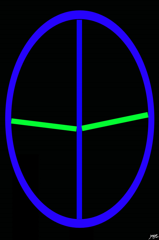
The Central Sulcus Separates the Frontal Lobes from the Parietal Lobes |
|
The diagram reflects the interhemispheric fissure as depicted in the axial plane dividing the brain into two halves. (blue). The sulci that separate the frontal lobes and the parietal lobes are called the central sulci (represented in green). Upward or forward of the central sulcus is the frontal lobe and backward or posterior are the parietal lobes. The occipital lobes and temporal lobes, midbrain and hindbrain are all mor caudal to this plane Davidoff art Courtesy Ashley Davidoff MD copyright 2010 93914.83s |
DOMElement Object
(
    [schemaTypeInfo] =>
    [tagName] => table
    [firstElementChild] => (object value omitted)
    [lastElementChild] => (object value omitted)
    [childElementCount] => 1
    [previousElementSibling] => (object value omitted)
    [nextElementSibling] =>
    [nodeName] => table
    [nodeValue] =>


The Central Sulcus
Separates the Frontal Lobes from the Parietal Lobes


The diagram reflects the interhemispheric fissure as depicted in the axial plane dividing the brain into two halves. (blue). The sulci that separate the frontal lobes and the parietal lobes are called the central sulci (represented in green). Upward or forward of the central sulcus is the frontal lobe and backward or posterior are the parietal lobes. The occipital lobes and temporal lobes, midbrain and hindbrain are all mor caudal to this plane
Davidoff art Courtesy Ashley Davidoff MD copyright 2010 93914.83s


    [nodeType] => 1
    [parentNode] => (object value omitted)
    [childNodes] => (object value omitted)
    [firstChild] => (object value omitted)
    [lastChild] => (object value omitted)
    [previousSibling] => (object value omitted)
    [nextSibling] => (object value omitted)
    [attributes] => (object value omitted)
    [ownerDocument] => (object value omitted)
    [namespaceURI] =>
    [prefix] =>
    [localName] => table
    [baseURI] =>
    [textContent] =>


The Central Sulcus
Separates the Frontal Lobes from the Parietal Lobes


The diagram reflects the interhemispheric fissure as depicted in the axial plane dividing the brain into two halves. (blue). The sulci that separate the frontal lobes and the parietal lobes are called the central sulci (represented in green). Upward or forward of the central sulcus is the frontal lobe and backward or posterior are the parietal lobes. The occipital lobes and temporal lobes, midbrain and hindbrain are all mor caudal to this plane
Davidoff art Courtesy Ashley Davidoff MD copyright 2010 93914.83s


)
DOMElement Object
(
    [schemaTypeInfo] =>
    [tagName] => td
    [firstElementChild] => (object value omitted)
    [lastElementChild] => (object value omitted)
    [childElementCount] => 2
    [previousElementSibling] =>
    [nextElementSibling] =>
    [nodeName] => td
    [nodeValue] =>
The diagram reflects the interhemispheric fissure as depicted in the axial plane dividing the brain into two halves. (blue). The sulci that separate the frontal lobes and the parietal lobes are called the central sulci (represented in green). Upward or forward of the central sulcus is the frontal lobe and backward or posterior are the parietal lobes. The occipital lobes and temporal lobes, midbrain and hindbrain are all mor caudal to this plane
Davidoff art Courtesy Ashley Davidoff MD copyright 2010 93914.83s

    [nodeType] => 1
    [parentNode] => (object value omitted)
    [childNodes] => (object value omitted)
    [firstChild] => (object value omitted)
    [lastChild] => (object value omitted)
    [previousSibling] => (object value omitted)
    [nextSibling] => (object value omitted)
    [attributes] => (object value omitted)
    [ownerDocument] => (object value omitted)
    [namespaceURI] =>
    [prefix] =>
    [localName] => td
    [baseURI] =>
    [textContent] =>
The diagram reflects the interhemispheric fissure as depicted in the axial plane dividing the brain into two halves. (blue). The sulci that separate the frontal lobes and the parietal lobes are called the central sulci (represented in green). Upward or forward of the central sulcus is the frontal lobe and backward or posterior are the parietal lobes. The occipital lobes and temporal lobes, midbrain and hindbrain are all mor caudal to this plane
Davidoff art Courtesy Ashley Davidoff MD copyright 2010 93914.83s

)
DOMElement Object
(
    [schemaTypeInfo] =>
    [tagName] => td
    [firstElementChild] => (object value omitted)
    [lastElementChild] => (object value omitted)
    [childElementCount] => 3
    [previousElementSibling] =>
    [nextElementSibling] =>
    [nodeName] => td
    [nodeValue] =>

The Central Sulcus
Separates the Frontal Lobes from the Parietal Lobes
    [nodeType] => 1
    [parentNode] => (object value omitted)
    [childNodes] => (object value omitted)
    [firstChild] => (object value omitted)
    [lastChild] => (object value omitted)
    [previousSibling] => (object value omitted)
    [nextSibling] => (object value omitted)
    [attributes] => (object value omitted)
    [ownerDocument] => (object value omitted)
    [namespaceURI] =>
    [prefix] =>
    [localName] => td
    [baseURI] =>
    [textContent] =>

The Central Sulcus
Separates the Frontal Lobes from the Parietal Lobes
)
DOMElement Object
(
    [schemaTypeInfo] =>
    [tagName] => table
    [firstElementChild] => (object value omitted)
    [lastElementChild] => (object value omitted)
    [childElementCount] => 1
    [previousElementSibling] => (object value omitted)
    [nextElementSibling] => (object value omitted)
    [nodeName] => table
    [nodeValue] =>


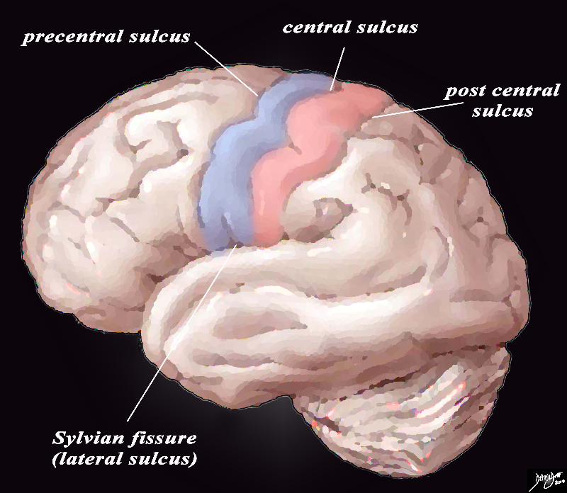
Introduction to the Central Sulci and Gyri
Characteristic Location and Shape


The lateral view of the brain shows the 4 important central sulci (aka fissures), 3 of which are vertically oriented and 1 of which is horizontal. The vertical sulci include the central sulcus, precentral sulcus and post central sulcus. The central sulcus is important because it separates the frontal lobe from the parietal lobe. The post central sulcus is important because together with the central sulcus it forms the borders of the post central gyrus (pink) which is the sensory cortex. The precentral sulcus together with the central sulcus forms the borders post central gyrus (blue) which is the motor cortex. The Sylvian fissure or lateral sulcus runs horizontally and separates the temporal lobe below from the frontal lobe and parietal lobe above.
Courtesy Ashley Davidoff copyright 2010 all rights reserved 83029b01b01b02g01L.8s


    [nodeType] => 1
    [parentNode] => (object value omitted)
    [childNodes] => (object value omitted)
    [firstChild] => (object value omitted)
    [lastChild] => (object value omitted)
    [previousSibling] => (object value omitted)
    [nextSibling] => (object value omitted)
    [attributes] => (object value omitted)
    [ownerDocument] => (object value omitted)
    [namespaceURI] =>
    [prefix] =>
    [localName] => table
    [baseURI] =>
    [textContent] =>


Introduction to the Central Sulci and Gyri
Characteristic Location and Shape


The lateral view of the brain shows the 4 important central sulci (aka fissures), 3 of which are vertically oriented and 1 of which is horizontal. The vertical sulci include the central sulcus, precentral sulcus and post central sulcus. The central sulcus is important because it separates the frontal lobe from the parietal lobe. The post central sulcus is important because together with the central sulcus it forms the borders of the post central gyrus (pink) which is the sensory cortex. The precentral sulcus together with the central sulcus forms the borders post central gyrus (blue) which is the motor cortex. The Sylvian fissure or lateral sulcus runs horizontally and separates the temporal lobe below from the frontal lobe and parietal lobe above.
Courtesy Ashley Davidoff copyright 2010 all rights reserved 83029b01b01b02g01L.8s


)
DOMElement Object
(
    [schemaTypeInfo] =>
    [tagName] => td
    [firstElementChild] => (object value omitted)
    [lastElementChild] => (object value omitted)
    [childElementCount] => 2
    [previousElementSibling] =>
    [nextElementSibling] =>
    [nodeName] => td
    [nodeValue] =>
The lateral view of the brain shows the 4 important central sulci (aka fissures), 3 of which are vertically oriented and 1 of which is horizontal. The vertical sulci include the central sulcus, precentral sulcus and post central sulcus. The central sulcus is important because it separates the frontal lobe from the parietal lobe. The post central sulcus is important because together with the central sulcus it forms the borders of the post central gyrus (pink) which is the sensory cortex. The precentral sulcus together with the central sulcus forms the borders post central gyrus (blue) which is the motor cortex. The Sylvian fissure or lateral sulcus runs horizontally and separates the temporal lobe below from the frontal lobe and parietal lobe above.
Courtesy Ashley Davidoff copyright 2010 all rights reserved 83029b01b01b02g01L.8s
    [nodeType] => 1
    [parentNode] => (object value omitted)
    [childNodes] => (object value omitted)
    [firstChild] => (object value omitted)
    [lastChild] => (object value omitted)
    [previousSibling] => (object value omitted)
    [nextSibling] => (object value omitted)
    [attributes] => (object value omitted)
    [ownerDocument] => (object value omitted)
    [namespaceURI] =>
    [prefix] =>
    [localName] => td
    [baseURI] =>
    [textContent] =>
The lateral view of the brain shows the 4 important central sulci (aka fissures), 3 of which are vertically oriented and 1 of which is horizontal. The vertical sulci include the central sulcus, precentral sulcus and post central sulcus. The central sulcus is important because it separates the frontal lobe from the parietal lobe. The post central sulcus is important because together with the central sulcus it forms the borders of the post central gyrus (pink) which is the sensory cortex. The precentral sulcus together with the central sulcus forms the borders post central gyrus (blue) which is the motor cortex. The Sylvian fissure or lateral sulcus runs horizontally and separates the temporal lobe below from the frontal lobe and parietal lobe above.
Courtesy Ashley Davidoff copyright 2010 all rights reserved 83029b01b01b02g01L.8s
)
DOMElement Object
(
    [schemaTypeInfo] =>
    [tagName] => td
    [firstElementChild] => (object value omitted)
    [lastElementChild] => (object value omitted)
    [childElementCount] => 3
    [previousElementSibling] =>
    [nextElementSibling] =>
    [nodeName] => td
    [nodeValue] =>
Introduction to the Central Sulci and Gyri
Characteristic Location and Shape
    [nodeType] => 1
    [parentNode] => (object value omitted)
    [childNodes] => (object value omitted)
    [firstChild] => (object value omitted)
    [lastChild] => (object value omitted)
    [previousSibling] => (object value omitted)
    [nextSibling] => (object value omitted)
    [attributes] => (object value omitted)
    [ownerDocument] => (object value omitted)
    [namespaceURI] =>
    [prefix] =>
    [localName] => td
    [baseURI] =>
    [textContent] =>
Introduction to the Central Sulci and Gyri
Characteristic Location and Shape
)
DOMElement Object
(
    [schemaTypeInfo] =>
    [tagName] => table
    [firstElementChild] => (object value omitted)
    [lastElementChild] => (object value omitted)
    [childElementCount] => 1
    [previousElementSibling] => (object value omitted)
    [nextElementSibling] => (object value omitted)
    [nodeName] => table
    [nodeValue] =>


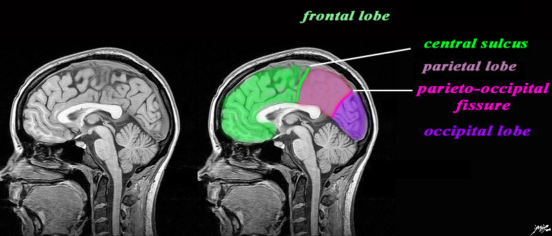
Central Sulcus in Sagittal MRI


The sagittal image is deeper in the brain near the midline through the interhemispheric fissure, and is intended to demonstrate the the parieto-occipital fissure (pink) in order to define the border between the parietall and occipital lobe. The central sulcus (pink) has also been inferred in to demonstrate the border between the frontal lobe and parietal lobe. It is not usually
Image Courtesy Philips Medical Systems rendered by Davidoff art 92141c01label.82s


    [nodeType] => 1
    [parentNode] => (object value omitted)
    [childNodes] => (object value omitted)
    [firstChild] => (object value omitted)
    [lastChild] => (object value omitted)
    [previousSibling] => (object value omitted)
    [nextSibling] => (object value omitted)
    [attributes] => (object value omitted)
    [ownerDocument] => (object value omitted)
    [namespaceURI] =>
    [prefix] =>
    [localName] => table
    [baseURI] =>
    [textContent] =>


Central Sulcus in Sagittal MRI


The sagittal image is deeper in the brain near the midline through the interhemispheric fissure, and is intended to demonstrate the the parieto-occipital fissure (pink) in order to define the border between the parietall and occipital lobe. The central sulcus (pink) has also been inferred in to demonstrate the border between the frontal lobe and parietal lobe. It is not usually
Image Courtesy Philips Medical Systems rendered by Davidoff art 92141c01label.82s


)
DOMElement Object
(
    [schemaTypeInfo] =>
    [tagName] => td
    [firstElementChild] => (object value omitted)
    [lastElementChild] => (object value omitted)
    [childElementCount] => 2
    [previousElementSibling] =>
    [nextElementSibling] =>
    [nodeName] => td
    [nodeValue] =>
The sagittal image is deeper in the brain near the midline through the interhemispheric fissure, and is intended to demonstrate the the parieto-occipital fissure (pink) in order to define the border between the parietall and occipital lobe. The central sulcus (pink) has also been inferred in to demonstrate the border between the frontal lobe and parietal lobe. It is not usually
Image Courtesy Philips Medical Systems rendered by Davidoff art 92141c01label.82s

    [nodeType] => 1
    [parentNode] => (object value omitted)
    [childNodes] => (object value omitted)
    [firstChild] => (object value omitted)
    [lastChild] => (object value omitted)
    [previousSibling] => (object value omitted)
    [nextSibling] => (object value omitted)
    [attributes] => (object value omitted)
    [ownerDocument] => (object value omitted)
    [namespaceURI] =>
    [prefix] =>
    [localName] => td
    [baseURI] =>
    [textContent] =>
The sagittal image is deeper in the brain near the midline through the interhemispheric fissure, and is intended to demonstrate the the parieto-occipital fissure (pink) in order to define the border between the parietall and occipital lobe. The central sulcus (pink) has also been inferred in to demonstrate the border between the frontal lobe and parietal lobe. It is not usually
Image Courtesy Philips Medical Systems rendered by Davidoff art 92141c01label.82s

)
DOMElement Object
(
    [schemaTypeInfo] =>
    [tagName] => td
    [firstElementChild] => (object value omitted)
    [lastElementChild] => (object value omitted)
    [childElementCount] => 2
    [previousElementSibling] =>
    [nextElementSibling] =>
    [nodeName] => td
    [nodeValue] =>

Central Sulcus in Sagittal MRI
    [nodeType] => 1
    [parentNode] => (object value omitted)
    [childNodes] => (object value omitted)
    [firstChild] => (object value omitted)
    [lastChild] => (object value omitted)
    [previousSibling] => (object value omitted)
    [nextSibling] => (object value omitted)
    [attributes] => (object value omitted)
    [ownerDocument] => (object value omitted)
    [namespaceURI] =>
    [prefix] =>
    [localName] => td
    [baseURI] =>
    [textContent] =>

Central Sulcus in Sagittal MRI
)
DOMElement Object
(
    [schemaTypeInfo] =>
    [tagName] => table
    [firstElementChild] => (object value omitted)
    [lastElementChild] => (object value omitted)
    [childElementCount] => 1
    [previousElementSibling] => (object value omitted)
    [nextElementSibling] => (object value omitted)
    [nodeName] => table
    [nodeValue] =>


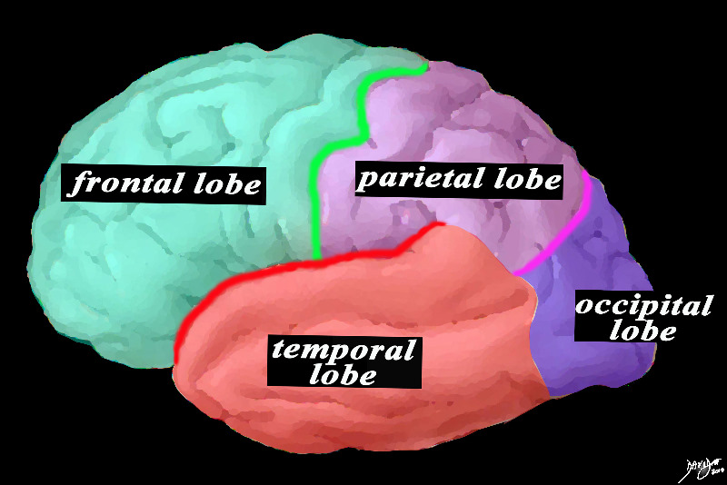
The Central Sulcus (Bright Green)


This is a diagram looking at the forebrain from the side showing the anteriorly placed frontal lobe, separated from the inferiorly placed temporal lobe by the Sylvian fissure (red), and separated from the parietal lobe by the central sulcus (green). The parietal lobe is also separated from the temporal lobe by the Sylvian fissure and from the occipital lobe by the parieto-occipital fissure (pink).
Courtesy Ashley DAvidoff MD copyright 2010 83029d13b01.8s


    [nodeType] => 1
    [parentNode] => (object value omitted)
    [childNodes] => (object value omitted)
    [firstChild] => (object value omitted)
    [lastChild] => (object value omitted)
    [previousSibling] => (object value omitted)
    [nextSibling] => (object value omitted)
    [attributes] => (object value omitted)
    [ownerDocument] => (object value omitted)
    [namespaceURI] =>
    [prefix] =>
    [localName] => table
    [baseURI] =>
    [textContent] =>


The Central Sulcus (Bright Green)


This is a diagram looking at the forebrain from the side showing the anteriorly placed frontal lobe, separated from the inferiorly placed temporal lobe by the Sylvian fissure (red), and separated from the parietal lobe by the central sulcus (green). The parietal lobe is also separated from the temporal lobe by the Sylvian fissure and from the occipital lobe by the parieto-occipital fissure (pink).
Courtesy Ashley DAvidoff MD copyright 2010 83029d13b01.8s


)
DOMElement Object
(
    [schemaTypeInfo] =>
    [tagName] => td
    [firstElementChild] => (object value omitted)
    [lastElementChild] => (object value omitted)
    [childElementCount] => 2
    [previousElementSibling] =>
    [nextElementSibling] =>
    [nodeName] => td
    [nodeValue] =>
This is a diagram looking at the forebrain from the side showing the anteriorly placed frontal lobe, separated from the inferiorly placed temporal lobe by the Sylvian fissure (red), and separated from the parietal lobe by the central sulcus (green). The parietal lobe is also separated from the temporal lobe by the Sylvian fissure and from the occipital lobe by the parieto-occipital fissure (pink).
Courtesy Ashley DAvidoff MD copyright 2010 83029d13b01.8s

    [nodeType] => 1
    [parentNode] => (object value omitted)
    [childNodes] => (object value omitted)
    [firstChild] => (object value omitted)
    [lastChild] => (object value omitted)
    [previousSibling] => (object value omitted)
    [nextSibling] => (object value omitted)
    [attributes] => (object value omitted)
    [ownerDocument] => (object value omitted)
    [namespaceURI] =>
    [prefix] =>
    [localName] => td
    [baseURI] =>
    [textContent] =>
This is a diagram looking at the forebrain from the side showing the anteriorly placed frontal lobe, separated from the inferiorly placed temporal lobe by the Sylvian fissure (red), and separated from the parietal lobe by the central sulcus (green). The parietal lobe is also separated from the temporal lobe by the Sylvian fissure and from the occipital lobe by the parieto-occipital fissure (pink).
Courtesy Ashley DAvidoff MD copyright 2010 83029d13b01.8s

)
DOMElement Object
(
    [schemaTypeInfo] =>
    [tagName] => td
    [firstElementChild] => (object value omitted)
    [lastElementChild] => (object value omitted)
    [childElementCount] => 2
    [previousElementSibling] =>
    [nextElementSibling] =>
    [nodeName] => td
    [nodeValue] =>
The Central Sulcus (Bright Green)
    [nodeType] => 1
    [parentNode] => (object value omitted)
    [childNodes] => (object value omitted)
    [firstChild] => (object value omitted)
    [lastChild] => (object value omitted)
    [previousSibling] => (object value omitted)
    [nextSibling] => (object value omitted)
    [attributes] => (object value omitted)
    [ownerDocument] => (object value omitted)
    [namespaceURI] =>
    [prefix] =>
    [localName] => td
    [baseURI] =>
    [textContent] =>
The Central Sulcus (Bright Green)
)
DOMElement Object
(
    [schemaTypeInfo] =>
    [tagName] => table
    [firstElementChild] => (object value omitted)
    [lastElementChild] => (object value omitted)
    [childElementCount] => 1
    [previousElementSibling] => (object value omitted)
    [nextElementSibling] => (object value omitted)
    [nodeName] => table
    [nodeValue] =>


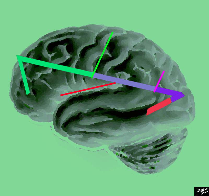
Central Sulcus in Bright Green


This artistic rendition of the brain reflects the vectors of the major parts of the brain revealing the major border forming fissures and sulci. The central sulcus (bright green line) divides the frontal lobe from the parietal lobe (light mauve). The region of the parieto-occipital fissure (pink line) divides the parietal lobe from the occipital lobe (purple). and the Sylvian fissure (thin red line)divides the temporal lobe from the frontal and parietal lobe
Courtesy Ashley Davidoff copyright 2010 all rights reserved 83029e04.87s


    [nodeType] => 1
    [parentNode] => (object value omitted)
    [childNodes] => (object value omitted)
    [firstChild] => (object value omitted)
    [lastChild] => (object value omitted)
    [previousSibling] => (object value omitted)
    [nextSibling] => (object value omitted)
    [attributes] => (object value omitted)
    [ownerDocument] => (object value omitted)
    [namespaceURI] =>
    [prefix] =>
    [localName] => table
    [baseURI] =>
    [textContent] =>


Central Sulcus in Bright Green


This artistic rendition of the brain reflects the vectors of the major parts of the brain revealing the major border forming fissures and sulci. The central sulcus (bright green line) divides the frontal lobe from the parietal lobe (light mauve). The region of the parieto-occipital fissure (pink line) divides the parietal lobe from the occipital lobe (purple). and the Sylvian fissure (thin red line)divides the temporal lobe from the frontal and parietal lobe
Courtesy Ashley Davidoff copyright 2010 all rights reserved 83029e04.87s


)
DOMElement Object
(
    [schemaTypeInfo] =>
    [tagName] => td
    [firstElementChild] => (object value omitted)
    [lastElementChild] => (object value omitted)
    [childElementCount] => 2
    [previousElementSibling] =>
    [nextElementSibling] =>
    [nodeName] => td
    [nodeValue] =>
This artistic rendition of the brain reflects the vectors of the major parts of the brain revealing the major border forming fissures and sulci. The central sulcus (bright green line) divides the frontal lobe from the parietal lobe (light mauve). The region of the parieto-occipital fissure (pink line) divides the parietal lobe from the occipital lobe (purple). and the Sylvian fissure (thin red line)divides the temporal lobe from the frontal and parietal lobe
Courtesy Ashley Davidoff copyright 2010 all rights reserved 83029e04.87s

    [nodeType] => 1
    [parentNode] => (object value omitted)
    [childNodes] => (object value omitted)
    [firstChild] => (object value omitted)
    [lastChild] => (object value omitted)
    [previousSibling] => (object value omitted)
    [nextSibling] => (object value omitted)
    [attributes] => (object value omitted)
    [ownerDocument] => (object value omitted)
    [namespaceURI] =>
    [prefix] =>
    [localName] => td
    [baseURI] =>
    [textContent] =>
This artistic rendition of the brain reflects the vectors of the major parts of the brain revealing the major border forming fissures and sulci. The central sulcus (bright green line) divides the frontal lobe from the parietal lobe (light mauve). The region of the parieto-occipital fissure (pink line) divides the parietal lobe from the occipital lobe (purple). and the Sylvian fissure (thin red line)divides the temporal lobe from the frontal and parietal lobe
Courtesy Ashley Davidoff copyright 2010 all rights reserved 83029e04.87s

)
DOMElement Object
(
    [schemaTypeInfo] =>
    [tagName] => td
    [firstElementChild] => (object value omitted)
    [lastElementChild] => (object value omitted)
    [childElementCount] => 2
    [previousElementSibling] =>
    [nextElementSibling] =>
    [nodeName] => td
    [nodeValue] =>
Central Sulcus in Bright Green
    [nodeType] => 1
    [parentNode] => (object value omitted)
    [childNodes] => (object value omitted)
    [firstChild] => (object value omitted)
    [lastChild] => (object value omitted)
    [previousSibling] => (object value omitted)
    [nextSibling] => (object value omitted)
    [attributes] => (object value omitted)
    [ownerDocument] => (object value omitted)
    [namespaceURI] =>
    [prefix] =>
    [localName] => td
    [baseURI] =>
    [textContent] =>
Central Sulcus in Bright Green
)

