Low Grade Astrocytoma
Author
The Common Vein Copyright 2010
Definition
DOMElement Object
(
    [schemaTypeInfo] =>
    [tagName] => table
    [firstElementChild] => (object value omitted)
    [lastElementChild] => (object value omitted)
    [childElementCount] => 1
    [previousElementSibling] => (object value omitted)
    [nextElementSibling] =>
    [nodeName] => table
    [nodeValue] =>


Low Grade Pericytoma in the Left Thalamus


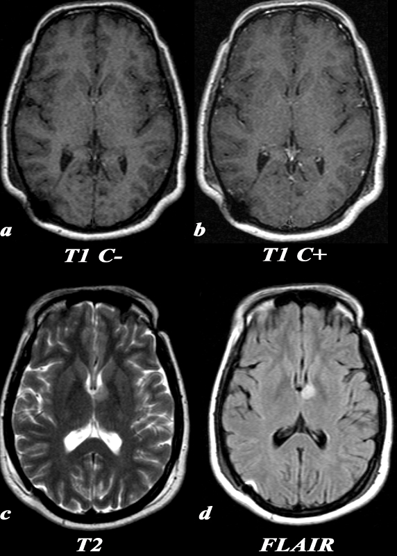
A 31 year old female presented with transient episodes of numbness and tingling in her right face, arm and leg. MRI: T1 C- (a) No definite signal abnormality is identified in the left thalamic low grade glioma. T1 post (T1 C+ b) : Post contrast images demonstrate no enhancement in the region of the known mass. This is typical of a low grade glioma. T2 (c): This T2 weighted image shows a unilateral well circumscribed area of increased signal in the anterior left thalamus. There is no significant mass effect on the adjacent structures. FLAIR (d): This FLAIR image from her MRI demonstrates (with slightly better clarity than the T2 weighted image) a unilateral well circumscribed area of increased signal in the anterior left thalamus. There is no significant mass effect on the adjacent structures.
Image Courtesy Elisa Flower MD and Asim Mian MD 97664c.8


    [nodeType] => 1
    [parentNode] => (object value omitted)
    [childNodes] => (object value omitted)
    [firstChild] => (object value omitted)
    [lastChild] => (object value omitted)
    [previousSibling] => (object value omitted)
    [nextSibling] => (object value omitted)
    [attributes] => (object value omitted)
    [ownerDocument] => (object value omitted)
    [namespaceURI] =>
    [prefix] =>
    [localName] => table
    [baseURI] =>
    [textContent] =>


Low Grade Pericytoma in the Left Thalamus


A 31 year old female presented with transient episodes of numbness and tingling in her right face, arm and leg. MRI: T1 C- (a) No definite signal abnormality is identified in the left thalamic low grade glioma. T1 post (T1 C+ b) : Post contrast images demonstrate no enhancement in the region of the known mass. This is typical of a low grade glioma. T2 (c): This T2 weighted image shows a unilateral well circumscribed area of increased signal in the anterior left thalamus. There is no significant mass effect on the adjacent structures. FLAIR (d): This FLAIR image from her MRI demonstrates (with slightly better clarity than the T2 weighted image) a unilateral well circumscribed area of increased signal in the anterior left thalamus. There is no significant mass effect on the adjacent structures.
Image Courtesy Elisa Flower MD and Asim Mian MD 97664c.8


)
DOMElement Object
(
    [schemaTypeInfo] =>
    [tagName] => td
    [firstElementChild] => (object value omitted)
    [lastElementChild] => (object value omitted)
    [childElementCount] => 2
    [previousElementSibling] =>
    [nextElementSibling] =>
    [nodeName] => td
    [nodeValue] =>
A 31 year old female presented with transient episodes of numbness and tingling in her right face, arm and leg. MRI: T1 C- (a) No definite signal abnormality is identified in the left thalamic low grade glioma. T1 post (T1 C+ b) : Post contrast images demonstrate no enhancement in the region of the known mass. This is typical of a low grade glioma. T2 (c): This T2 weighted image shows a unilateral well circumscribed area of increased signal in the anterior left thalamus. There is no significant mass effect on the adjacent structures. FLAIR (d): This FLAIR image from her MRI demonstrates (with slightly better clarity than the T2 weighted image) a unilateral well circumscribed area of increased signal in the anterior left thalamus. There is no significant mass effect on the adjacent structures.
Image Courtesy Elisa Flower MD and Asim Mian MD 97664c.8

    [nodeType] => 1
    [parentNode] => (object value omitted)
    [childNodes] => (object value omitted)
    [firstChild] => (object value omitted)
    [lastChild] => (object value omitted)
    [previousSibling] => (object value omitted)
    [nextSibling] => (object value omitted)
    [attributes] => (object value omitted)
    [ownerDocument] => (object value omitted)
    [namespaceURI] =>
    [prefix] =>
    [localName] => td
    [baseURI] =>
    [textContent] =>
A 31 year old female presented with transient episodes of numbness and tingling in her right face, arm and leg. MRI: T1 C- (a) No definite signal abnormality is identified in the left thalamic low grade glioma. T1 post (T1 C+ b) : Post contrast images demonstrate no enhancement in the region of the known mass. This is typical of a low grade glioma. T2 (c): This T2 weighted image shows a unilateral well circumscribed area of increased signal in the anterior left thalamus. There is no significant mass effect on the adjacent structures. FLAIR (d): This FLAIR image from her MRI demonstrates (with slightly better clarity than the T2 weighted image) a unilateral well circumscribed area of increased signal in the anterior left thalamus. There is no significant mass effect on the adjacent structures.
Image Courtesy Elisa Flower MD and Asim Mian MD 97664c.8

)
DOMElement Object
(
    [schemaTypeInfo] =>
    [tagName] => td
    [firstElementChild] => (object value omitted)
    [lastElementChild] => (object value omitted)
    [childElementCount] => 2
    [previousElementSibling] =>
    [nextElementSibling] =>
    [nodeName] => td
    [nodeValue] =>
Low Grade Pericytoma in the Left Thalamus
    [nodeType] => 1
    [parentNode] => (object value omitted)
    [childNodes] => (object value omitted)
    [firstChild] => (object value omitted)
    [lastChild] => (object value omitted)
    [previousSibling] => (object value omitted)
    [nextSibling] => (object value omitted)
    [attributes] => (object value omitted)
    [ownerDocument] => (object value omitted)
    [namespaceURI] =>
    [prefix] =>
    [localName] => td
    [baseURI] =>
    [textContent] =>
Low Grade Pericytoma in the Left Thalamus
)

