Acute Hypertensive Encephalopathy
Ashley Davidoff MD
The Common vein Copyright 2010
Definition
Hypertensive encephalopathy is a reversible circulatory disorder caused by sudden and sustained elevation of systolic blood pressure (above 130 mm Hg) associated with diseases such as eclampsia, acute nephritis and resulting in a neurological disorder.
Structural changes are usually manifest by changes in the occipital lobe and symptoms include headache, restlessness, nausea, disturbances of consciousness and seizures. Signs on clinical examination include retinal hemorrhage and papilledema.
Imaging by MRI or CT usually show change in the occipital lobes.
The condition is treated by antihypertensives.
 
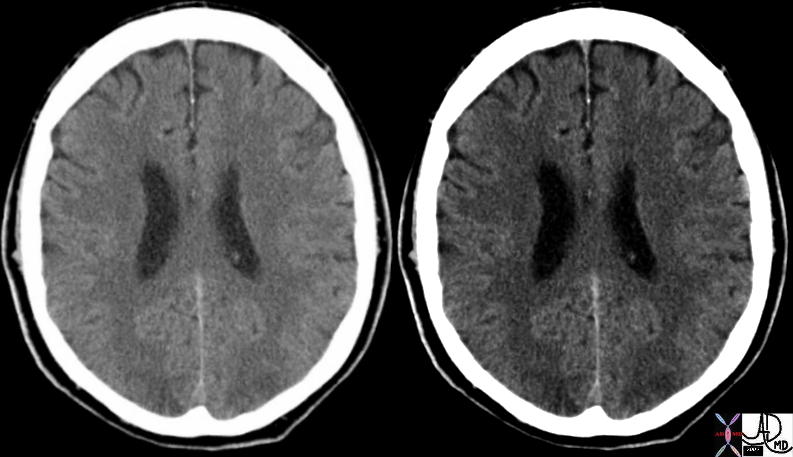
Subtle Occipital Lobe Loss of Gray White Differentiation |
|
This hypertensive patient presents with visual disturbance and the axial CT scan shows subtle changes in the occipital lobe characterised by vague low density regions. There is loss of gray-white matter differentiation consistent with acute ischemia. These findings are consistent with acute hypertensive encephalopathy
Courtesyt Ashley Davidoff MD 73422
|

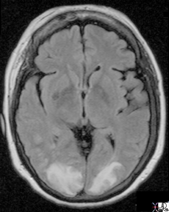
FLAIR Hyperintensity in the Occipital Lobes |
|
This hypertensive patient presents with visual disturbance and the MRI using a FLAIR sequence shows obvious FLAIR hyperintensity in the occipital lobes and extending into the parietal lobes. These findings are consistent with acute hypertensive encephalopathy and ischemia of the occipital lobes characteristic of acute hypertensive encephalopathy
Courtesy Ashley Davidoff MD Copyright 2010 All rights reserved 73422
|
DOMElement Object
(
    [schemaTypeInfo] =>
    [tagName] => table
    [firstElementChild] => (object value omitted)
    [lastElementChild] => (object value omitted)
    [childElementCount] => 1
    [previousElementSibling] => (object value omitted)
    [nextElementSibling] =>
    [nodeName] => table
    [nodeValue] =>


FLAIR Hyperintensity in the Occipital Lobes


This hypertensive patient presents with visual disturbance and the MRI using a FLAIR sequence shows obvious FLAIR hyperintensity in the occipital lobes and extending into the parietal lobes. These findings are consistent with acute hypertensive encephalopathy and ischemia of the occipital lobes characteristic of acute hypertensive encephalopathy
Courtesy Ashley Davidoff MD Copyright 2010 All rights reserved 73422


    [nodeType] => 1
    [parentNode] => (object value omitted)
    [childNodes] => (object value omitted)
    [firstChild] => (object value omitted)
    [lastChild] => (object value omitted)
    [previousSibling] => (object value omitted)
    [nextSibling] => (object value omitted)
    [attributes] => (object value omitted)
    [ownerDocument] => (object value omitted)
    [namespaceURI] =>
    [prefix] =>
    [localName] => table
    [baseURI] =>
    [textContent] =>


FLAIR Hyperintensity in the Occipital Lobes


This hypertensive patient presents with visual disturbance and the MRI using a FLAIR sequence shows obvious FLAIR hyperintensity in the occipital lobes and extending into the parietal lobes. These findings are consistent with acute hypertensive encephalopathy and ischemia of the occipital lobes characteristic of acute hypertensive encephalopathy
Courtesy Ashley Davidoff MD Copyright 2010 All rights reserved 73422


)
DOMElement Object
(
    [schemaTypeInfo] =>
    [tagName] => td
    [firstElementChild] => (object value omitted)
    [lastElementChild] => (object value omitted)
    [childElementCount] => 2
    [previousElementSibling] =>
    [nextElementSibling] =>
    [nodeName] => td
    [nodeValue] =>
This hypertensive patient presents with visual disturbance and the MRI using a FLAIR sequence shows obvious FLAIR hyperintensity in the occipital lobes and extending into the parietal lobes. These findings are consistent with acute hypertensive encephalopathy and ischemia of the occipital lobes characteristic of acute hypertensive encephalopathy
Courtesy Ashley Davidoff MD Copyright 2010 All rights reserved 73422

    [nodeType] => 1
    [parentNode] => (object value omitted)
    [childNodes] => (object value omitted)
    [firstChild] => (object value omitted)
    [lastChild] => (object value omitted)
    [previousSibling] => (object value omitted)
    [nextSibling] => (object value omitted)
    [attributes] => (object value omitted)
    [ownerDocument] => (object value omitted)
    [namespaceURI] =>
    [prefix] =>
    [localName] => td
    [baseURI] =>
    [textContent] =>
This hypertensive patient presents with visual disturbance and the MRI using a FLAIR sequence shows obvious FLAIR hyperintensity in the occipital lobes and extending into the parietal lobes. These findings are consistent with acute hypertensive encephalopathy and ischemia of the occipital lobes characteristic of acute hypertensive encephalopathy
Courtesy Ashley Davidoff MD Copyright 2010 All rights reserved 73422

)
DOMElement Object
(
    [schemaTypeInfo] =>
    [tagName] => td
    [firstElementChild] => (object value omitted)
    [lastElementChild] => (object value omitted)
    [childElementCount] => 2
    [previousElementSibling] =>
    [nextElementSibling] =>
    [nodeName] => td
    [nodeValue] =>
FLAIR Hyperintensity in the Occipital Lobes
    [nodeType] => 1
    [parentNode] => (object value omitted)
    [childNodes] => (object value omitted)
    [firstChild] => (object value omitted)
    [lastChild] => (object value omitted)
    [previousSibling] => (object value omitted)
    [nextSibling] => (object value omitted)
    [attributes] => (object value omitted)
    [ownerDocument] => (object value omitted)
    [namespaceURI] =>
    [prefix] =>
    [localName] => td
    [baseURI] =>
    [textContent] =>
FLAIR Hyperintensity in the Occipital Lobes
)
DOMElement Object
(
    [schemaTypeInfo] =>
    [tagName] => table
    [firstElementChild] => (object value omitted)
    [lastElementChild] => (object value omitted)
    [childElementCount] => 1
    [previousElementSibling] => (object value omitted)
    [nextElementSibling] => (object value omitted)
    [nodeName] => table
    [nodeValue] =>


Subtle Occipital Lobe Loss of Gray White Differentiation


This hypertensive patient presents with visual disturbance and the axial CT scan shows subtle changes in the occipital lobe characterised by vague low density regions. There is loss of gray-white matter differentiation consistent with acute ischemia. These findings are consistent with acute hypertensive encephalopathy
Courtesyt Ashley Davidoff MD 73422


    [nodeType] => 1
    [parentNode] => (object value omitted)
    [childNodes] => (object value omitted)
    [firstChild] => (object value omitted)
    [lastChild] => (object value omitted)
    [previousSibling] => (object value omitted)
    [nextSibling] => (object value omitted)
    [attributes] => (object value omitted)
    [ownerDocument] => (object value omitted)
    [namespaceURI] =>
    [prefix] =>
    [localName] => table
    [baseURI] =>
    [textContent] =>


Subtle Occipital Lobe Loss of Gray White Differentiation


This hypertensive patient presents with visual disturbance and the axial CT scan shows subtle changes in the occipital lobe characterised by vague low density regions. There is loss of gray-white matter differentiation consistent with acute ischemia. These findings are consistent with acute hypertensive encephalopathy
Courtesyt Ashley Davidoff MD 73422


)
DOMElement Object
(
    [schemaTypeInfo] =>
    [tagName] => td
    [firstElementChild] => (object value omitted)
    [lastElementChild] => (object value omitted)
    [childElementCount] => 2
    [previousElementSibling] =>
    [nextElementSibling] =>
    [nodeName] => td
    [nodeValue] =>
This hypertensive patient presents with visual disturbance and the axial CT scan shows subtle changes in the occipital lobe characterised by vague low density regions. There is loss of gray-white matter differentiation consistent with acute ischemia. These findings are consistent with acute hypertensive encephalopathy
Courtesyt Ashley Davidoff MD 73422

    [nodeType] => 1
    [parentNode] => (object value omitted)
    [childNodes] => (object value omitted)
    [firstChild] => (object value omitted)
    [lastChild] => (object value omitted)
    [previousSibling] => (object value omitted)
    [nextSibling] => (object value omitted)
    [attributes] => (object value omitted)
    [ownerDocument] => (object value omitted)
    [namespaceURI] =>
    [prefix] =>
    [localName] => td
    [baseURI] =>
    [textContent] =>
This hypertensive patient presents with visual disturbance and the axial CT scan shows subtle changes in the occipital lobe characterised by vague low density regions. There is loss of gray-white matter differentiation consistent with acute ischemia. These findings are consistent with acute hypertensive encephalopathy
Courtesyt Ashley Davidoff MD 73422

)
DOMElement Object
(
    [schemaTypeInfo] =>
    [tagName] => td
    [firstElementChild] => (object value omitted)
    [lastElementChild] => (object value omitted)
    [childElementCount] => 2
    [previousElementSibling] =>
    [nextElementSibling] =>
    [nodeName] => td
    [nodeValue] =>
Subtle Occipital Lobe Loss of Gray White Differentiation
    [nodeType] => 1
    [parentNode] => (object value omitted)
    [childNodes] => (object value omitted)
    [firstChild] => (object value omitted)
    [lastChild] => (object value omitted)
    [previousSibling] => (object value omitted)
    [nextSibling] => (object value omitted)
    [attributes] => (object value omitted)
    [ownerDocument] => (object value omitted)
    [namespaceURI] =>
    [prefix] =>
    [localName] => td
    [baseURI] =>
    [textContent] =>
Subtle Occipital Lobe Loss of Gray White Differentiation
)
https://beta.thecommonvein.net/wp-content/uploads/2023/06/73422.jpg
http://www.thecommonvein.net/media/73467c01.jpg


