Neurons
Sumit Karia MD
The Common Vein Copyright 2010
Definition
Neurons are the core components of the nervous system and are found in the brain spinal cord and nerves. It typically consists of a cell body, axon and dendrites. There may be many dendrites but only one axon. They are electrically excitable. In general the neurons of the brain cannot undergo cell division.
Although there are different types of neurons and variations from one region of the CNS to another, neurons have common features.
They are cells with a large cell body which contains a nucleus with a prominent nucleolus and dispersed chromatin.
The peri-nuclear cytoplasm is characterized by the presence of a large endoplasmic reticulum in clusters, making the Nissl bodies. The cytoplasm also contains numerous mitochondria and a cytoskeleton, composed of intermediate filaments and microfilaments.
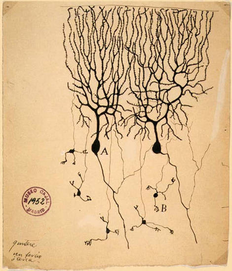
The unique feature of neurons is represented by their cell processes, initially described in the early twentieth century by two pioneers in neurohistology (Golgi and Cajal). Neurons have two types of processes: dendrites and axons. Dendrites are usually short, multiple and highly branched, while the axon is a single extension, often with multiple endings. The axon is surrounded by a sheath ? myelin (composed of lipids) – created by a type of glial cells, the oligodendrocytes.
Neurons are separated by highly specialized intercellular junctions: the synapses, which allow communications between neurons or effector cells.

Neuron |
| This image is a drawing by Santiago Ramón y Cajal depicting Purkinje cells (A) and granule cells (B) from pigeon cerebellum (1899); Instituto Santiago Ramón y Cajal, Madrid, Spain
The copyright has expired and applies to the United States, Australia, the European Union and those countries with a copyright term of life of the author plus 70 years (Wapedia) 54956
|
The human brain contains roughly 84.6 billion glia and 86.1 billion neurons. (Azevedo)
The glia/neuron ratio in the
cerebral cortex gray matter is 1.48
basal ganglia, thalamic structures, and brainstem combined is 11.35.
cerebellum is only 0.23

The cerebral Cortex Exemplifying the Neuron
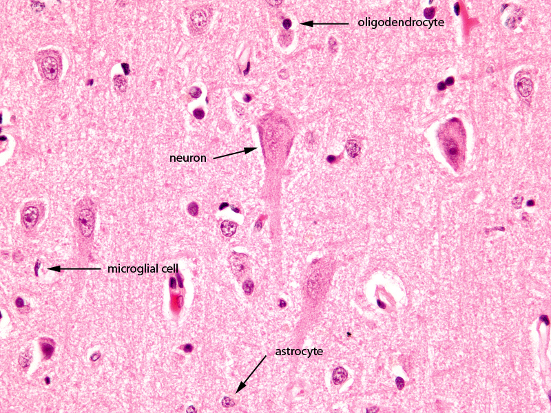
Magnification 40X |
|
The image shows a histological section of the brain at 40X magnification. The section shows the 3 of the common basic types of brain cells; the largest is the neuronal cell which is only found in gray matter. It is exemplified in this image. They are 10 times larger than the other cells but less common than the second type of cell called the glial cell. The astrocyte is the second largest cell and often lies in close association with the blood vessels. The third type of cell seen in this section, also a glial cell is the oligodendrocyte. It is smaller than the other type of glial cell the astrocyte has a darker nucleus and it is responsible to myelinate the white matter. It is in fact the predominant cell of white matter.
Image Courtesy of Thomas W.Smith, MD; Department of Pathology, University of Massachusetts Medical School. 97802
|

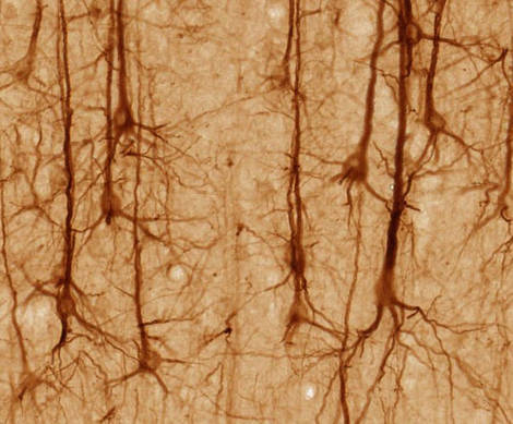
Pyramidal Neurons |
|
Pyramidal neurons stained SMI32 showing the centrl body axon and dendrites
54957 SMI32-stained pyramidal neurons in cerebral cortex from http://brainmaps.org. Please note: UC Regents Davis campus maintains copyright of BrainMaps.org and licenses content under a form of a non-commercial license as per http://brainmaps.org/index.php?p=termsofuse. They have, however, released this and other select screenshots under the Creative Commons Attribution 2.5 License, as per http://brainmaps.org/index.php?p=termsofuse and http://brainmaps.org/index.php?p=screenshots. Wapedia
|
References
Azevedo FA, Carvalho LR, Grinberg LT, Farfel JM, Ferretti RE, Leite RE, Jacob Filho W, Lent R, Herculano-Houzel S. (2009). Equal numbers of neuronal and nonneuronal cells make the human brain an isometrically scaled-up primate brain. J Comp Neurol. 513(5):532-41. PubMed
Pelvig DP, Pakkenberg H, Stark AK, Pakkenberg B. (2008). Neocortical glial cell numbers in human brains. Neurobiol Aging. 29(11):1754-62.(figures given are those for females) PubMed
DOMElement Object
(
    [schemaTypeInfo] =>
    [tagName] => table
    [firstElementChild] => (object value omitted)
    [lastElementChild] => (object value omitted)
    [childElementCount] => 1
    [previousElementSibling] => (object value omitted)
    [nextElementSibling] => (object value omitted)
    [nodeName] => table
    [nodeValue] =>


Pyramidal Neurons


Pyramidal neurons stained SMI32 showing the centrl body axon and dendrites
54957 SMI32-stained pyramidal neurons in cerebral cortex from http://brainmaps.org. Please note: UC Regents Davis campus maintains copyright of BrainMaps.org and licenses content under a form of a non-commercial license as per http://brainmaps.org/index.php?p=termsofuse. They have, however, released this and other select screenshots under the Creative Commons Attribution 2.5 License, as per http://brainmaps.org/index.php?p=termsofuse and http://brainmaps.org/index.php?p=screenshots. Wapedia


    [nodeType] => 1
    [parentNode] => (object value omitted)
    [childNodes] => (object value omitted)
    [firstChild] => (object value omitted)
    [lastChild] => (object value omitted)
    [previousSibling] => (object value omitted)
    [nextSibling] => (object value omitted)
    [attributes] => (object value omitted)
    [ownerDocument] => (object value omitted)
    [namespaceURI] =>
    [prefix] =>
    [localName] => table
    [baseURI] =>
    [textContent] =>


Pyramidal Neurons


Pyramidal neurons stained SMI32 showing the centrl body axon and dendrites
54957 SMI32-stained pyramidal neurons in cerebral cortex from http://brainmaps.org. Please note: UC Regents Davis campus maintains copyright of BrainMaps.org and licenses content under a form of a non-commercial license as per http://brainmaps.org/index.php?p=termsofuse. They have, however, released this and other select screenshots under the Creative Commons Attribution 2.5 License, as per http://brainmaps.org/index.php?p=termsofuse and http://brainmaps.org/index.php?p=screenshots. Wapedia


)
DOMElement Object
(
    [schemaTypeInfo] =>
    [tagName] => td
    [firstElementChild] => (object value omitted)
    [lastElementChild] => (object value omitted)
    [childElementCount] => 2
    [previousElementSibling] =>
    [nextElementSibling] =>
    [nodeName] => td
    [nodeValue] =>
Pyramidal neurons stained SMI32 showing the centrl body axon and dendrites
54957 SMI32-stained pyramidal neurons in cerebral cortex from http://brainmaps.org. Please note: UC Regents Davis campus maintains copyright of BrainMaps.org and licenses content under a form of a non-commercial license as per http://brainmaps.org/index.php?p=termsofuse. They have, however, released this and other select screenshots under the Creative Commons Attribution 2.5 License, as per http://brainmaps.org/index.php?p=termsofuse and http://brainmaps.org/index.php?p=screenshots. Wapedia

    [nodeType] => 1
    [parentNode] => (object value omitted)
    [childNodes] => (object value omitted)
    [firstChild] => (object value omitted)
    [lastChild] => (object value omitted)
    [previousSibling] => (object value omitted)
    [nextSibling] => (object value omitted)
    [attributes] => (object value omitted)
    [ownerDocument] => (object value omitted)
    [namespaceURI] =>
    [prefix] =>
    [localName] => td
    [baseURI] =>
    [textContent] =>
Pyramidal neurons stained SMI32 showing the centrl body axon and dendrites
54957 SMI32-stained pyramidal neurons in cerebral cortex from http://brainmaps.org. Please note: UC Regents Davis campus maintains copyright of BrainMaps.org and licenses content under a form of a non-commercial license as per http://brainmaps.org/index.php?p=termsofuse. They have, however, released this and other select screenshots under the Creative Commons Attribution 2.5 License, as per http://brainmaps.org/index.php?p=termsofuse and http://brainmaps.org/index.php?p=screenshots. Wapedia

)
DOMElement Object
(
    [schemaTypeInfo] =>
    [tagName] => td
    [firstElementChild] => (object value omitted)
    [lastElementChild] => (object value omitted)
    [childElementCount] => 2
    [previousElementSibling] =>
    [nextElementSibling] =>
    [nodeName] => td
    [nodeValue] =>
Pyramidal Neurons
    [nodeType] => 1
    [parentNode] => (object value omitted)
    [childNodes] => (object value omitted)
    [firstChild] => (object value omitted)
    [lastChild] => (object value omitted)
    [previousSibling] => (object value omitted)
    [nextSibling] => (object value omitted)
    [attributes] => (object value omitted)
    [ownerDocument] => (object value omitted)
    [namespaceURI] =>
    [prefix] =>
    [localName] => td
    [baseURI] =>
    [textContent] =>
Pyramidal Neurons
)
DOMElement Object
(
    [schemaTypeInfo] =>
    [tagName] => table
    [firstElementChild] => (object value omitted)
    [lastElementChild] => (object value omitted)
    [childElementCount] => 1
    [previousElementSibling] => (object value omitted)
    [nextElementSibling] => (object value omitted)
    [nodeName] => table
    [nodeValue] =>


The cerebral Cortex Exemplifying the Neuron
Magnification 40X


The image shows a histological section of the brain at 40X magnification. The section shows the 3 of the common basic types of brain cells; the largest is the neuronal cell which is only found in gray matter. It is exemplified in this image. They are 10 times larger than the other cells but less common than the second type of cell called the glial cell. The astrocyte is the second largest cell and often lies in close association with the blood vessels. The third type of cell seen in this section, also a glial cell is the oligodendrocyte. It is smaller than the other type of glial cell the astrocyte has a darker nucleus and it is responsible to myelinate the white matter. It is in fact the predominant cell of white matter.
Image Courtesy of Thomas W.Smith, MD; Department of Pathology, University of Massachusetts Medical School. 97802


    [nodeType] => 1
    [parentNode] => (object value omitted)
    [childNodes] => (object value omitted)
    [firstChild] => (object value omitted)
    [lastChild] => (object value omitted)
    [previousSibling] => (object value omitted)
    [nextSibling] => (object value omitted)
    [attributes] => (object value omitted)
    [ownerDocument] => (object value omitted)
    [namespaceURI] =>
    [prefix] =>
    [localName] => table
    [baseURI] =>
    [textContent] =>


The cerebral Cortex Exemplifying the Neuron
Magnification 40X


The image shows a histological section of the brain at 40X magnification. The section shows the 3 of the common basic types of brain cells; the largest is the neuronal cell which is only found in gray matter. It is exemplified in this image. They are 10 times larger than the other cells but less common than the second type of cell called the glial cell. The astrocyte is the second largest cell and often lies in close association with the blood vessels. The third type of cell seen in this section, also a glial cell is the oligodendrocyte. It is smaller than the other type of glial cell the astrocyte has a darker nucleus and it is responsible to myelinate the white matter. It is in fact the predominant cell of white matter.
Image Courtesy of Thomas W.Smith, MD; Department of Pathology, University of Massachusetts Medical School. 97802


)
DOMElement Object
(
    [schemaTypeInfo] =>
    [tagName] => td
    [firstElementChild] => (object value omitted)
    [lastElementChild] => (object value omitted)
    [childElementCount] => 2
    [previousElementSibling] =>
    [nextElementSibling] =>
    [nodeName] => td
    [nodeValue] =>
The image shows a histological section of the brain at 40X magnification. The section shows the 3 of the common basic types of brain cells; the largest is the neuronal cell which is only found in gray matter. It is exemplified in this image. They are 10 times larger than the other cells but less common than the second type of cell called the glial cell. The astrocyte is the second largest cell and often lies in close association with the blood vessels. The third type of cell seen in this section, also a glial cell is the oligodendrocyte. It is smaller than the other type of glial cell the astrocyte has a darker nucleus and it is responsible to myelinate the white matter. It is in fact the predominant cell of white matter.
Image Courtesy of Thomas W.Smith, MD; Department of Pathology, University of Massachusetts Medical School. 97802

    [nodeType] => 1
    [parentNode] => (object value omitted)
    [childNodes] => (object value omitted)
    [firstChild] => (object value omitted)
    [lastChild] => (object value omitted)
    [previousSibling] => (object value omitted)
    [nextSibling] => (object value omitted)
    [attributes] => (object value omitted)
    [ownerDocument] => (object value omitted)
    [namespaceURI] =>
    [prefix] =>
    [localName] => td
    [baseURI] =>
    [textContent] =>
The image shows a histological section of the brain at 40X magnification. The section shows the 3 of the common basic types of brain cells; the largest is the neuronal cell which is only found in gray matter. It is exemplified in this image. They are 10 times larger than the other cells but less common than the second type of cell called the glial cell. The astrocyte is the second largest cell and often lies in close association with the blood vessels. The third type of cell seen in this section, also a glial cell is the oligodendrocyte. It is smaller than the other type of glial cell the astrocyte has a darker nucleus and it is responsible to myelinate the white matter. It is in fact the predominant cell of white matter.
Image Courtesy of Thomas W.Smith, MD; Department of Pathology, University of Massachusetts Medical School. 97802

)
DOMElement Object
(
    [schemaTypeInfo] =>
    [tagName] => td
    [firstElementChild] => (object value omitted)
    [lastElementChild] => (object value omitted)
    [childElementCount] => 3
    [previousElementSibling] =>
    [nextElementSibling] =>
    [nodeName] => td
    [nodeValue] =>
The cerebral Cortex Exemplifying the Neuron
Magnification 40X
    [nodeType] => 1
    [parentNode] => (object value omitted)
    [childNodes] => (object value omitted)
    [firstChild] => (object value omitted)
    [lastChild] => (object value omitted)
    [previousSibling] => (object value omitted)
    [nextSibling] => (object value omitted)
    [attributes] => (object value omitted)
    [ownerDocument] => (object value omitted)
    [namespaceURI] =>
    [prefix] =>
    [localName] => td
    [baseURI] =>
    [textContent] =>
The cerebral Cortex Exemplifying the Neuron
Magnification 40X
)
DOMElement Object
(
    [schemaTypeInfo] =>
    [tagName] => table
    [firstElementChild] => (object value omitted)
    [lastElementChild] => (object value omitted)
    [childElementCount] => 1
    [previousElementSibling] => (object value omitted)
    [nextElementSibling] => (object value omitted)
    [nodeName] => table
    [nodeValue] =>


Neuron


This image is a drawing by Santiago Ramón y Cajal depicting Purkinje cells (A) and granule cells (B) from pigeon cerebellum (1899); Instituto Santiago Ramón y Cajal, Madrid, Spain
The copyright has expired and applies to the United States, Australia, the European Union and those countries with a copyright term of life of the author plus 70 years (Wapedia) 54956


    [nodeType] => 1
    [parentNode] => (object value omitted)
    [childNodes] => (object value omitted)
    [firstChild] => (object value omitted)
    [lastChild] => (object value omitted)
    [previousSibling] => (object value omitted)
    [nextSibling] => (object value omitted)
    [attributes] => (object value omitted)
    [ownerDocument] => (object value omitted)
    [namespaceURI] =>
    [prefix] =>
    [localName] => table
    [baseURI] =>
    [textContent] =>


Neuron


This image is a drawing by Santiago Ramón y Cajal depicting Purkinje cells (A) and granule cells (B) from pigeon cerebellum (1899); Instituto Santiago Ramón y Cajal, Madrid, Spain
The copyright has expired and applies to the United States, Australia, the European Union and those countries with a copyright term of life of the author plus 70 years (Wapedia) 54956


)
DOMElement Object
(
    [schemaTypeInfo] =>
    [tagName] => td
    [firstElementChild] => (object value omitted)
    [lastElementChild] => (object value omitted)
    [childElementCount] => 1
    [previousElementSibling] =>
    [nextElementSibling] =>
    [nodeName] => td
    [nodeValue] => This image is a drawing by Santiago Ramón y Cajal depicting Purkinje cells (A) and granule cells (B) from pigeon cerebellum (1899); Instituto Santiago Ramón y Cajal, Madrid, Spain
The copyright has expired and applies to the United States, Australia, the European Union and those countries with a copyright term of life of the author plus 70 years (Wapedia) 54956

    [nodeType] => 1
    [parentNode] => (object value omitted)
    [childNodes] => (object value omitted)
    [firstChild] => (object value omitted)
    [lastChild] => (object value omitted)
    [previousSibling] => (object value omitted)
    [nextSibling] => (object value omitted)
    [attributes] => (object value omitted)
    [ownerDocument] => (object value omitted)
    [namespaceURI] =>
    [prefix] =>
    [localName] => td
    [baseURI] =>
    [textContent] => This image is a drawing by Santiago Ramón y Cajal depicting Purkinje cells (A) and granule cells (B) from pigeon cerebellum (1899); Instituto Santiago Ramón y Cajal, Madrid, Spain
The copyright has expired and applies to the United States, Australia, the European Union and those countries with a copyright term of life of the author plus 70 years (Wapedia) 54956

)
DOMElement Object
(
    [schemaTypeInfo] =>
    [tagName] => td
    [firstElementChild] => (object value omitted)
    [lastElementChild] => (object value omitted)
    [childElementCount] => 2
    [previousElementSibling] =>
    [nextElementSibling] =>
    [nodeName] => td
    [nodeValue] =>
Neuron
    [nodeType] => 1
    [parentNode] => (object value omitted)
    [childNodes] => (object value omitted)
    [firstChild] => (object value omitted)
    [lastChild] => (object value omitted)
    [previousSibling] => (object value omitted)
    [nextSibling] => (object value omitted)
    [attributes] => (object value omitted)
    [ownerDocument] => (object value omitted)
    [namespaceURI] =>
    [prefix] =>
    [localName] => td
    [baseURI] =>
    [textContent] =>
Neuron
)


