Concepts of the Forebrain
Ashley Davidoff MD
The Common Vein Copyright 2010
Introduction
This section is designed to frame the forebrain to enable basic concepts to start at ground level. It is easiest to conceive the forebrain in three views;- The sagittal, axial and coronal view.
Sagittal View
The Axial View
DOMElement Object
(
    [schemaTypeInfo] =>
    [tagName] => table
    [firstElementChild] => (object value omitted)
    [lastElementChild] => (object value omitted)
    [childElementCount] => 1
    [previousElementSibling] => (object value omitted)
    [nextElementSibling] =>
    [nodeName] => table
    [nodeValue] =>


MRI Coronal View


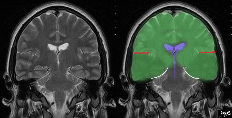
In this T2 weighted MRI image – the forebrain (green is seen centered aroubnd the ventriclar system. The red linees indicate the Sylvian fissures which in this instance in a relatively posterior cut reflect the parietal lobe superiorly and the temporal lobes inferiorly (darker green) The temporal lobes rest of the tentorium (white curvilinear convex lines). below the tenntorium is the midbrain and hind brain
Courtesy Ashley Davidoff MD copyright 2010 all rights reserved 89721c02b03.8s


    [nodeType] => 1
    [parentNode] => (object value omitted)
    [childNodes] => (object value omitted)
    [firstChild] => (object value omitted)
    [lastChild] => (object value omitted)
    [previousSibling] => (object value omitted)
    [nextSibling] => (object value omitted)
    [attributes] => (object value omitted)
    [ownerDocument] => (object value omitted)
    [namespaceURI] =>
    [prefix] =>
    [localName] => table
    [baseURI] =>
    [textContent] =>


MRI Coronal View


In this T2 weighted MRI image – the forebrain (green is seen centered aroubnd the ventriclar system. The red linees indicate the Sylvian fissures which in this instance in a relatively posterior cut reflect the parietal lobe superiorly and the temporal lobes inferiorly (darker green) The temporal lobes rest of the tentorium (white curvilinear convex lines). below the tenntorium is the midbrain and hind brain
Courtesy Ashley Davidoff MD copyright 2010 all rights reserved 89721c02b03.8s


)
DOMElement Object
(
    [schemaTypeInfo] =>
    [tagName] => td
    [firstElementChild] => (object value omitted)
    [lastElementChild] => (object value omitted)
    [childElementCount] => 2
    [previousElementSibling] =>
    [nextElementSibling] =>
    [nodeName] => td
    [nodeValue] =>
In this T2 weighted MRI image – the forebrain (green is seen centered aroubnd the ventriclar system. The red linees indicate the Sylvian fissures which in this instance in a relatively posterior cut reflect the parietal lobe superiorly and the temporal lobes inferiorly (darker green) The temporal lobes rest of the tentorium (white curvilinear convex lines). below the tenntorium is the midbrain and hind brain
Courtesy Ashley Davidoff MD copyright 2010 all rights reserved 89721c02b03.8s

    [nodeType] => 1
    [parentNode] => (object value omitted)
    [childNodes] => (object value omitted)
    [firstChild] => (object value omitted)
    [lastChild] => (object value omitted)
    [previousSibling] => (object value omitted)
    [nextSibling] => (object value omitted)
    [attributes] => (object value omitted)
    [ownerDocument] => (object value omitted)
    [namespaceURI] =>
    [prefix] =>
    [localName] => td
    [baseURI] =>
    [textContent] =>
In this T2 weighted MRI image – the forebrain (green is seen centered aroubnd the ventriclar system. The red linees indicate the Sylvian fissures which in this instance in a relatively posterior cut reflect the parietal lobe superiorly and the temporal lobes inferiorly (darker green) The temporal lobes rest of the tentorium (white curvilinear convex lines). below the tenntorium is the midbrain and hind brain
Courtesy Ashley Davidoff MD copyright 2010 all rights reserved 89721c02b03.8s

)
DOMElement Object
(
    [schemaTypeInfo] =>
    [tagName] => td
    [firstElementChild] => (object value omitted)
    [lastElementChild] => (object value omitted)
    [childElementCount] => 2
    [previousElementSibling] =>
    [nextElementSibling] =>
    [nodeName] => td
    [nodeValue] =>
MRI Coronal View
    [nodeType] => 1
    [parentNode] => (object value omitted)
    [childNodes] => (object value omitted)
    [firstChild] => (object value omitted)
    [lastChild] => (object value omitted)
    [previousSibling] => (object value omitted)
    [nextSibling] => (object value omitted)
    [attributes] => (object value omitted)
    [ownerDocument] => (object value omitted)
    [namespaceURI] =>
    [prefix] =>
    [localName] => td
    [baseURI] =>
    [textContent] =>
MRI Coronal View
)
DOMElement Object
(
    [schemaTypeInfo] =>
    [tagName] => table
    [firstElementChild] => (object value omitted)
    [lastElementChild] => (object value omitted)
    [childElementCount] => 1
    [previousElementSibling] => (object value omitted)
    [nextElementSibling] => (object value omitted)
    [nodeName] => table
    [nodeValue] =>


Forebrain – The Axial View


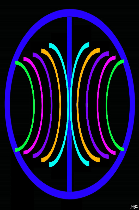
The anchoring concept of the brain in the axial plane is two cerebral hemispheres with a series of structures reminiscent of backward and downward facing “C’s” symettrically positioned around the center. The orange ring represents the other major portion of the forebrain.
Courtesy Ashley Davidoff MD copyright 2010 93914.3ka09.8sd01


    [nodeType] => 1
    [parentNode] => (object value omitted)
    [childNodes] => (object value omitted)
    [firstChild] => (object value omitted)
    [lastChild] => (object value omitted)
    [previousSibling] => (object value omitted)
    [nextSibling] => (object value omitted)
    [attributes] => (object value omitted)
    [ownerDocument] => (object value omitted)
    [namespaceURI] =>
    [prefix] =>
    [localName] => table
    [baseURI] =>
    [textContent] =>


Forebrain – The Axial View


The anchoring concept of the brain in the axial plane is two cerebral hemispheres with a series of structures reminiscent of backward and downward facing “C’s” symettrically positioned around the center. The orange ring represents the other major portion of the forebrain.
Courtesy Ashley Davidoff MD copyright 2010 93914.3ka09.8sd01


)
DOMElement Object
(
    [schemaTypeInfo] =>
    [tagName] => td
    [firstElementChild] => (object value omitted)
    [lastElementChild] => (object value omitted)
    [childElementCount] => 2
    [previousElementSibling] =>
    [nextElementSibling] =>
    [nodeName] => td
    [nodeValue] =>
The anchoring concept of the brain in the axial plane is two cerebral hemispheres with a series of structures reminiscent of backward and downward facing “C’s” symettrically positioned around the center. The orange ring represents the other major portion of the forebrain.
Courtesy Ashley Davidoff MD copyright 2010 93914.3ka09.8sd01

    [nodeType] => 1
    [parentNode] => (object value omitted)
    [childNodes] => (object value omitted)
    [firstChild] => (object value omitted)
    [lastChild] => (object value omitted)
    [previousSibling] => (object value omitted)
    [nextSibling] => (object value omitted)
    [attributes] => (object value omitted)
    [ownerDocument] => (object value omitted)
    [namespaceURI] =>
    [prefix] =>
    [localName] => td
    [baseURI] =>
    [textContent] =>
The anchoring concept of the brain in the axial plane is two cerebral hemispheres with a series of structures reminiscent of backward and downward facing “C’s” symettrically positioned around the center. The orange ring represents the other major portion of the forebrain.
Courtesy Ashley Davidoff MD copyright 2010 93914.3ka09.8sd01

)
DOMElement Object
(
    [schemaTypeInfo] =>
    [tagName] => td
    [firstElementChild] => (object value omitted)
    [lastElementChild] => (object value omitted)
    [childElementCount] => 2
    [previousElementSibling] =>
    [nextElementSibling] =>
    [nodeName] => td
    [nodeValue] =>
Forebrain – The Axial View
    [nodeType] => 1
    [parentNode] => (object value omitted)
    [childNodes] => (object value omitted)
    [firstChild] => (object value omitted)
    [lastChild] => (object value omitted)
    [previousSibling] => (object value omitted)
    [nextSibling] => (object value omitted)
    [attributes] => (object value omitted)
    [ownerDocument] => (object value omitted)
    [namespaceURI] =>
    [prefix] =>
    [localName] => td
    [baseURI] =>
    [textContent] =>
Forebrain – The Axial View
)
DOMElement Object
(
    [schemaTypeInfo] =>
    [tagName] => table
    [firstElementChild] => (object value omitted)
    [lastElementChild] => (object value omitted)
    [childElementCount] => 1
    [previousElementSibling] => (object value omitted)
    [nextElementSibling] => (object value omitted)
    [nodeName] => table
    [nodeValue] =>


The External View of the Brain


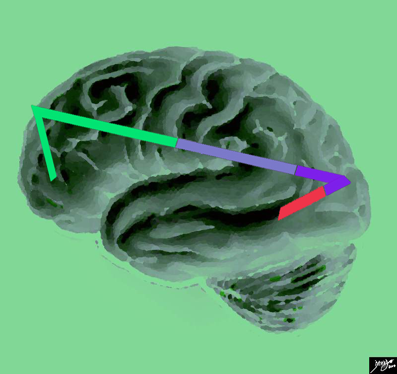
This artistic rendition of the brain reflects the vectors of the major parts of the brain with the stick diagram overlaid over a sagittal external view of the brain. In the stick diagram, the forebrain has been divided into the frontal lobe (bright green), parietal lobe (light mauve) occipital lobe (purple) and temporal lobe (red).
Courtesy Ashley Davidoff copyright 2010 all rights reserved 83029e04.86s


    [nodeType] => 1
    [parentNode] => (object value omitted)
    [childNodes] => (object value omitted)
    [firstChild] => (object value omitted)
    [lastChild] => (object value omitted)
    [previousSibling] => (object value omitted)
    [nextSibling] => (object value omitted)
    [attributes] => (object value omitted)
    [ownerDocument] => (object value omitted)
    [namespaceURI] =>
    [prefix] =>
    [localName] => table
    [baseURI] =>
    [textContent] =>


The External View of the Brain


This artistic rendition of the brain reflects the vectors of the major parts of the brain with the stick diagram overlaid over a sagittal external view of the brain. In the stick diagram, the forebrain has been divided into the frontal lobe (bright green), parietal lobe (light mauve) occipital lobe (purple) and temporal lobe (red).
Courtesy Ashley Davidoff copyright 2010 all rights reserved 83029e04.86s


)
DOMElement Object
(
    [schemaTypeInfo] =>
    [tagName] => td
    [firstElementChild] => (object value omitted)
    [lastElementChild] => (object value omitted)
    [childElementCount] => 2
    [previousElementSibling] =>
    [nextElementSibling] =>
    [nodeName] => td
    [nodeValue] =>
This artistic rendition of the brain reflects the vectors of the major parts of the brain with the stick diagram overlaid over a sagittal external view of the brain. In the stick diagram, the forebrain has been divided into the frontal lobe (bright green), parietal lobe (light mauve) occipital lobe (purple) and temporal lobe (red).
Courtesy Ashley Davidoff copyright 2010 all rights reserved 83029e04.86s

    [nodeType] => 1
    [parentNode] => (object value omitted)
    [childNodes] => (object value omitted)
    [firstChild] => (object value omitted)
    [lastChild] => (object value omitted)
    [previousSibling] => (object value omitted)
    [nextSibling] => (object value omitted)
    [attributes] => (object value omitted)
    [ownerDocument] => (object value omitted)
    [namespaceURI] =>
    [prefix] =>
    [localName] => td
    [baseURI] =>
    [textContent] =>
This artistic rendition of the brain reflects the vectors of the major parts of the brain with the stick diagram overlaid over a sagittal external view of the brain. In the stick diagram, the forebrain has been divided into the frontal lobe (bright green), parietal lobe (light mauve) occipital lobe (purple) and temporal lobe (red).
Courtesy Ashley Davidoff copyright 2010 all rights reserved 83029e04.86s

)
DOMElement Object
(
    [schemaTypeInfo] =>
    [tagName] => td
    [firstElementChild] => (object value omitted)
    [lastElementChild] => (object value omitted)
    [childElementCount] => 2
    [previousElementSibling] =>
    [nextElementSibling] =>
    [nodeName] => td
    [nodeValue] =>
The External View of the Brain
    [nodeType] => 1
    [parentNode] => (object value omitted)
    [childNodes] => (object value omitted)
    [firstChild] => (object value omitted)
    [lastChild] => (object value omitted)
    [previousSibling] => (object value omitted)
    [nextSibling] => (object value omitted)
    [attributes] => (object value omitted)
    [ownerDocument] => (object value omitted)
    [namespaceURI] =>
    [prefix] =>
    [localName] => td
    [baseURI] =>
    [textContent] =>
The External View of the Brain
)


