Basal Ganglia
Ashley Davidoff MD
The Common Vein Copyright 2010
Definition
The basal ganglia are a group of nuclii in the brain that are situated deep in the white matter of the cerebral cortex.
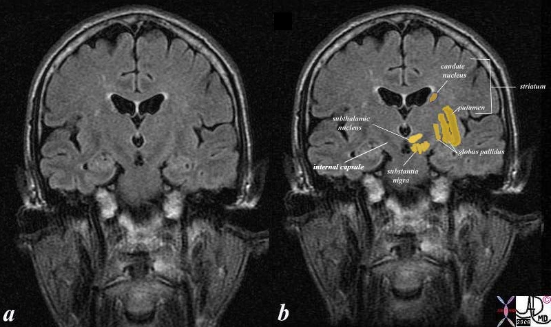
From a structural point of view there are 5 major components including the caudate nucleus, putamen, globus pallidus, subthalamic nucleus and the substantia nigra. The nucleus accumbens is also considered part of the basal ganglia while the claustrum and amygdala which were previously considered part of the system, have been demoted. Additionally other names to combinations of the major parts have been applied including striatum (caudate nucleus, putamen and nucleus accumbens), corpus striatum (striatum and globus pallidus) and lenticular nucleus (putamen and globus pallidus).
From a functional point of view thebasal ganglia are mostly thought of as an inhibitory mechanism of motor function opposing, balancing and complementing the excitatory function of the cerebellum to enable smooth movement. The disease of either will change the balance and result in loss of smooth movement. They also have a role in the sensory system and in the context of pain are involved in the affective dimension, in modulation and sensory gating of nociceptive information, and possiblly on the sensory-discriminative cognitive dimension. (Chudler), The sensory aspect of the basal ganglia is a relatively new development in the neurosciences, and the exact mechanisms and manifestations are yet to be established.
The caudate nucleus and the putamen are the doorway to the basal ganglia and they receive input from both the sensory cortex and motor cortex. They distribute the signals to the globus pallidus substantia nigra and subthalamic nuclii. The latter (two subthalamic nuclii and substantia nigra) process the signal and send the result back to the globus pallidus which in turn sends the signal back to the thalamus.
The transmitters in the basal ganglia include Acetyl choline, gamma amino butyric acid (GABA) and dopamine
|
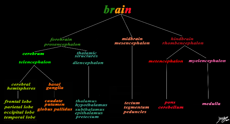
The Basal Ganglia (deep orange)
Part of the Forebrain (Prosencephalon)
Member of the Cerebrum (Telencephalon)
Head of the Basal Ganglia Family
|
|
The basic and simplest classification of the brain into forebrain midbrain and hindbrain is shown in this diagram and advanced to a more complex tree using the embryological and evolutionary terminologies. The forebrain consists of the cerebrum also called the prosencephalon, which contains the more advanced form of the brain and the thalamic structures which contain more basic structures. The cerebrum (telencephalon) itself consists of two cerebral hemispheres and paired basal ganglial structures. Each cerebral hemisphere will have gray and white matter distributed in the frontal parietal temporal and occipital lobe, with the basal ganglia being part of the gray matter deep in the cerebral hemispheres. The most important thalamic structures arising from the diencephalons include the thalamus itself and the hypothalamus. The midbrain (mesencepaholon) consists of the tectum tegmentum and cerebral peduncles. The hindbrain has two major branch points based on the evolutionary development. The pons and cerebellum(part of the metencephalon) are grouped and the medulla (part of the myelencephalon is the second branch.
Courtesy Ashley Davidoff MD Copyright 2010 All rights reserved 97686.8s
|

The Inner Orange C- Ring
|
|
The orange c shaped ring lies lateral and to some extent inferior to the lateral ventricles typified by the light blue ring and consists of the thalamus and basal ganglia including the globus pallidus, putamen, head of the caudate nucleus, tail of the caudate nucleus and the amydala. This diagram infers the concept of recurring inverted C shaped structures that are layered both from deep to superficial but also to some extent medial to lateral.
Courtesy Ashley Davidoff MD Copyright 2010 All rights reserved 93907d13b05b02.8s
|
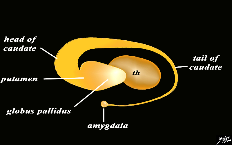
The Basal Ganglia of the Forebrain in Sagittal Projection |
|
The basal ganglia lie both in the forebrain and in the midbrain. In the forebrain they are distributed in paraventricular fashion almost in an inverted C fashion. In the conceptual diagrams we have colored the inverted ?C? that abuts the ventricular system orange, and this includes the thalamus and the basal ganglia. The basal ganglia that lie in the forebrain include the globus pallidus, putamen, head of the caudate nucleus, tail of the caudate nucleus and the amygdala. The amygdala appears to be part of the limbic system and the basal ganglia.
Courtesy Ashley Davidoff MD Copyright 2010 All rights reserved 93907d13b05dL3.8s
|
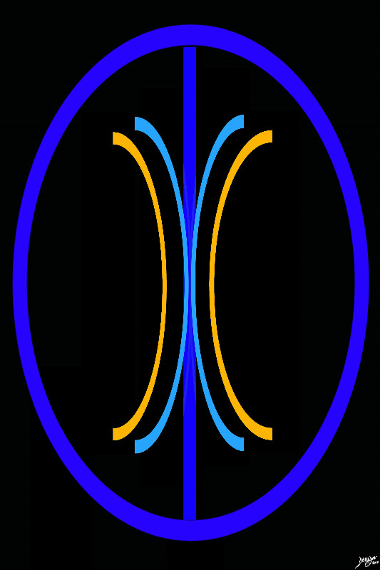
Basal Ganglia in the Forebrain Lie within the The Orange Paraventricular Layer
|
|
The paraventricular orange layer contains the basal ganglia and the thalamus. The paraventricular structures in general conform to the axis of the lateral ventricles superiorly. In this instance we will highlight the components of the basal ganglia that lie along this plane.
Courtesy Ashley DAvidoff MD Copyright 2010 all rights reserved 93914.3ka04b01.8s
|
|

Forebrain Basal Ganglia in Axial Projection
|
|
The basal ganglia in the forebrain in the axial projection are seen as a continuous inverted C as well. In this diagram their relationship to the ventricles distributed in paraventricular fashion is exemplified. In the conceptual diagrams we have colored the inverted ?C? that abuts the ventricular system orange, and this includes the putamen, globus pallidus, head of the caudate nucleus, tail of the caudate nucleus and the amygdala. The amygdala appears to be part of the limbic system and the basal ganglia.
Courtesy Ashley Davidoff MD Copyright 2010 All rights reserved 93914.3ka04b01e05.8s
|
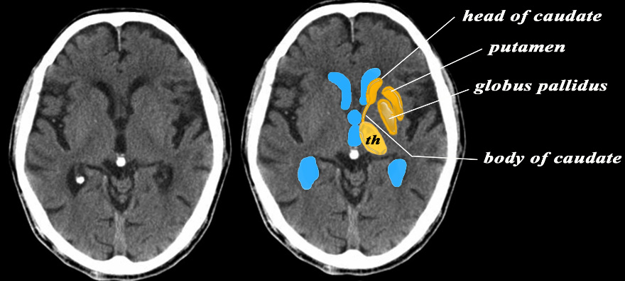
CTscan in the Axial Plane Shows the Major components of the Basal Ganglia of the Forebrain |
|
The basal ganglia in the forebrain in the axial projection on this CTscan are overlaid in dark orange while the thalamus is in lighter orange At a single axial level the major components of the basal ganglia of the forebrain are seen including the head and tail of the caudate nucleus, the putamen and the globus pallidus In this diagram their relationship to the ventricles distributed in paraventricular fashion (ventricles in blue) is also seen.
Courtesy Ashley Davidoff MD Copyright 2010 All rights reserved 38568c12b08L
|
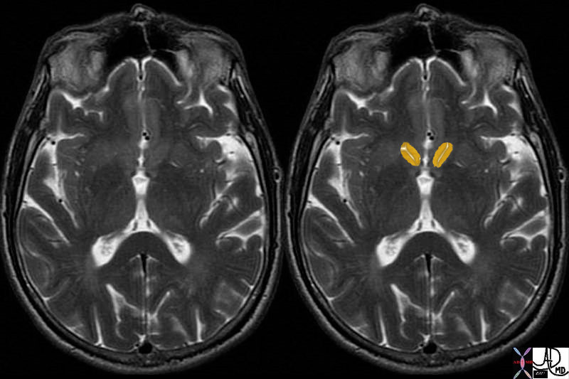
Caudate Nucleus MRI T2 Weighted Image in Transverse Plane |
| brain basal ganglia basal ganglion caudate nucleus part of the dorsal striatum involved in learning learning, language control, regulation of the cerebral cortex threshold, and affection for loved ones MRI T2 weighted Davidoff MD 38693c01 |

Putamen |
|
brain basal ganglia basal ganglion putamen part of the dorsal striatum inner seed involved in learning MRI T2 weighted Davidoff MD 38693c02
|
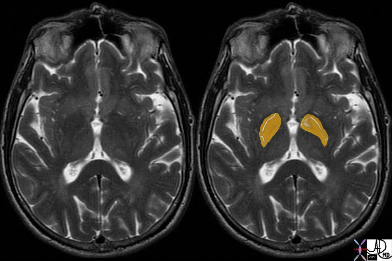
Globus Pallidus |
| brain basal ganglia basal ganglion globus pallidus (meaning pale globe) two parts external and internal divided by medial medullary lamina involved as a pacemaker rate of discharge and pattern receiving afferents from the striatum (caudate nucleus and putamen) MRI T2 weighted Davidoff MD 38693c03 |

Subthalamic Nucleus |
| 38610c02b brain forebrain basal ganglia basal ganglion subthalamic nucleus subthalamic nuclii MRI coronal FLAIR Davidoff MD |
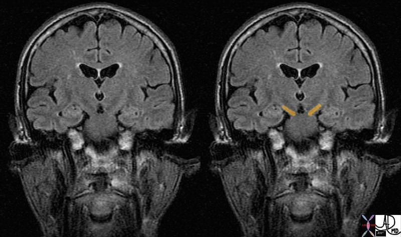
Substantia Nigra |
| 38610c03 brain midbrain basal ganglia basal ganglion substantia nigra MRI coronal FLAIR Davidoff MD |
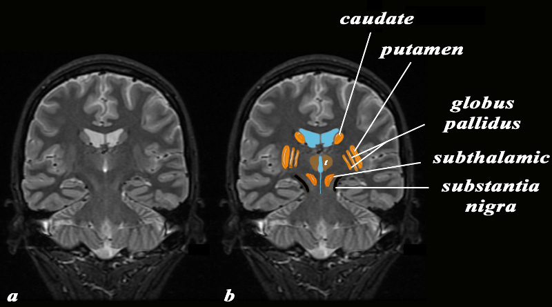
The Basal Ganglia in Coronal Projection
MRI Inversion Recovery STIR Technique |
|
The basal ganglia in the forebrain and midbrain are displayed in the coronal projection on this MRI STIR sequence In the forebrain the basal ganglia are overlaid in orange while the basal ganglia in the midbrain ? th substntia nigra are overlaid in black. The thalamus is overlaid in light brown. The basal ganglia of the forebrain are seen including the caudate nucleus, the putamen, globus pallidus and subthalamic nuclii. In this diagram the relationship of the basal ganglia to the (ventricles in blue) is demonstrated.
Courtesy Ashley Davidoff MD Copyright 2010 All rights reserved 95110c04b01.8
|
|
Structural and Functional relationships of the Basal Ganglia |
|
The coronal T1 weighted image reveals the structures that are functionally related to basal ganglial function. These include the caudate nucleus, globus pallidus, putamen, substantia nigra and subthalamus. The caudate nucleus and the putamen are the doorway to the basal ganglia and they receive input from both the sensory cortex and motor cortex. They distribute the signals to the globus pallidus substantia nigra and subthalamic nuclii. The latter (two subthalamic nuclii and substantia nigra) process the signal and send the result back to the globus pallidus which in turn sends the signal back to the thalamus.
Courtesy Ashley Davidoff Md Copyright 2010 All rights reserved 38610c06c06d.81s
|
Function
The basal ganglia function to help control control fine and gross motor movement. They select and maintain purposeful movement, and suppress unwanted and unintentional movement. They are involved in slow and sustained contractions required for posture and body support.. Since muscle tone is a balance between excitatory and inhibitory inputs the muscular system requires a control for each. They are also responsible for speech prosody.
Together with limbic system, ( amygdala, hippocampus, and septum pellucidum) the basal ganglia help regulate emotion, and memory.
The striatum receives input from the cerebral cortex while the aglobus pallidus is the source of output to the thalamus. The substantia nigra is a midbrain structures well as the thalamic nuclei utilizing GABA and cholinergic hormones in signaling. The substantia nigra affects the motor system through the production of dopamine. Dopamine is self regulating in the basal ganglia. Efferents pathways are connected to the thalamus and cortex.
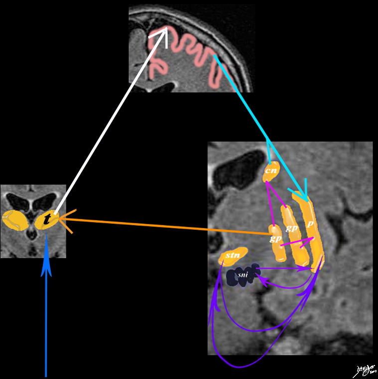
Interaction of the Basal Ganglia with the Thalamus, each other and the Cortex |
|
A simplified drawing of the connections between the caudate nucleus (orange c), the sensory cortex (salmon pink) and the basal ganglia is shown. After the stimulus has reached the sensory cortex for quantification and qualification it connects to the basal ganglia through the caudate nucleus and putamen. Each of these connects with the two parts of the globus pallidus (gp) which feed back to the thalamus. The caudate nucleus also feeds back and forth to the substantia nigra (sni) and the subthalamic nucleus (stn)
Courtesy Ashley Davidoff MD Copyright 2010 38610d09e02.8s
|
Disease
The most common diseae affecting the basal ganglia is Parkinson’s disease. It is characterized by hypokinesia, monotonous voice, shuffkling gait, and repertitive pill rollers tremor. Parkinson?s disease (PD) is primarily caused by the degeneration of dopamine secreting neurons in the substantia nigra. Vascular Parkinsonism (VP) is caused by lesions in the basal ganglia. Some success in mitigating symptoms in PD and VP has been achieved through the administration of levodopa, the chemical precursor to dopamine.
Huntington’s disease is also a result of disease of the basal ganglia and is characterized by hyperkinetic muscular movements. The loss of GABA receptors in the globus pallidus results in inhibition of subthalamic nucleus neurons responsible for suppression of hyperkinesia.
Fabry?s Disease is an X linked disorder of lysosomal storage manifesting in childhood and adolescence characterized by pain in the extremities. The lesions are more prevalent in males with the disease.
Neurofibromatosis, neoplasms, and dementia occur with calcification of the basal ganglia. Subcortical aphasia is caused by lesions in the basal ganglia.
Diagnosis
The diagnosis of these diseases is mostly clinical manifesting with aberrance in muscular control. Neuroimaging can detect basal ganglia calcification.
Treatment
Treatment is often medicinal and therapeutic.
Levodopa, the chemical precursor to dopamine, has been demonstrated to have moderate success in mitigating effects of dopamine loss.
Surgical intervention is also necessary in some instances.

Age Related Benign Calcification |
|
Calcification in the basal ganglia in the region of the globus pallidus is shown in axial projection in this CTscan. Aside from mild brain atrophy the scan is normal Age related dystrophic calcification of the basal ganglia is usually a benign finding in the elderly
Courtesy Ashley Davidoff MD Copyright 2010 All rights reserved 77120.8
|

Basal Ganglia Calcification in Sarcoidosis |
|
Calcification in the basal ganglia in the region of the globus pallidus is shown in axial projection in this CTscan of a 29 year old female with sarcoidosis. The scan is otherwise normal. It is likely that there is granulomatous involvement of the basal ganglia with sarcoidosis
Courtesy Ashley Davidoff MD Copyright 2010 All rights reserved 89070.8
|
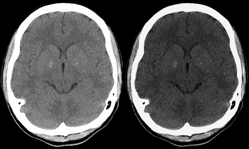
Basal Ganglial Calcification in Psychiatric Disease |
|
Calcification in the basal ganglia in the region of the globus pallidus is shown in axial projection in this CTscan of a 26 year old female with history of psychiatric disease. The scan is otherwise normal. The windows have been narrowed in the image on the right to accentuate the calcification The association of premature calcification of the basal ganglia with psychiatric illness is well established
Courtesy Ashley Davidoff MD Copyright 2010 All rights reserved 89070c.8
|
|
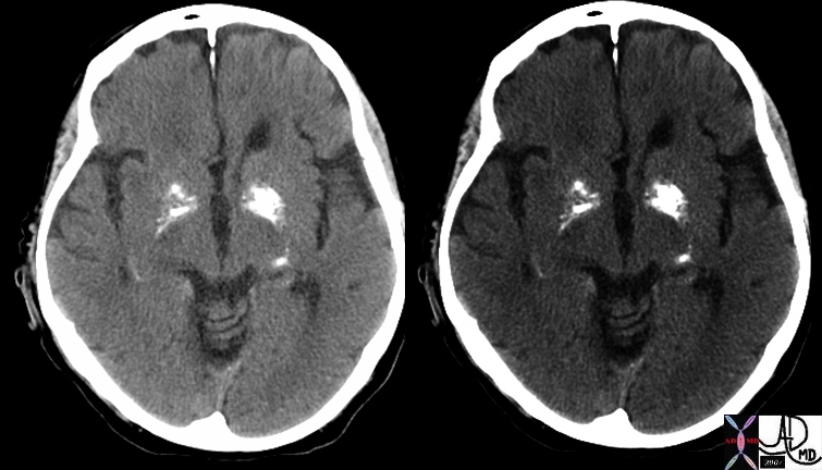
Basal Ganglia Calcification |
|
In this patient there is more than the usual calcification in the basal ganglia, and more specifically the globus pallidus.
Calcification is usually considered as dystrophic or metastatic. Most benign cases are age related and are due to dystrophic calcification. When calcification is heavy then metastatic calcification should be considered and diseases such as hyperparathyroidism should be considered.
Heavy calcification is also seen with Fabry?s disease which is a rare genetic abnormality resulting in deposition of glycolipids in tissues
Courtesy Ashley Davidoff MD 48702c01
|
|

Acute Hemorrhagic Stroke of the BAsal Ganglia
Rupture into the Choroid Plexus |
|
The CT is from a 33year old male with an acute left basal ganglial hemorrhagic stroke. The CT shows a hyperdense accumulation of hemorrhage(d) complicated by extension or rupture of the hemorrhage into the ipsilateral choroid plexus (green arrows a,b,c) and hemorrhage into the ventricles with blood lying dependently in the occipital horns (maroon arrows in c) and midline shift with septum pellucidum (white arrow of the eyes (lenses overlaid in d) mass effect on the left frontal horn (d) and midline shift exemplified by the shift of the septum pellucidum (white arrow c).
Image Courtesy Ashley Davidoff MD Copyright 2010 98551cL.8s
|
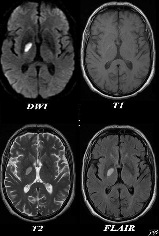
Acute Infarction Right Globus Pallidus |
|
The basal ganglia with an abnormality in the right globus pallidus are shown in axial projection in the MRI of a 64 year old male who presents with acute neurological deficit. In the first image a high intensity region in the right globus pallidus is shown in axial projection on DWI consistent with an acute infarction. The second image is a T1 weighted image without contrast and shows a rim of high intensity suggesting rim hemorrhage.. The third T2 weighted image shows the lesion to be bright but not as bright as CSF a finding consistent with the acute infarction. The fourth image is a FLAIR image and also shows increased intensity of the lesion. The constellation of findings is consistent with an acute infarction of the right globus pallidus with a rim of acute hemorrhage.
Courtesy Ashley Davidoff MD Copyright 2010 All rights reserved 89087c01.8s
|

Basal Ganglia infarction due to Lenticulostriate Disease |
|
The CT and MRI images are from an 82 year old make with acute neurological deficit. An acute infarction is shown that involves territory within and beyond the basal ganglia. In the CTscan two low density regions are seen medial to the Sylvian fissure and medial to the insular cortex, likely involving the putamen and the part of the right caudate nucleus as well as some white matter in right middle cerebra;l territory. In the second image a high intensity region in the putamen and part of the caudate nucleus is shown together with an area that does not involve the basal ganglia more posteriorly, likely part of the white matter of the right parietal lobe shown in an axial projection on DWI consistent with an acute infarction. The third image is an axial FLAIR sequence and shows the area of parenchymal edema with presumed central necrosis, while the image at low right shows the periventricular disease to better advantage. The constellation of findings is consistent with an acute infarction with distribution of the lenticulostriate vessels arising from the right middle cerebral territory
Courtesy Ashley Davidoff MD Copyright 2010 All rights reserved 89367c01.8s
|
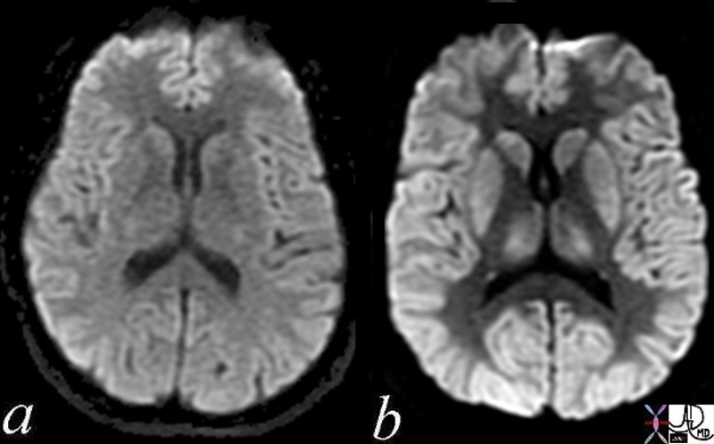
Normal (a) and Acute Global Ischemia (b) |
|
The two images represent a diffusion weighted MRI image which measures Brownian motion of molecules. In acute infarction there is restricted Brownian motion of the affected area and the image can be manipulated to present this as a bright region. In this case the acute infarction or ischemia (b) is relatively bright compared to the white namatter and compared to the gray matterof the normal (a)
Courtesy Ashley Davidoff MD Copyright 2010 All rights reserved 49433c02.800
|
 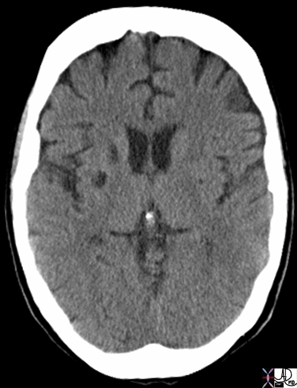
Chronic Infarcts |
| 71562 brain caudate nucleus caudate nuclii region of basal ganglion putamen claustrum subinsula region and internal capsule lacunes lacunar infarct circulatory disease chronic infarction head CT Davidoff MD 71561 |
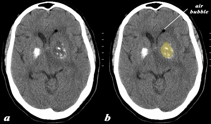
Calcification and Mass in the Basal Ganglia on the left
Evolving Abscess |
|
The basal ganglia in the region of the caudate nucleus and globus pallidus are shown in axial projection in this 60 year old female who presents with neurological deficit and a fever. The CT scan shows asymmetric calcification in the region of the caudate nucleus and globus pallidus. The calcifications on the left are expanded by a low density presumably fluid collection (yellow). There is associated surrounding edema and mass effect on the ipsilateral ventricle. A small air bubble is noted in the anterior most portion of the left frontal horn. There is mild midline shift An MRI confirmed the presence of a complex fluid in the left basal ganglion and significant surrounding edema. The patient had a fever and the constellation of findings were consistent with an abscess of the basal ganglia on the left. In this diagram the intimate relationship of the basal ganglia to the ventricles is exemplified by the ipsilateral mass effect and the presence of air presumably from gas forming organisms.
Courtesy Ashley Davidoff MD Copyright 2010 All rights reserved 89065c01.8
|

Abscess Left Basal Ganglia |
|
The basal ganglia in the region of the caudate nucleus and globus pallidus are shown in axial projection in this 60 year old female who presents with neurological deficit and a fever. In the first image the focal ill defined mass with mild mass effect is shown in axial projection on a T1 weighted image without contrast (a). The second image with gadolinium shows rim enhancement with mass effect and obstruction of the frontal horn as seen by asymmetric dilatation (b). The third T2 weighted image (c) shows the fluid nature of the cavity and the surrounding edema, mass effect, and accumulation of fluid in the dependant portion of the occipital horn. The fourth image is a FLAIR image and also shows th extent of the edema in the brain The patient had a fever and the constellation of findings were consistent with an abscess of the basal ganglia on the left.
Courtesy Ashley Davidoff MD Copyright 2010 All rights reserved 89054c.8s
|
References
Farr G Basic Review of the Brain (good introduction – defines amygdala )
DOMElement Object
(
    [schemaTypeInfo] =>
    [tagName] => table
    [firstElementChild] => (object value omitted)
    [lastElementChild] => (object value omitted)
    [childElementCount] => 1
    [previousElementSibling] => (object value omitted)
    [nextElementSibling] => (object value omitted)
    [nodeName] => table
    [nodeValue] =>


Abscess Left Basal Ganglia


The basal ganglia in the region of the caudate nucleus and globus pallidus are shown in axial projection in this 60 year old female who presents with neurological deficit and a fever. In the first image the focal ill defined mass with mild mass effect is shown in axial projection on a T1 weighted image without contrast (a). The second image with gadolinium shows rim enhancement with mass effect and obstruction of the frontal horn as seen by asymmetric dilatation (b). The third T2 weighted image (c) shows the fluid nature of the cavity and the surrounding edema, mass effect, and accumulation of fluid in the dependant portion of the occipital horn. The fourth image is a FLAIR image and also shows th extent of the edema in the brain The patient had a fever and the constellation of findings were consistent with an abscess of the basal ganglia on the left.
Courtesy Ashley Davidoff MD Copyright 2010 All rights reserved 89054c.8s


    [nodeType] => 1
    [parentNode] => (object value omitted)
    [childNodes] => (object value omitted)
    [firstChild] => (object value omitted)
    [lastChild] => (object value omitted)
    [previousSibling] => (object value omitted)
    [nextSibling] => (object value omitted)
    [attributes] => (object value omitted)
    [ownerDocument] => (object value omitted)
    [namespaceURI] =>
    [prefix] =>
    [localName] => table
    [baseURI] =>
    [textContent] =>


Abscess Left Basal Ganglia


The basal ganglia in the region of the caudate nucleus and globus pallidus are shown in axial projection in this 60 year old female who presents with neurological deficit and a fever. In the first image the focal ill defined mass with mild mass effect is shown in axial projection on a T1 weighted image without contrast (a). The second image with gadolinium shows rim enhancement with mass effect and obstruction of the frontal horn as seen by asymmetric dilatation (b). The third T2 weighted image (c) shows the fluid nature of the cavity and the surrounding edema, mass effect, and accumulation of fluid in the dependant portion of the occipital horn. The fourth image is a FLAIR image and also shows th extent of the edema in the brain The patient had a fever and the constellation of findings were consistent with an abscess of the basal ganglia on the left.
Courtesy Ashley Davidoff MD Copyright 2010 All rights reserved 89054c.8s


)
DOMElement Object
(
    [schemaTypeInfo] =>
    [tagName] => td
    [firstElementChild] => (object value omitted)
    [lastElementChild] => (object value omitted)
    [childElementCount] => 2
    [previousElementSibling] =>
    [nextElementSibling] =>
    [nodeName] => td
    [nodeValue] =>
The basal ganglia in the region of the caudate nucleus and globus pallidus are shown in axial projection in this 60 year old female who presents with neurological deficit and a fever. In the first image the focal ill defined mass with mild mass effect is shown in axial projection on a T1 weighted image without contrast (a). The second image with gadolinium shows rim enhancement with mass effect and obstruction of the frontal horn as seen by asymmetric dilatation (b). The third T2 weighted image (c) shows the fluid nature of the cavity and the surrounding edema, mass effect, and accumulation of fluid in the dependant portion of the occipital horn. The fourth image is a FLAIR image and also shows th extent of the edema in the brain The patient had a fever and the constellation of findings were consistent with an abscess of the basal ganglia on the left.
Courtesy Ashley Davidoff MD Copyright 2010 All rights reserved 89054c.8s

    [nodeType] => 1
    [parentNode] => (object value omitted)
    [childNodes] => (object value omitted)
    [firstChild] => (object value omitted)
    [lastChild] => (object value omitted)
    [previousSibling] => (object value omitted)
    [nextSibling] => (object value omitted)
    [attributes] => (object value omitted)
    [ownerDocument] => (object value omitted)
    [namespaceURI] =>
    [prefix] =>
    [localName] => td
    [baseURI] =>
    [textContent] =>
The basal ganglia in the region of the caudate nucleus and globus pallidus are shown in axial projection in this 60 year old female who presents with neurological deficit and a fever. In the first image the focal ill defined mass with mild mass effect is shown in axial projection on a T1 weighted image without contrast (a). The second image with gadolinium shows rim enhancement with mass effect and obstruction of the frontal horn as seen by asymmetric dilatation (b). The third T2 weighted image (c) shows the fluid nature of the cavity and the surrounding edema, mass effect, and accumulation of fluid in the dependant portion of the occipital horn. The fourth image is a FLAIR image and also shows th extent of the edema in the brain The patient had a fever and the constellation of findings were consistent with an abscess of the basal ganglia on the left.
Courtesy Ashley Davidoff MD Copyright 2010 All rights reserved 89054c.8s

)
DOMElement Object
(
    [schemaTypeInfo] =>
    [tagName] => td
    [firstElementChild] => (object value omitted)
    [lastElementChild] => (object value omitted)
    [childElementCount] => 2
    [previousElementSibling] =>
    [nextElementSibling] =>
    [nodeName] => td
    [nodeValue] =>
Abscess Left Basal Ganglia
    [nodeType] => 1
    [parentNode] => (object value omitted)
    [childNodes] => (object value omitted)
    [firstChild] => (object value omitted)
    [lastChild] => (object value omitted)
    [previousSibling] => (object value omitted)
    [nextSibling] => (object value omitted)
    [attributes] => (object value omitted)
    [ownerDocument] => (object value omitted)
    [namespaceURI] =>
    [prefix] =>
    [localName] => td
    [baseURI] =>
    [textContent] =>
Abscess Left Basal Ganglia
)
DOMElement Object
(
    [schemaTypeInfo] =>
    [tagName] => table
    [firstElementChild] => (object value omitted)
    [lastElementChild] => (object value omitted)
    [childElementCount] => 1
    [previousElementSibling] => (object value omitted)
    [nextElementSibling] => (object value omitted)
    [nodeName] => table
    [nodeValue] =>


Calcification and Mass in the Basal Ganglia on the left
Evolving Abscess


The basal ganglia in the region of the caudate nucleus and globus pallidus are shown in axial projection in this 60 year old female who presents with neurological deficit and a fever. The CT scan shows asymmetric calcification in the region of the caudate nucleus and globus pallidus. The calcifications on the left are expanded by a low density presumably fluid collection (yellow). There is associated surrounding edema and mass effect on the ipsilateral ventricle. A small air bubble is noted in the anterior most portion of the left frontal horn. There is mild midline shift An MRI confirmed the presence of a complex fluid in the left basal ganglion and significant surrounding edema. The patient had a fever and the constellation of findings were consistent with an abscess of the basal ganglia on the left. In this diagram the intimate relationship of the basal ganglia to the ventricles is exemplified by the ipsilateral mass effect and the presence of air presumably from gas forming organisms.
Courtesy Ashley Davidoff MD Copyright 2010 All rights reserved 89065c01.8


    [nodeType] => 1
    [parentNode] => (object value omitted)
    [childNodes] => (object value omitted)
    [firstChild] => (object value omitted)
    [lastChild] => (object value omitted)
    [previousSibling] => (object value omitted)
    [nextSibling] => (object value omitted)
    [attributes] => (object value omitted)
    [ownerDocument] => (object value omitted)
    [namespaceURI] =>
    [prefix] =>
    [localName] => table
    [baseURI] =>
    [textContent] =>


Calcification and Mass in the Basal Ganglia on the left
Evolving Abscess


The basal ganglia in the region of the caudate nucleus and globus pallidus are shown in axial projection in this 60 year old female who presents with neurological deficit and a fever. The CT scan shows asymmetric calcification in the region of the caudate nucleus and globus pallidus. The calcifications on the left are expanded by a low density presumably fluid collection (yellow). There is associated surrounding edema and mass effect on the ipsilateral ventricle. A small air bubble is noted in the anterior most portion of the left frontal horn. There is mild midline shift An MRI confirmed the presence of a complex fluid in the left basal ganglion and significant surrounding edema. The patient had a fever and the constellation of findings were consistent with an abscess of the basal ganglia on the left. In this diagram the intimate relationship of the basal ganglia to the ventricles is exemplified by the ipsilateral mass effect and the presence of air presumably from gas forming organisms.
Courtesy Ashley Davidoff MD Copyright 2010 All rights reserved 89065c01.8


)
DOMElement Object
(
    [schemaTypeInfo] =>
    [tagName] => td
    [firstElementChild] => (object value omitted)
    [lastElementChild] => (object value omitted)
    [childElementCount] => 2
    [previousElementSibling] =>
    [nextElementSibling] =>
    [nodeName] => td
    [nodeValue] =>
The basal ganglia in the region of the caudate nucleus and globus pallidus are shown in axial projection in this 60 year old female who presents with neurological deficit and a fever. The CT scan shows asymmetric calcification in the region of the caudate nucleus and globus pallidus. The calcifications on the left are expanded by a low density presumably fluid collection (yellow). There is associated surrounding edema and mass effect on the ipsilateral ventricle. A small air bubble is noted in the anterior most portion of the left frontal horn. There is mild midline shift An MRI confirmed the presence of a complex fluid in the left basal ganglion and significant surrounding edema. The patient had a fever and the constellation of findings were consistent with an abscess of the basal ganglia on the left. In this diagram the intimate relationship of the basal ganglia to the ventricles is exemplified by the ipsilateral mass effect and the presence of air presumably from gas forming organisms.
Courtesy Ashley Davidoff MD Copyright 2010 All rights reserved 89065c01.8

    [nodeType] => 1
    [parentNode] => (object value omitted)
    [childNodes] => (object value omitted)
    [firstChild] => (object value omitted)
    [lastChild] => (object value omitted)
    [previousSibling] => (object value omitted)
    [nextSibling] => (object value omitted)
    [attributes] => (object value omitted)
    [ownerDocument] => (object value omitted)
    [namespaceURI] =>
    [prefix] =>
    [localName] => td
    [baseURI] =>
    [textContent] =>
The basal ganglia in the region of the caudate nucleus and globus pallidus are shown in axial projection in this 60 year old female who presents with neurological deficit and a fever. The CT scan shows asymmetric calcification in the region of the caudate nucleus and globus pallidus. The calcifications on the left are expanded by a low density presumably fluid collection (yellow). There is associated surrounding edema and mass effect on the ipsilateral ventricle. A small air bubble is noted in the anterior most portion of the left frontal horn. There is mild midline shift An MRI confirmed the presence of a complex fluid in the left basal ganglion and significant surrounding edema. The patient had a fever and the constellation of findings were consistent with an abscess of the basal ganglia on the left. In this diagram the intimate relationship of the basal ganglia to the ventricles is exemplified by the ipsilateral mass effect and the presence of air presumably from gas forming organisms.
Courtesy Ashley Davidoff MD Copyright 2010 All rights reserved 89065c01.8

)
DOMElement Object
(
    [schemaTypeInfo] =>
    [tagName] => td
    [firstElementChild] => (object value omitted)
    [lastElementChild] => (object value omitted)
    [childElementCount] => 3
    [previousElementSibling] =>
    [nextElementSibling] =>
    [nodeName] => td
    [nodeValue] =>
Calcification and Mass in the Basal Ganglia on the left
Evolving Abscess
    [nodeType] => 1
    [parentNode] => (object value omitted)
    [childNodes] => (object value omitted)
    [firstChild] => (object value omitted)
    [lastChild] => (object value omitted)
    [previousSibling] => (object value omitted)
    [nextSibling] => (object value omitted)
    [attributes] => (object value omitted)
    [ownerDocument] => (object value omitted)
    [namespaceURI] =>
    [prefix] =>
    [localName] => td
    [baseURI] =>
    [textContent] =>
Calcification and Mass in the Basal Ganglia on the left
Evolving Abscess
)
DOMElement Object
(
    [schemaTypeInfo] =>
    [tagName] => table
    [firstElementChild] => (object value omitted)
    [lastElementChild] => (object value omitted)
    [childElementCount] => 1
    [previousElementSibling] => (object value omitted)
    [nextElementSibling] => (object value omitted)
    [nodeName] => table
    [nodeValue] =>


Chronic Infarcts


71562 brain caudate nucleus caudate nuclii region of basal ganglion putamen claustrum subinsula region and internal capsule lacunes lacunar infarct circulatory disease chronic infarction head CT Davidoff MD 71561


    [nodeType] => 1
    [parentNode] => (object value omitted)
    [childNodes] => (object value omitted)
    [firstChild] => (object value omitted)
    [lastChild] => (object value omitted)
    [previousSibling] => (object value omitted)
    [nextSibling] => (object value omitted)
    [attributes] => (object value omitted)
    [ownerDocument] => (object value omitted)
    [namespaceURI] =>
    [prefix] =>
    [localName] => table
    [baseURI] =>
    [textContent] =>


Chronic Infarcts


71562 brain caudate nucleus caudate nuclii region of basal ganglion putamen claustrum subinsula region and internal capsule lacunes lacunar infarct circulatory disease chronic infarction head CT Davidoff MD 71561


)
DOMElement Object
(
    [schemaTypeInfo] =>
    [tagName] => td
    [firstElementChild] =>
    [lastElementChild] =>
    [childElementCount] => 0
    [previousElementSibling] =>
    [nextElementSibling] =>
    [nodeName] => td
    [nodeValue] => 71562 brain caudate nucleus caudate nuclii region of basal ganglion putamen claustrum subinsula region and internal capsule lacunes lacunar infarct circulatory disease chronic infarction head CT Davidoff MD 71561
    [nodeType] => 1
    [parentNode] => (object value omitted)
    [childNodes] => (object value omitted)
    [firstChild] => (object value omitted)
    [lastChild] => (object value omitted)
    [previousSibling] => (object value omitted)
    [nextSibling] => (object value omitted)
    [attributes] => (object value omitted)
    [ownerDocument] => (object value omitted)
    [namespaceURI] =>
    [prefix] =>
    [localName] => td
    [baseURI] =>
    [textContent] => 71562 brain caudate nucleus caudate nuclii region of basal ganglion putamen claustrum subinsula region and internal capsule lacunes lacunar infarct circulatory disease chronic infarction head CT Davidoff MD 71561
)
DOMElement Object
(
    [schemaTypeInfo] =>
    [tagName] => td
    [firstElementChild] => (object value omitted)
    [lastElementChild] => (object value omitted)
    [childElementCount] => 2
    [previousElementSibling] =>
    [nextElementSibling] =>
    [nodeName] => td
    [nodeValue] =>
Chronic Infarcts
    [nodeType] => 1
    [parentNode] => (object value omitted)
    [childNodes] => (object value omitted)
    [firstChild] => (object value omitted)
    [lastChild] => (object value omitted)
    [previousSibling] => (object value omitted)
    [nextSibling] => (object value omitted)
    [attributes] => (object value omitted)
    [ownerDocument] => (object value omitted)
    [namespaceURI] =>
    [prefix] =>
    [localName] => td
    [baseURI] =>
    [textContent] =>
Chronic Infarcts
)
https://beta.thecommonvein.net/wp-content/uploads/2023/06/71562.jpg https://beta.thecommonvein.net/wp-content/uploads/2023/06/71561.jpg
http://www.thecommonvein.net/media/71562.jpg
DOMElement Object
(
    [schemaTypeInfo] =>
    [tagName] => table
    [firstElementChild] => (object value omitted)
    [lastElementChild] => (object value omitted)
    [childElementCount] => 1
    [previousElementSibling] => (object value omitted)
    [nextElementSibling] => (object value omitted)
    [nodeName] => table
    [nodeValue] =>


Normal (a) and Acute Global Ischemia (b)


The two images represent a diffusion weighted MRI image which measures Brownian motion of molecules. In acute infarction there is restricted Brownian motion of the affected area and the image can be manipulated to present this as a bright region. In this case the acute infarction or ischemia (b) is relatively bright compared to the white namatter and compared to the gray matterof the normal (a)
Courtesy Ashley Davidoff MD Copyright 2010 All rights reserved 49433c02.800


    [nodeType] => 1
    [parentNode] => (object value omitted)
    [childNodes] => (object value omitted)
    [firstChild] => (object value omitted)
    [lastChild] => (object value omitted)
    [previousSibling] => (object value omitted)
    [nextSibling] => (object value omitted)
    [attributes] => (object value omitted)
    [ownerDocument] => (object value omitted)
    [namespaceURI] =>
    [prefix] =>
    [localName] => table
    [baseURI] =>
    [textContent] =>


Normal (a) and Acute Global Ischemia (b)


The two images represent a diffusion weighted MRI image which measures Brownian motion of molecules. In acute infarction there is restricted Brownian motion of the affected area and the image can be manipulated to present this as a bright region. In this case the acute infarction or ischemia (b) is relatively bright compared to the white namatter and compared to the gray matterof the normal (a)
Courtesy Ashley Davidoff MD Copyright 2010 All rights reserved 49433c02.800


)
DOMElement Object
(
    [schemaTypeInfo] =>
    [tagName] => td
    [firstElementChild] => (object value omitted)
    [lastElementChild] => (object value omitted)
    [childElementCount] => 2
    [previousElementSibling] =>
    [nextElementSibling] =>
    [nodeName] => td
    [nodeValue] =>
The two images represent a diffusion weighted MRI image which measures Brownian motion of molecules. In acute infarction there is restricted Brownian motion of the affected area and the image can be manipulated to present this as a bright region. In this case the acute infarction or ischemia (b) is relatively bright compared to the white namatter and compared to the gray matterof the normal (a)
Courtesy Ashley Davidoff MD Copyright 2010 All rights reserved 49433c02.800

    [nodeType] => 1
    [parentNode] => (object value omitted)
    [childNodes] => (object value omitted)
    [firstChild] => (object value omitted)
    [lastChild] => (object value omitted)
    [previousSibling] => (object value omitted)
    [nextSibling] => (object value omitted)
    [attributes] => (object value omitted)
    [ownerDocument] => (object value omitted)
    [namespaceURI] =>
    [prefix] =>
    [localName] => td
    [baseURI] =>
    [textContent] =>
The two images represent a diffusion weighted MRI image which measures Brownian motion of molecules. In acute infarction there is restricted Brownian motion of the affected area and the image can be manipulated to present this as a bright region. In this case the acute infarction or ischemia (b) is relatively bright compared to the white namatter and compared to the gray matterof the normal (a)
Courtesy Ashley Davidoff MD Copyright 2010 All rights reserved 49433c02.800

)
DOMElement Object
(
    [schemaTypeInfo] =>
    [tagName] => td
    [firstElementChild] => (object value omitted)
    [lastElementChild] => (object value omitted)
    [childElementCount] => 2
    [previousElementSibling] =>
    [nextElementSibling] =>
    [nodeName] => td
    [nodeValue] =>
Normal (a) and Acute Global Ischemia (b)
    [nodeType] => 1
    [parentNode] => (object value omitted)
    [childNodes] => (object value omitted)
    [firstChild] => (object value omitted)
    [lastChild] => (object value omitted)
    [previousSibling] => (object value omitted)
    [nextSibling] => (object value omitted)
    [attributes] => (object value omitted)
    [ownerDocument] => (object value omitted)
    [namespaceURI] =>
    [prefix] =>
    [localName] => td
    [baseURI] =>
    [textContent] =>
Normal (a) and Acute Global Ischemia (b)
)
DOMElement Object
(
    [schemaTypeInfo] =>
    [tagName] => table
    [firstElementChild] => (object value omitted)
    [lastElementChild] => (object value omitted)
    [childElementCount] => 1
    [previousElementSibling] => (object value omitted)
    [nextElementSibling] => (object value omitted)
    [nodeName] => table
    [nodeValue] =>


Basal Ganglia infarction due to Lenticulostriate Disease


The CT and MRI images are from an 82 year old make with acute neurological deficit. An acute infarction is shown that involves territory within and beyond the basal ganglia. In the CTscan two low density regions are seen medial to the Sylvian fissure and medial to the insular cortex, likely involving the putamen and the part of the right caudate nucleus as well as some white matter in right middle cerebra;l territory. In the second image a high intensity region in the putamen and part of the caudate nucleus is shown together with an area that does not involve the basal ganglia more posteriorly, likely part of the white matter of the right parietal lobe shown in an axial projection on DWI consistent with an acute infarction. The third image is an axial FLAIR sequence and shows the area of parenchymal edema with presumed central necrosis, while the image at low right shows the periventricular disease to better advantage. The constellation of findings is consistent with an acute infarction with distribution of the lenticulostriate vessels arising from the right middle cerebral territory
Courtesy Ashley Davidoff MD Copyright 2010 All rights reserved 89367c01.8s


    [nodeType] => 1
    [parentNode] => (object value omitted)
    [childNodes] => (object value omitted)
    [firstChild] => (object value omitted)
    [lastChild] => (object value omitted)
    [previousSibling] => (object value omitted)
    [nextSibling] => (object value omitted)
    [attributes] => (object value omitted)
    [ownerDocument] => (object value omitted)
    [namespaceURI] =>
    [prefix] =>
    [localName] => table
    [baseURI] =>
    [textContent] =>


Basal Ganglia infarction due to Lenticulostriate Disease


The CT and MRI images are from an 82 year old make with acute neurological deficit. An acute infarction is shown that involves territory within and beyond the basal ganglia. In the CTscan two low density regions are seen medial to the Sylvian fissure and medial to the insular cortex, likely involving the putamen and the part of the right caudate nucleus as well as some white matter in right middle cerebra;l territory. In the second image a high intensity region in the putamen and part of the caudate nucleus is shown together with an area that does not involve the basal ganglia more posteriorly, likely part of the white matter of the right parietal lobe shown in an axial projection on DWI consistent with an acute infarction. The third image is an axial FLAIR sequence and shows the area of parenchymal edema with presumed central necrosis, while the image at low right shows the periventricular disease to better advantage. The constellation of findings is consistent with an acute infarction with distribution of the lenticulostriate vessels arising from the right middle cerebral territory
Courtesy Ashley Davidoff MD Copyright 2010 All rights reserved 89367c01.8s


)
DOMElement Object
(
    [schemaTypeInfo] =>
    [tagName] => td
    [firstElementChild] => (object value omitted)
    [lastElementChild] => (object value omitted)
    [childElementCount] => 2
    [previousElementSibling] =>
    [nextElementSibling] =>
    [nodeName] => td
    [nodeValue] =>
The CT and MRI images are from an 82 year old make with acute neurological deficit. An acute infarction is shown that involves territory within and beyond the basal ganglia. In the CTscan two low density regions are seen medial to the Sylvian fissure and medial to the insular cortex, likely involving the putamen and the part of the right caudate nucleus as well as some white matter in right middle cerebra;l territory. In the second image a high intensity region in the putamen and part of the caudate nucleus is shown together with an area that does not involve the basal ganglia more posteriorly, likely part of the white matter of the right parietal lobe shown in an axial projection on DWI consistent with an acute infarction. The third image is an axial FLAIR sequence and shows the area of parenchymal edema with presumed central necrosis, while the image at low right shows the periventricular disease to better advantage. The constellation of findings is consistent with an acute infarction with distribution of the lenticulostriate vessels arising from the right middle cerebral territory
Courtesy Ashley Davidoff MD Copyright 2010 All rights reserved 89367c01.8s

    [nodeType] => 1
    [parentNode] => (object value omitted)
    [childNodes] => (object value omitted)
    [firstChild] => (object value omitted)
    [lastChild] => (object value omitted)
    [previousSibling] => (object value omitted)
    [nextSibling] => (object value omitted)
    [attributes] => (object value omitted)
    [ownerDocument] => (object value omitted)
    [namespaceURI] =>
    [prefix] =>
    [localName] => td
    [baseURI] =>
    [textContent] =>
The CT and MRI images are from an 82 year old make with acute neurological deficit. An acute infarction is shown that involves territory within and beyond the basal ganglia. In the CTscan two low density regions are seen medial to the Sylvian fissure and medial to the insular cortex, likely involving the putamen and the part of the right caudate nucleus as well as some white matter in right middle cerebra;l territory. In the second image a high intensity region in the putamen and part of the caudate nucleus is shown together with an area that does not involve the basal ganglia more posteriorly, likely part of the white matter of the right parietal lobe shown in an axial projection on DWI consistent with an acute infarction. The third image is an axial FLAIR sequence and shows the area of parenchymal edema with presumed central necrosis, while the image at low right shows the periventricular disease to better advantage. The constellation of findings is consistent with an acute infarction with distribution of the lenticulostriate vessels arising from the right middle cerebral territory
Courtesy Ashley Davidoff MD Copyright 2010 All rights reserved 89367c01.8s

)
DOMElement Object
(
    [schemaTypeInfo] =>
    [tagName] => td
    [firstElementChild] => (object value omitted)
    [lastElementChild] => (object value omitted)
    [childElementCount] => 2
    [previousElementSibling] =>
    [nextElementSibling] =>
    [nodeName] => td
    [nodeValue] =>
Basal Ganglia infarction due to Lenticulostriate Disease
    [nodeType] => 1
    [parentNode] => (object value omitted)
    [childNodes] => (object value omitted)
    [firstChild] => (object value omitted)
    [lastChild] => (object value omitted)
    [previousSibling] => (object value omitted)
    [nextSibling] => (object value omitted)
    [attributes] => (object value omitted)
    [ownerDocument] => (object value omitted)
    [namespaceURI] =>
    [prefix] =>
    [localName] => td
    [baseURI] =>
    [textContent] =>
Basal Ganglia infarction due to Lenticulostriate Disease
)
DOMElement Object
(
    [schemaTypeInfo] =>
    [tagName] => table
    [firstElementChild] => (object value omitted)
    [lastElementChild] => (object value omitted)
    [childElementCount] => 1
    [previousElementSibling] => (object value omitted)
    [nextElementSibling] => (object value omitted)
    [nodeName] => table
    [nodeValue] =>


Acute Infarction Right Globus Pallidus


The basal ganglia with an abnormality in the right globus pallidus are shown in axial projection in the MRI of a 64 year old male who presents with acute neurological deficit. In the first image a high intensity region in the right globus pallidus is shown in axial projection on DWI consistent with an acute infarction. The second image is a T1 weighted image without contrast and shows a rim of high intensity suggesting rim hemorrhage.. The third T2 weighted image shows the lesion to be bright but not as bright as CSF a finding consistent with the acute infarction. The fourth image is a FLAIR image and also shows increased intensity of the lesion. The constellation of findings is consistent with an acute infarction of the right globus pallidus with a rim of acute hemorrhage.
Courtesy Ashley Davidoff MD Copyright 2010 All rights reserved 89087c01.8s


    [nodeType] => 1
    [parentNode] => (object value omitted)
    [childNodes] => (object value omitted)
    [firstChild] => (object value omitted)
    [lastChild] => (object value omitted)
    [previousSibling] => (object value omitted)
    [nextSibling] => (object value omitted)
    [attributes] => (object value omitted)
    [ownerDocument] => (object value omitted)
    [namespaceURI] =>
    [prefix] =>
    [localName] => table
    [baseURI] =>
    [textContent] =>


Acute Infarction Right Globus Pallidus


The basal ganglia with an abnormality in the right globus pallidus are shown in axial projection in the MRI of a 64 year old male who presents with acute neurological deficit. In the first image a high intensity region in the right globus pallidus is shown in axial projection on DWI consistent with an acute infarction. The second image is a T1 weighted image without contrast and shows a rim of high intensity suggesting rim hemorrhage.. The third T2 weighted image shows the lesion to be bright but not as bright as CSF a finding consistent with the acute infarction. The fourth image is a FLAIR image and also shows increased intensity of the lesion. The constellation of findings is consistent with an acute infarction of the right globus pallidus with a rim of acute hemorrhage.
Courtesy Ashley Davidoff MD Copyright 2010 All rights reserved 89087c01.8s


)
DOMElement Object
(
    [schemaTypeInfo] =>
    [tagName] => td
    [firstElementChild] => (object value omitted)
    [lastElementChild] => (object value omitted)
    [childElementCount] => 2
    [previousElementSibling] =>
    [nextElementSibling] =>
    [nodeName] => td
    [nodeValue] =>
The basal ganglia with an abnormality in the right globus pallidus are shown in axial projection in the MRI of a 64 year old male who presents with acute neurological deficit. In the first image a high intensity region in the right globus pallidus is shown in axial projection on DWI consistent with an acute infarction. The second image is a T1 weighted image without contrast and shows a rim of high intensity suggesting rim hemorrhage.. The third T2 weighted image shows the lesion to be bright but not as bright as CSF a finding consistent with the acute infarction. The fourth image is a FLAIR image and also shows increased intensity of the lesion. The constellation of findings is consistent with an acute infarction of the right globus pallidus with a rim of acute hemorrhage.
Courtesy Ashley Davidoff MD Copyright 2010 All rights reserved 89087c01.8s

    [nodeType] => 1
    [parentNode] => (object value omitted)
    [childNodes] => (object value omitted)
    [firstChild] => (object value omitted)
    [lastChild] => (object value omitted)
    [previousSibling] => (object value omitted)
    [nextSibling] => (object value omitted)
    [attributes] => (object value omitted)
    [ownerDocument] => (object value omitted)
    [namespaceURI] =>
    [prefix] =>
    [localName] => td
    [baseURI] =>
    [textContent] =>
The basal ganglia with an abnormality in the right globus pallidus are shown in axial projection in the MRI of a 64 year old male who presents with acute neurological deficit. In the first image a high intensity region in the right globus pallidus is shown in axial projection on DWI consistent with an acute infarction. The second image is a T1 weighted image without contrast and shows a rim of high intensity suggesting rim hemorrhage.. The third T2 weighted image shows the lesion to be bright but not as bright as CSF a finding consistent with the acute infarction. The fourth image is a FLAIR image and also shows increased intensity of the lesion. The constellation of findings is consistent with an acute infarction of the right globus pallidus with a rim of acute hemorrhage.
Courtesy Ashley Davidoff MD Copyright 2010 All rights reserved 89087c01.8s

)
DOMElement Object
(
    [schemaTypeInfo] =>
    [tagName] => td
    [firstElementChild] => (object value omitted)
    [lastElementChild] => (object value omitted)
    [childElementCount] => 2
    [previousElementSibling] =>
    [nextElementSibling] =>
    [nodeName] => td
    [nodeValue] =>
Acute Infarction Right Globus Pallidus
    [nodeType] => 1
    [parentNode] => (object value omitted)
    [childNodes] => (object value omitted)
    [firstChild] => (object value omitted)
    [lastChild] => (object value omitted)
    [previousSibling] => (object value omitted)
    [nextSibling] => (object value omitted)
    [attributes] => (object value omitted)
    [ownerDocument] => (object value omitted)
    [namespaceURI] =>
    [prefix] =>
    [localName] => td
    [baseURI] =>
    [textContent] =>
Acute Infarction Right Globus Pallidus
)
DOMElement Object
(
    [schemaTypeInfo] =>
    [tagName] => table
    [firstElementChild] => (object value omitted)
    [lastElementChild] => (object value omitted)
    [childElementCount] => 1
    [previousElementSibling] => (object value omitted)
    [nextElementSibling] => (object value omitted)
    [nodeName] => table
    [nodeValue] =>


Acute Hemorrhagic Stroke of the BAsal Ganglia
Rupture into the Choroid Plexus


The CT is from a 33year old male with an acute left basal ganglial hemorrhagic stroke. The CT shows a hyperdense accumulation of hemorrhage(d) complicated by extension or rupture of the hemorrhage into the ipsilateral choroid plexus (green arrows a,b,c) and hemorrhage into the ventricles with blood lying dependently in the occipital horns (maroon arrows in c) and midline shift with septum pellucidum (white arrow of the eyes (lenses overlaid in d) mass effect on the left frontal horn (d) and midline shift exemplified by the shift of the septum pellucidum (white arrow c).
Image Courtesy Ashley Davidoff MD Copyright 2010 98551cL.8s


    [nodeType] => 1
    [parentNode] => (object value omitted)
    [childNodes] => (object value omitted)
    [firstChild] => (object value omitted)
    [lastChild] => (object value omitted)
    [previousSibling] => (object value omitted)
    [nextSibling] => (object value omitted)
    [attributes] => (object value omitted)
    [ownerDocument] => (object value omitted)
    [namespaceURI] =>
    [prefix] =>
    [localName] => table
    [baseURI] =>
    [textContent] =>


Acute Hemorrhagic Stroke of the BAsal Ganglia
Rupture into the Choroid Plexus


The CT is from a 33year old male with an acute left basal ganglial hemorrhagic stroke. The CT shows a hyperdense accumulation of hemorrhage(d) complicated by extension or rupture of the hemorrhage into the ipsilateral choroid plexus (green arrows a,b,c) and hemorrhage into the ventricles with blood lying dependently in the occipital horns (maroon arrows in c) and midline shift with septum pellucidum (white arrow of the eyes (lenses overlaid in d) mass effect on the left frontal horn (d) and midline shift exemplified by the shift of the septum pellucidum (white arrow c).
Image Courtesy Ashley Davidoff MD Copyright 2010 98551cL.8s


)
DOMElement Object
(
    [schemaTypeInfo] =>
    [tagName] => td
    [firstElementChild] => (object value omitted)
    [lastElementChild] => (object value omitted)
    [childElementCount] => 2
    [previousElementSibling] =>
    [nextElementSibling] =>
    [nodeName] => td
    [nodeValue] =>
The CT is from a 33year old male with an acute left basal ganglial hemorrhagic stroke. The CT shows a hyperdense accumulation of hemorrhage(d) complicated by extension or rupture of the hemorrhage into the ipsilateral choroid plexus (green arrows a,b,c) and hemorrhage into the ventricles with blood lying dependently in the occipital horns (maroon arrows in c) and midline shift with septum pellucidum (white arrow of the eyes (lenses overlaid in d) mass effect on the left frontal horn (d) and midline shift exemplified by the shift of the septum pellucidum (white arrow c).
Image Courtesy Ashley Davidoff MD Copyright 2010 98551cL.8s

    [nodeType] => 1
    [parentNode] => (object value omitted)
    [childNodes] => (object value omitted)
    [firstChild] => (object value omitted)
    [lastChild] => (object value omitted)
    [previousSibling] => (object value omitted)
    [nextSibling] => (object value omitted)
    [attributes] => (object value omitted)
    [ownerDocument] => (object value omitted)
    [namespaceURI] =>
    [prefix] =>
    [localName] => td
    [baseURI] =>
    [textContent] =>
The CT is from a 33year old male with an acute left basal ganglial hemorrhagic stroke. The CT shows a hyperdense accumulation of hemorrhage(d) complicated by extension or rupture of the hemorrhage into the ipsilateral choroid plexus (green arrows a,b,c) and hemorrhage into the ventricles with blood lying dependently in the occipital horns (maroon arrows in c) and midline shift with septum pellucidum (white arrow of the eyes (lenses overlaid in d) mass effect on the left frontal horn (d) and midline shift exemplified by the shift of the septum pellucidum (white arrow c).
Image Courtesy Ashley Davidoff MD Copyright 2010 98551cL.8s

)
DOMElement Object
(
    [schemaTypeInfo] =>
    [tagName] => td
    [firstElementChild] => (object value omitted)
    [lastElementChild] => (object value omitted)
    [childElementCount] => 3
    [previousElementSibling] =>
    [nextElementSibling] =>
    [nodeName] => td
    [nodeValue] =>

Acute Hemorrhagic Stroke of the BAsal Ganglia
Rupture into the Choroid Plexus
    [nodeType] => 1
    [parentNode] => (object value omitted)
    [childNodes] => (object value omitted)
    [firstChild] => (object value omitted)
    [lastChild] => (object value omitted)
    [previousSibling] => (object value omitted)
    [nextSibling] => (object value omitted)
    [attributes] => (object value omitted)
    [ownerDocument] => (object value omitted)
    [namespaceURI] =>
    [prefix] =>
    [localName] => td
    [baseURI] =>
    [textContent] =>

Acute Hemorrhagic Stroke of the BAsal Ganglia
Rupture into the Choroid Plexus
)
DOMElement Object
(
    [schemaTypeInfo] =>
    [tagName] => table
    [firstElementChild] => (object value omitted)
    [lastElementChild] => (object value omitted)
    [childElementCount] => 1
    [previousElementSibling] => (object value omitted)
    [nextElementSibling] => (object value omitted)
    [nodeName] => table
    [nodeValue] =>


Basal Ganglia Calcification


In this patient there is more than the usual calcification in the basal ganglia, and more specifically the globus pallidus.
Calcification is usually considered as dystrophic or metastatic.  Most benign cases are age related and are due to dystrophic calcification.  When calcification is heavy then metastatic calcification should be considered and diseases such as hyperparathyroidism should be considered.
Heavy calcification is also seen with Fabry?s disease which is a rare genetic abnormality resulting in deposition of glycolipids in tissues
Courtesy Ashley Davidoff MD 48702c01


    [nodeType] => 1
    [parentNode] => (object value omitted)
    [childNodes] => (object value omitted)
    [firstChild] => (object value omitted)
    [lastChild] => (object value omitted)
    [previousSibling] => (object value omitted)
    [nextSibling] => (object value omitted)
    [attributes] => (object value omitted)
    [ownerDocument] => (object value omitted)
    [namespaceURI] =>
    [prefix] =>
    [localName] => table
    [baseURI] =>
    [textContent] =>


Basal Ganglia Calcification


In this patient there is more than the usual calcification in the basal ganglia, and more specifically the globus pallidus.
Calcification is usually considered as dystrophic or metastatic.  Most benign cases are age related and are due to dystrophic calcification.  When calcification is heavy then metastatic calcification should be considered and diseases such as hyperparathyroidism should be considered.
Heavy calcification is also seen with Fabry?s disease which is a rare genetic abnormality resulting in deposition of glycolipids in tissues
Courtesy Ashley Davidoff MD 48702c01


)
DOMElement Object
(
    [schemaTypeInfo] =>
    [tagName] => td
    [firstElementChild] => (object value omitted)
    [lastElementChild] => (object value omitted)
    [childElementCount] => 4
    [previousElementSibling] =>
    [nextElementSibling] =>
    [nodeName] => td
    [nodeValue] =>
In this patient there is more than the usual calcification in the basal ganglia, and more specifically the globus pallidus.
Calcification is usually considered as dystrophic or metastatic.  Most benign cases are age related and are due to dystrophic calcification.  When calcification is heavy then metastatic calcification should be considered and diseases such as hyperparathyroidism should be considered.
Heavy calcification is also seen with Fabry?s disease which is a rare genetic abnormality resulting in deposition of glycolipids in tissues
Courtesy Ashley Davidoff MD 48702c01

    [nodeType] => 1
    [parentNode] => (object value omitted)
    [childNodes] => (object value omitted)
    [firstChild] => (object value omitted)
    [lastChild] => (object value omitted)
    [previousSibling] => (object value omitted)
    [nextSibling] => (object value omitted)
    [attributes] => (object value omitted)
    [ownerDocument] => (object value omitted)
    [namespaceURI] =>
    [prefix] =>
    [localName] => td
    [baseURI] =>
    [textContent] =>
In this patient there is more than the usual calcification in the basal ganglia, and more specifically the globus pallidus.
Calcification is usually considered as dystrophic or metastatic.  Most benign cases are age related and are due to dystrophic calcification.  When calcification is heavy then metastatic calcification should be considered and diseases such as hyperparathyroidism should be considered.
Heavy calcification is also seen with Fabry?s disease which is a rare genetic abnormality resulting in deposition of glycolipids in tissues
Courtesy Ashley Davidoff MD 48702c01

)
DOMElement Object
(
    [schemaTypeInfo] =>
    [tagName] => td
    [firstElementChild] => (object value omitted)
    [lastElementChild] => (object value omitted)
    [childElementCount] => 2
    [previousElementSibling] =>
    [nextElementSibling] =>
    [nodeName] => td
    [nodeValue] =>

Basal Ganglia Calcification
    [nodeType] => 1
    [parentNode] => (object value omitted)
    [childNodes] => (object value omitted)
    [firstChild] => (object value omitted)
    [lastChild] => (object value omitted)
    [previousSibling] => (object value omitted)
    [nextSibling] => (object value omitted)
    [attributes] => (object value omitted)
    [ownerDocument] => (object value omitted)
    [namespaceURI] =>
    [prefix] =>
    [localName] => td
    [baseURI] =>
    [textContent] =>

Basal Ganglia Calcification
)
DOMElement Object
(
    [schemaTypeInfo] =>
    [tagName] => table
    [firstElementChild] => (object value omitted)
    [lastElementChild] => (object value omitted)
    [childElementCount] => 1
    [previousElementSibling] => (object value omitted)
    [nextElementSibling] => (object value omitted)
    [nodeName] => table
    [nodeValue] =>


Basal Ganglial Calcification in Psychiatric Disease


Calcification in the basal ganglia in the region of the globus pallidus is shown in axial projection in this CTscan of a 26 year old female with history of psychiatric disease. The scan is otherwise normal. The windows have been narrowed in the image on the right to accentuate the calcification The association of premature calcification of the basal ganglia with psychiatric illness is well established
Courtesy Ashley Davidoff MD Copyright 2010 All rights reserved 89070c.8


    [nodeType] => 1
    [parentNode] => (object value omitted)
    [childNodes] => (object value omitted)
    [firstChild] => (object value omitted)
    [lastChild] => (object value omitted)
    [previousSibling] => (object value omitted)
    [nextSibling] => (object value omitted)
    [attributes] => (object value omitted)
    [ownerDocument] => (object value omitted)
    [namespaceURI] =>
    [prefix] =>
    [localName] => table
    [baseURI] =>
    [textContent] =>


Basal Ganglial Calcification in Psychiatric Disease


Calcification in the basal ganglia in the region of the globus pallidus is shown in axial projection in this CTscan of a 26 year old female with history of psychiatric disease. The scan is otherwise normal. The windows have been narrowed in the image on the right to accentuate the calcification The association of premature calcification of the basal ganglia with psychiatric illness is well established
Courtesy Ashley Davidoff MD Copyright 2010 All rights reserved 89070c.8


)
DOMElement Object
(
    [schemaTypeInfo] =>
    [tagName] => td
    [firstElementChild] => (object value omitted)
    [lastElementChild] => (object value omitted)
    [childElementCount] => 2
    [previousElementSibling] =>
    [nextElementSibling] =>
    [nodeName] => td
    [nodeValue] =>
Calcification in the basal ganglia in the region of the globus pallidus is shown in axial projection in this CTscan of a 26 year old female with history of psychiatric disease. The scan is otherwise normal. The windows have been narrowed in the image on the right to accentuate the calcification The association of premature calcification of the basal ganglia with psychiatric illness is well established
Courtesy Ashley Davidoff MD Copyright 2010 All rights reserved 89070c.8

    [nodeType] => 1
    [parentNode] => (object value omitted)
    [childNodes] => (object value omitted)
    [firstChild] => (object value omitted)
    [lastChild] => (object value omitted)
    [previousSibling] => (object value omitted)
    [nextSibling] => (object value omitted)
    [attributes] => (object value omitted)
    [ownerDocument] => (object value omitted)
    [namespaceURI] =>
    [prefix] =>
    [localName] => td
    [baseURI] =>
    [textContent] =>
Calcification in the basal ganglia in the region of the globus pallidus is shown in axial projection in this CTscan of a 26 year old female with history of psychiatric disease. The scan is otherwise normal. The windows have been narrowed in the image on the right to accentuate the calcification The association of premature calcification of the basal ganglia with psychiatric illness is well established
Courtesy Ashley Davidoff MD Copyright 2010 All rights reserved 89070c.8

)
DOMElement Object
(
    [schemaTypeInfo] =>
    [tagName] => td
    [firstElementChild] => (object value omitted)
    [lastElementChild] => (object value omitted)
    [childElementCount] => 2
    [previousElementSibling] =>
    [nextElementSibling] =>
    [nodeName] => td
    [nodeValue] =>
Basal Ganglial Calcification in Psychiatric Disease
    [nodeType] => 1
    [parentNode] => (object value omitted)
    [childNodes] => (object value omitted)
    [firstChild] => (object value omitted)
    [lastChild] => (object value omitted)
    [previousSibling] => (object value omitted)
    [nextSibling] => (object value omitted)
    [attributes] => (object value omitted)
    [ownerDocument] => (object value omitted)
    [namespaceURI] =>
    [prefix] =>
    [localName] => td
    [baseURI] =>
    [textContent] =>
Basal Ganglial Calcification in Psychiatric Disease
)
DOMElement Object
(
    [schemaTypeInfo] =>
    [tagName] => table
    [firstElementChild] => (object value omitted)
    [lastElementChild] => (object value omitted)
    [childElementCount] => 1
    [previousElementSibling] => (object value omitted)
    [nextElementSibling] => (object value omitted)
    [nodeName] => table
    [nodeValue] =>


Basal Ganglia Calcification in Sarcoidosis


Calcification in the basal ganglia in the region of the globus pallidus is shown in axial projection in this CTscan of a 29 year old female with sarcoidosis. The scan is otherwise normal. It is likely that there is granulomatous involvement of the basal ganglia with sarcoidosis
Courtesy Ashley Davidoff MD Copyright 2010 All rights reserved 89070.8


    [nodeType] => 1
    [parentNode] => (object value omitted)
    [childNodes] => (object value omitted)
    [firstChild] => (object value omitted)
    [lastChild] => (object value omitted)
    [previousSibling] => (object value omitted)
    [nextSibling] => (object value omitted)
    [attributes] => (object value omitted)
    [ownerDocument] => (object value omitted)
    [namespaceURI] =>
    [prefix] =>
    [localName] => table
    [baseURI] =>
    [textContent] =>


Basal Ganglia Calcification in Sarcoidosis


Calcification in the basal ganglia in the region of the globus pallidus is shown in axial projection in this CTscan of a 29 year old female with sarcoidosis. The scan is otherwise normal. It is likely that there is granulomatous involvement of the basal ganglia with sarcoidosis
Courtesy Ashley Davidoff MD Copyright 2010 All rights reserved 89070.8


)
DOMElement Object
(
    [schemaTypeInfo] =>
    [tagName] => td
    [firstElementChild] => (object value omitted)
    [lastElementChild] => (object value omitted)
    [childElementCount] => 2
    [previousElementSibling] =>
    [nextElementSibling] =>
    [nodeName] => td
    [nodeValue] =>
Calcification in the basal ganglia in the region of the globus pallidus is shown in axial projection in this CTscan of a 29 year old female with sarcoidosis. The scan is otherwise normal. It is likely that there is granulomatous involvement of the basal ganglia with sarcoidosis
Courtesy Ashley Davidoff MD Copyright 2010 All rights reserved 89070.8

    [nodeType] => 1
    [parentNode] => (object value omitted)
    [childNodes] => (object value omitted)
    [firstChild] => (object value omitted)
    [lastChild] => (object value omitted)
    [previousSibling] => (object value omitted)
    [nextSibling] => (object value omitted)
    [attributes] => (object value omitted)
    [ownerDocument] => (object value omitted)
    [namespaceURI] =>
    [prefix] =>
    [localName] => td
    [baseURI] =>
    [textContent] =>
Calcification in the basal ganglia in the region of the globus pallidus is shown in axial projection in this CTscan of a 29 year old female with sarcoidosis. The scan is otherwise normal. It is likely that there is granulomatous involvement of the basal ganglia with sarcoidosis
Courtesy Ashley Davidoff MD Copyright 2010 All rights reserved 89070.8

)
DOMElement Object
(
    [schemaTypeInfo] =>
    [tagName] => td
    [firstElementChild] => (object value omitted)
    [lastElementChild] => (object value omitted)
    [childElementCount] => 2
    [previousElementSibling] =>
    [nextElementSibling] =>
    [nodeName] => td
    [nodeValue] =>
Basal Ganglia Calcification in Sarcoidosis
    [nodeType] => 1
    [parentNode] => (object value omitted)
    [childNodes] => (object value omitted)
    [firstChild] => (object value omitted)
    [lastChild] => (object value omitted)
    [previousSibling] => (object value omitted)
    [nextSibling] => (object value omitted)
    [attributes] => (object value omitted)
    [ownerDocument] => (object value omitted)
    [namespaceURI] =>
    [prefix] =>
    [localName] => td
    [baseURI] =>
    [textContent] =>
Basal Ganglia Calcification in Sarcoidosis
)
DOMElement Object
(
    [schemaTypeInfo] =>
    [tagName] => table
    [firstElementChild] => (object value omitted)
    [lastElementChild] => (object value omitted)
    [childElementCount] => 1
    [previousElementSibling] => (object value omitted)
    [nextElementSibling] => (object value omitted)
    [nodeName] => table
    [nodeValue] =>


Age Related Benign Calcification


Calcification in the basal ganglia in the region of the globus pallidus is shown in axial projection in this CTscan. Aside from mild brain atrophy the scan is normal Age related dystrophic calcification of the basal ganglia is usually a benign finding in the elderly
Courtesy Ashley Davidoff MD Copyright 2010 All rights reserved 77120.8


    [nodeType] => 1
    [parentNode] => (object value omitted)
    [childNodes] => (object value omitted)
    [firstChild] => (object value omitted)
    [lastChild] => (object value omitted)
    [previousSibling] => (object value omitted)
    [nextSibling] => (object value omitted)
    [attributes] => (object value omitted)
    [ownerDocument] => (object value omitted)
    [namespaceURI] =>
    [prefix] =>
    [localName] => table
    [baseURI] =>
    [textContent] =>


Age Related Benign Calcification


Calcification in the basal ganglia in the region of the globus pallidus is shown in axial projection in this CTscan. Aside from mild brain atrophy the scan is normal Age related dystrophic calcification of the basal ganglia is usually a benign finding in the elderly
Courtesy Ashley Davidoff MD Copyright 2010 All rights reserved 77120.8


)
DOMElement Object
(
    [schemaTypeInfo] =>
    [tagName] => td
    [firstElementChild] => (object value omitted)
    [lastElementChild] => (object value omitted)
    [childElementCount] => 2
    [previousElementSibling] =>
    [nextElementSibling] =>
    [nodeName] => td
    [nodeValue] =>
Calcification in the basal ganglia in the region of the globus pallidus is shown in axial projection in this CTscan. Aside from mild brain atrophy the scan is normal Age related dystrophic calcification of the basal ganglia is usually a benign finding in the elderly
Courtesy Ashley Davidoff MD Copyright 2010 All rights reserved 77120.8

    [nodeType] => 1
    [parentNode] => (object value omitted)
    [childNodes] => (object value omitted)
    [firstChild] => (object value omitted)
    [lastChild] => (object value omitted)
    [previousSibling] => (object value omitted)
    [nextSibling] => (object value omitted)
    [attributes] => (object value omitted)
    [ownerDocument] => (object value omitted)
    [namespaceURI] =>
    [prefix] =>
    [localName] => td
    [baseURI] =>
    [textContent] =>
Calcification in the basal ganglia in the region of the globus pallidus is shown in axial projection in this CTscan. Aside from mild brain atrophy the scan is normal Age related dystrophic calcification of the basal ganglia is usually a benign finding in the elderly
Courtesy Ashley Davidoff MD Copyright 2010 All rights reserved 77120.8

)
DOMElement Object
(
    [schemaTypeInfo] =>
    [tagName] => td
    [firstElementChild] => (object value omitted)
    [lastElementChild] => (object value omitted)
    [childElementCount] => 2
    [previousElementSibling] =>
    [nextElementSibling] =>
    [nodeName] => td
    [nodeValue] =>
Age Related Benign Calcification
    [nodeType] => 1
    [parentNode] => (object value omitted)
    [childNodes] => (object value omitted)
    [firstChild] => (object value omitted)
    [lastChild] => (object value omitted)
    [previousSibling] => (object value omitted)
    [nextSibling] => (object value omitted)
    [attributes] => (object value omitted)
    [ownerDocument] => (object value omitted)
    [namespaceURI] =>
    [prefix] =>
    [localName] => td
    [baseURI] =>
    [textContent] =>
Age Related Benign Calcification
)
DOMElement Object
(
    [schemaTypeInfo] =>
    [tagName] => table
    [firstElementChild] => (object value omitted)
    [lastElementChild] => (object value omitted)
    [childElementCount] => 1
    [previousElementSibling] => (object value omitted)
    [nextElementSibling] => (object value omitted)
    [nodeName] => table
    [nodeValue] =>


Interaction of the Basal Ganglia with the Thalamus, each other and the Cortex


A simplified drawing of the connections between the caudate nucleus (orange c), the sensory cortex (salmon pink) and the basal ganglia is shown. After the stimulus has reached the sensory cortex for quantification and qualification it connects to the basal ganglia through the caudate nucleus and putamen. Each of these connects with the two parts of the globus pallidus (gp) which feed back to the thalamus. The caudate nucleus also feeds back and forth to the substantia nigra (sni) and the subthalamic nucleus (stn)
Courtesy Ashley Davidoff MD Copyright 2010 38610d09e02.8s


    [nodeType] => 1
    [parentNode] => (object value omitted)
    [childNodes] => (object value omitted)
    [firstChild] => (object value omitted)
    [lastChild] => (object value omitted)
    [previousSibling] => (object value omitted)
    [nextSibling] => (object value omitted)
    [attributes] => (object value omitted)
    [ownerDocument] => (object value omitted)
    [namespaceURI] =>
    [prefix] =>
    [localName] => table
    [baseURI] =>
    [textContent] =>


Interaction of the Basal Ganglia with the Thalamus, each other and the Cortex


A simplified drawing of the connections between the caudate nucleus (orange c), the sensory cortex (salmon pink) and the basal ganglia is shown. After the stimulus has reached the sensory cortex for quantification and qualification it connects to the basal ganglia through the caudate nucleus and putamen. Each of these connects with the two parts of the globus pallidus (gp) which feed back to the thalamus. The caudate nucleus also feeds back and forth to the substantia nigra (sni) and the subthalamic nucleus (stn)
Courtesy Ashley Davidoff MD Copyright 2010 38610d09e02.8s


)
DOMElement Object
(
    [schemaTypeInfo] =>
    [tagName] => td
    [firstElementChild] => (object value omitted)
    [lastElementChild] => (object value omitted)
    [childElementCount] => 2
    [previousElementSibling] =>
    [nextElementSibling] =>
    [nodeName] => td
    [nodeValue] =>
A simplified drawing of the connections between the caudate nucleus (orange c), the sensory cortex (salmon pink) and the basal ganglia is shown. After the stimulus has reached the sensory cortex for quantification and qualification it connects to the basal ganglia through the caudate nucleus and putamen. Each of these connects with the two parts of the globus pallidus (gp) which feed back to the thalamus. The caudate nucleus also feeds back and forth to the substantia nigra (sni) and the subthalamic nucleus (stn)
Courtesy Ashley Davidoff MD Copyright 2010 38610d09e02.8s

    [nodeType] => 1
    [parentNode] => (object value omitted)
    [childNodes] => (object value omitted)
    [firstChild] => (object value omitted)
    [lastChild] => (object value omitted)
    [previousSibling] => (object value omitted)
    [nextSibling] => (object value omitted)
    [attributes] => (object value omitted)
    [ownerDocument] => (object value omitted)
    [namespaceURI] =>
    [prefix] =>
    [localName] => td
    [baseURI] =>
    [textContent] =>
A simplified drawing of the connections between the caudate nucleus (orange c), the sensory cortex (salmon pink) and the basal ganglia is shown. After the stimulus has reached the sensory cortex for quantification and qualification it connects to the basal ganglia through the caudate nucleus and putamen. Each of these connects with the two parts of the globus pallidus (gp) which feed back to the thalamus. The caudate nucleus also feeds back and forth to the substantia nigra (sni) and the subthalamic nucleus (stn)
Courtesy Ashley Davidoff MD Copyright 2010 38610d09e02.8s

)
DOMElement Object
(
    [schemaTypeInfo] =>
    [tagName] => td
    [firstElementChild] => (object value omitted)
    [lastElementChild] => (object value omitted)
    [childElementCount] => 2
    [previousElementSibling] =>
    [nextElementSibling] =>
    [nodeName] => td
    [nodeValue] =>
Interaction of the Basal Ganglia with the Thalamus, each other and the Cortex
    [nodeType] => 1
    [parentNode] => (object value omitted)
    [childNodes] => (object value omitted)
    [firstChild] => (object value omitted)
    [lastChild] => (object value omitted)
    [previousSibling] => (object value omitted)
    [nextSibling] => (object value omitted)
    [attributes] => (object value omitted)
    [ownerDocument] => (object value omitted)
    [namespaceURI] =>
    [prefix] =>
    [localName] => td
    [baseURI] =>
    [textContent] =>
Interaction of the Basal Ganglia with the Thalamus, each other and the Cortex
)
DOMElement Object
(
    [schemaTypeInfo] =>
    [tagName] => table
    [firstElementChild] => (object value omitted)
    [lastElementChild] => (object value omitted)
    [childElementCount] => 1
    [previousElementSibling] => (object value omitted)
    [nextElementSibling] => (object value omitted)
    [nodeName] => table
    [nodeValue] =>


Structural and Functional relationships of the Basal Ganglia


The coronal T1 weighted image reveals the structures that are functionally related to basal ganglial function. These include the caudate nucleus, globus pallidus, putamen, substantia nigra and subthalamus. The caudate nucleus and the putamen are the doorway to the basal ganglia and they receive input from both the sensory cortex and motor cortex. They distribute the signals to the globus pallidus substantia nigra and subthalamic nuclii. The latter (two subthalamic nuclii and substantia nigra) process the signal and send the result back to the globus pallidus which in turn sends the signal back to the thalamus.
Courtesy Ashley Davidoff Md Copyright 2010 All rights reserved 38610c06c06d.81s


    [nodeType] => 1
    [parentNode] => (object value omitted)
    [childNodes] => (object value omitted)
    [firstChild] => (object value omitted)
    [lastChild] => (object value omitted)
    [previousSibling] => (object value omitted)
    [nextSibling] => (object value omitted)
    [attributes] => (object value omitted)
    [ownerDocument] => (object value omitted)
    [namespaceURI] =>
    [prefix] =>
    [localName] => table
    [baseURI] =>
    [textContent] =>


Structural and Functional relationships of the Basal Ganglia


The coronal T1 weighted image reveals the structures that are functionally related to basal ganglial function. These include the caudate nucleus, globus pallidus, putamen, substantia nigra and subthalamus. The caudate nucleus and the putamen are the doorway to the basal ganglia and they receive input from both the sensory cortex and motor cortex. They distribute the signals to the globus pallidus substantia nigra and subthalamic nuclii. The latter (two subthalamic nuclii and substantia nigra) process the signal and send the result back to the globus pallidus which in turn sends the signal back to the thalamus.
Courtesy Ashley Davidoff Md Copyright 2010 All rights reserved 38610c06c06d.81s


)
DOMElement Object
(
    [schemaTypeInfo] =>
    [tagName] => td
    [firstElementChild] => (object value omitted)
    [lastElementChild] => (object value omitted)
    [childElementCount] => 2
    [previousElementSibling] =>
    [nextElementSibling] =>
    [nodeName] => td
    [nodeValue] =>
The coronal T1 weighted image reveals the structures that are functionally related to basal ganglial function. These include the caudate nucleus, globus pallidus, putamen, substantia nigra and subthalamus. The caudate nucleus and the putamen are the doorway to the basal ganglia and they receive input from both the sensory cortex and motor cortex. They distribute the signals to the globus pallidus substantia nigra and subthalamic nuclii. The latter (two subthalamic nuclii and substantia nigra) process the signal and send the result back to the globus pallidus which in turn sends the signal back to the thalamus.
Courtesy Ashley Davidoff Md Copyright 2010 All rights reserved 38610c06c06d.81s

    [nodeType] => 1
    [parentNode] => (object value omitted)
    [childNodes] => (object value omitted)
    [firstChild] => (object value omitted)
    [lastChild] => (object value omitted)
    [previousSibling] => (object value omitted)
    [nextSibling] => (object value omitted)
    [attributes] => (object value omitted)
    [ownerDocument] => (object value omitted)
    [namespaceURI] =>
    [prefix] =>
    [localName] => td
    [baseURI] =>
    [textContent] =>
The coronal T1 weighted image reveals the structures that are functionally related to basal ganglial function. These include the caudate nucleus, globus pallidus, putamen, substantia nigra and subthalamus. The caudate nucleus and the putamen are the doorway to the basal ganglia and they receive input from both the sensory cortex and motor cortex. They distribute the signals to the globus pallidus substantia nigra and subthalamic nuclii. The latter (two subthalamic nuclii and substantia nigra) process the signal and send the result back to the globus pallidus which in turn sends the signal back to the thalamus.
Courtesy Ashley Davidoff Md Copyright 2010 All rights reserved 38610c06c06d.81s

)
DOMElement Object
(
    [schemaTypeInfo] =>
    [tagName] => td
    [firstElementChild] => (object value omitted)
    [lastElementChild] => (object value omitted)
    [childElementCount] => 2
    [previousElementSibling] =>
    [nextElementSibling] =>
    [nodeName] => td
    [nodeValue] =>

Structural and Functional relationships of the Basal Ganglia
    [nodeType] => 1
    [parentNode] => (object value omitted)
    [childNodes] => (object value omitted)
    [firstChild] => (object value omitted)
    [lastChild] => (object value omitted)
    [previousSibling] => (object value omitted)
    [nextSibling] => (object value omitted)
    [attributes] => (object value omitted)
    [ownerDocument] => (object value omitted)
    [namespaceURI] =>
    [prefix] =>
    [localName] => td
    [baseURI] =>
    [textContent] =>

Structural and Functional relationships of the Basal Ganglia
)
DOMElement Object
(
    [schemaTypeInfo] =>
    [tagName] => table
    [firstElementChild] => (object value omitted)
    [lastElementChild] => (object value omitted)
    [childElementCount] => 1
    [previousElementSibling] => (object value omitted)
    [nextElementSibling] => (object value omitted)
    [nodeName] => table
    [nodeValue] =>


The Basal Ganglia in Coronal Projection
MRI Inversion Recovery STIR Technique


The basal ganglia in the forebrain and midbrain are displayed in the coronal projection on this MRI STIR sequence In the forebrain the basal ganglia are overlaid in orange while the basal ganglia in the midbrain ? th substntia nigra are overlaid in black. The thalamus is overlaid in light brown. The basal ganglia of the forebrain are seen including the caudate nucleus, the putamen, globus pallidus and subthalamic nuclii. In this diagram the relationship of the basal ganglia to the (ventricles in blue) is demonstrated.
Courtesy Ashley Davidoff MD Copyright 2010 All rights reserved 95110c04b01.8


    [nodeType] => 1
    [parentNode] => (object value omitted)
    [childNodes] => (object value omitted)
    [firstChild] => (object value omitted)
    [lastChild] => (object value omitted)
    [previousSibling] => (object value omitted)
    [nextSibling] => (object value omitted)
    [attributes] => (object value omitted)
    [ownerDocument] => (object value omitted)
    [namespaceURI] =>
    [prefix] =>
    [localName] => table
    [baseURI] =>
    [textContent] =>


The Basal Ganglia in Coronal Projection
MRI Inversion Recovery STIR Technique


The basal ganglia in the forebrain and midbrain are displayed in the coronal projection on this MRI STIR sequence In the forebrain the basal ganglia are overlaid in orange while the basal ganglia in the midbrain ? th substntia nigra are overlaid in black. The thalamus is overlaid in light brown. The basal ganglia of the forebrain are seen including the caudate nucleus, the putamen, globus pallidus and subthalamic nuclii. In this diagram the relationship of the basal ganglia to the (ventricles in blue) is demonstrated.
Courtesy Ashley Davidoff MD Copyright 2010 All rights reserved 95110c04b01.8


)
DOMElement Object
(
    [schemaTypeInfo] =>
    [tagName] => td
    [firstElementChild] => (object value omitted)
    [lastElementChild] => (object value omitted)
    [childElementCount] => 2
    [previousElementSibling] =>
    [nextElementSibling] =>
    [nodeName] => td
    [nodeValue] =>
The basal ganglia in the forebrain and midbrain are displayed in the coronal projection on this MRI STIR sequence In the forebrain the basal ganglia are overlaid in orange while the basal ganglia in the midbrain ? th substntia nigra are overlaid in black. The thalamus is overlaid in light brown. The basal ganglia of the forebrain are seen including the caudate nucleus, the putamen, globus pallidus and subthalamic nuclii. In this diagram the relationship of the basal ganglia to the (ventricles in blue) is demonstrated.
Courtesy Ashley Davidoff MD Copyright 2010 All rights reserved 95110c04b01.8

    [nodeType] => 1
    [parentNode] => (object value omitted)
    [childNodes] => (object value omitted)
    [firstChild] => (object value omitted)
    [lastChild] => (object value omitted)
    [previousSibling] => (object value omitted)
    [nextSibling] => (object value omitted)
    [attributes] => (object value omitted)
    [ownerDocument] => (object value omitted)
    [namespaceURI] =>
    [prefix] =>
    [localName] => td
    [baseURI] =>
    [textContent] =>
The basal ganglia in the forebrain and midbrain are displayed in the coronal projection on this MRI STIR sequence In the forebrain the basal ganglia are overlaid in orange while the basal ganglia in the midbrain ? th substntia nigra are overlaid in black. The thalamus is overlaid in light brown. The basal ganglia of the forebrain are seen including the caudate nucleus, the putamen, globus pallidus and subthalamic nuclii. In this diagram the relationship of the basal ganglia to the (ventricles in blue) is demonstrated.
Courtesy Ashley Davidoff MD Copyright 2010 All rights reserved 95110c04b01.8

)
DOMElement Object
(
    [schemaTypeInfo] =>
    [tagName] => td
    [firstElementChild] => (object value omitted)
    [lastElementChild] => (object value omitted)
    [childElementCount] => 3
    [previousElementSibling] =>
    [nextElementSibling] =>
    [nodeName] => td
    [nodeValue] =>
The Basal Ganglia in Coronal Projection
MRI Inversion Recovery STIR Technique
    [nodeType] => 1
    [parentNode] => (object value omitted)
    [childNodes] => (object value omitted)
    [firstChild] => (object value omitted)
    [lastChild] => (object value omitted)
    [previousSibling] => (object value omitted)
    [nextSibling] => (object value omitted)
    [attributes] => (object value omitted)
    [ownerDocument] => (object value omitted)
    [namespaceURI] =>
    [prefix] =>
    [localName] => td
    [baseURI] =>
    [textContent] =>
The Basal Ganglia in Coronal Projection
MRI Inversion Recovery STIR Technique
)
DOMElement Object
(
    [schemaTypeInfo] =>
    [tagName] => table
    [firstElementChild] => (object value omitted)
    [lastElementChild] => (object value omitted)
    [childElementCount] => 1
    [previousElementSibling] => (object value omitted)
    [nextElementSibling] => (object value omitted)
    [nodeName] => table
    [nodeValue] =>


Substantia Nigra


38610c03 brain midbrain basal ganglia basal ganglion substantia nigra MRI coronal FLAIR Davidoff MD


    [nodeType] => 1
    [parentNode] => (object value omitted)
    [childNodes] => (object value omitted)
    [firstChild] => (object value omitted)
    [lastChild] => (object value omitted)
    [previousSibling] => (object value omitted)
    [nextSibling] => (object value omitted)
    [attributes] => (object value omitted)
    [ownerDocument] => (object value omitted)
    [namespaceURI] =>
    [prefix] =>
    [localName] => table
    [baseURI] =>
    [textContent] =>


Substantia Nigra


38610c03 brain midbrain basal ganglia basal ganglion substantia nigra MRI coronal FLAIR Davidoff MD


)
DOMElement Object
(
    [schemaTypeInfo] =>
    [tagName] => td
    [firstElementChild] =>
    [lastElementChild] =>
    [childElementCount] => 0
    [previousElementSibling] =>
    [nextElementSibling] =>
    [nodeName] => td
    [nodeValue] => 38610c03 brain midbrain basal ganglia basal ganglion substantia nigra MRI coronal FLAIR Davidoff MD
    [nodeType] => 1
    [parentNode] => (object value omitted)
    [childNodes] => (object value omitted)
    [firstChild] => (object value omitted)
    [lastChild] => (object value omitted)
    [previousSibling] => (object value omitted)
    [nextSibling] => (object value omitted)
    [attributes] => (object value omitted)
    [ownerDocument] => (object value omitted)
    [namespaceURI] =>
    [prefix] =>
    [localName] => td
    [baseURI] =>
    [textContent] => 38610c03 brain midbrain basal ganglia basal ganglion substantia nigra MRI coronal FLAIR Davidoff MD
)
DOMElement Object
(
    [schemaTypeInfo] =>
    [tagName] => td
    [firstElementChild] => (object value omitted)
    [lastElementChild] => (object value omitted)
    [childElementCount] => 2
    [previousElementSibling] =>
    [nextElementSibling] =>
    [nodeName] => td
    [nodeValue] =>
Substantia Nigra
    [nodeType] => 1
    [parentNode] => (object value omitted)
    [childNodes] => (object value omitted)
    [firstChild] => (object value omitted)
    [lastChild] => (object value omitted)
    [previousSibling] => (object value omitted)
    [nextSibling] => (object value omitted)
    [attributes] => (object value omitted)
    [ownerDocument] => (object value omitted)
    [namespaceURI] =>
    [prefix] =>
    [localName] => td
    [baseURI] =>
    [textContent] =>
Substantia Nigra
)
DOMElement Object
(
    [schemaTypeInfo] =>
    [tagName] => table
    [firstElementChild] => (object value omitted)
    [lastElementChild] => (object value omitted)
    [childElementCount] => 1
    [previousElementSibling] => (object value omitted)
    [nextElementSibling] => (object value omitted)
    [nodeName] => table
    [nodeValue] =>


Subthalamic Nucleus


38610c02b brain forebrain basal ganglia basal ganglion subthalamic nucleus subthalamic nuclii MRI coronal FLAIR Davidoff MD


    [nodeType] => 1
    [parentNode] => (object value omitted)
    [childNodes] => (object value omitted)
    [firstChild] => (object value omitted)
    [lastChild] => (object value omitted)
    [previousSibling] => (object value omitted)
    [nextSibling] => (object value omitted)
    [attributes] => (object value omitted)
    [ownerDocument] => (object value omitted)
    [namespaceURI] =>
    [prefix] =>
    [localName] => table
    [baseURI] =>
    [textContent] =>


Subthalamic Nucleus


38610c02b brain forebrain basal ganglia basal ganglion subthalamic nucleus subthalamic nuclii MRI coronal FLAIR Davidoff MD


)
DOMElement Object
(
    [schemaTypeInfo] =>
    [tagName] => td
    [firstElementChild] =>
    [lastElementChild] =>
    [childElementCount] => 0
    [previousElementSibling] =>
    [nextElementSibling] =>
    [nodeName] => td
    [nodeValue] => 38610c02b brain forebrain basal ganglia basal ganglion subthalamic nucleus subthalamic nuclii MRI coronal FLAIR Davidoff MD
    [nodeType] => 1
    [parentNode] => (object value omitted)
    [childNodes] => (object value omitted)
    [firstChild] => (object value omitted)
    [lastChild] => (object value omitted)
    [previousSibling] => (object value omitted)
    [nextSibling] => (object value omitted)
    [attributes] => (object value omitted)
    [ownerDocument] => (object value omitted)
    [namespaceURI] =>
    [prefix] =>
    [localName] => td
    [baseURI] =>
    [textContent] => 38610c02b brain forebrain basal ganglia basal ganglion subthalamic nucleus subthalamic nuclii MRI coronal FLAIR Davidoff MD
)
DOMElement Object
(
    [schemaTypeInfo] =>
    [tagName] => td
    [firstElementChild] => (object value omitted)
    [lastElementChild] => (object value omitted)
    [childElementCount] => 2
    [previousElementSibling] =>
    [nextElementSibling] =>
    [nodeName] => td
    [nodeValue] =>
Subthalamic Nucleus
    [nodeType] => 1
    [parentNode] => (object value omitted)
    [childNodes] => (object value omitted)
    [firstChild] => (object value omitted)
    [lastChild] => (object value omitted)
    [previousSibling] => (object value omitted)
    [nextSibling] => (object value omitted)
    [attributes] => (object value omitted)
    [ownerDocument] => (object value omitted)
    [namespaceURI] =>
    [prefix] =>
    [localName] => td
    [baseURI] =>
    [textContent] =>
Subthalamic Nucleus
)
DOMElement Object
(
    [schemaTypeInfo] =>
    [tagName] => table
    [firstElementChild] => (object value omitted)
    [lastElementChild] => (object value omitted)
    [childElementCount] => 1
    [previousElementSibling] => (object value omitted)
    [nextElementSibling] => (object value omitted)
    [nodeName] => table
    [nodeValue] =>


Globus Pallidus


brain basal ganglia basal ganglion globus pallidus (meaning pale globe) two parts external and internal divided by medial medullary lamina involved as a pacemaker rate of discharge and pattern receiving afferents from the striatum (caudate nucleus and putamen) MRI T2 weighted Davidoff MD 38693c03


    [nodeType] => 1
    [parentNode] => (object value omitted)
    [childNodes] => (object value omitted)
    [firstChild] => (object value omitted)
    [lastChild] => (object value omitted)
    [previousSibling] => (object value omitted)
    [nextSibling] => (object value omitted)
    [attributes] => (object value omitted)
    [ownerDocument] => (object value omitted)
    [namespaceURI] =>
    [prefix] =>
    [localName] => table
    [baseURI] =>
    [textContent] =>


Globus Pallidus


brain basal ganglia basal ganglion globus pallidus (meaning pale globe) two parts external and internal divided by medial medullary lamina involved as a pacemaker rate of discharge and pattern receiving afferents from the striatum (caudate nucleus and putamen) MRI T2 weighted Davidoff MD 38693c03


)
DOMElement Object
(
    [schemaTypeInfo] =>
    [tagName] => td
    [firstElementChild] =>
    [lastElementChild] =>
    [childElementCount] => 0
    [previousElementSibling] =>
    [nextElementSibling] =>
    [nodeName] => td
    [nodeValue] => brain basal ganglia basal ganglion globus pallidus (meaning pale globe) two parts external and internal divided by medial medullary lamina involved as a pacemaker rate of discharge and pattern receiving afferents from the striatum (caudate nucleus and putamen) MRI T2 weighted Davidoff MD 38693c03
    [nodeType] => 1
    [parentNode] => (object value omitted)
    [childNodes] => (object value omitted)
    [firstChild] => (object value omitted)
    [lastChild] => (object value omitted)
    [previousSibling] => (object value omitted)
    [nextSibling] => (object value omitted)
    [attributes] => (object value omitted)
    [ownerDocument] => (object value omitted)
    [namespaceURI] =>
    [prefix] =>
    [localName] => td
    [baseURI] =>
    [textContent] => brain basal ganglia basal ganglion globus pallidus (meaning pale globe) two parts external and internal divided by medial medullary lamina involved as a pacemaker rate of discharge and pattern receiving afferents from the striatum (caudate nucleus and putamen) MRI T2 weighted Davidoff MD 38693c03
)
DOMElement Object
(
    [schemaTypeInfo] =>
    [tagName] => td
    [firstElementChild] => (object value omitted)
    [lastElementChild] => (object value omitted)
    [childElementCount] => 2
    [previousElementSibling] =>
    [nextElementSibling] =>
    [nodeName] => td
    [nodeValue] =>
Globus Pallidus
    [nodeType] => 1
    [parentNode] => (object value omitted)
    [childNodes] => (object value omitted)
    [firstChild] => (object value omitted)
    [lastChild] => (object value omitted)
    [previousSibling] => (object value omitted)
    [nextSibling] => (object value omitted)
    [attributes] => (object value omitted)
    [ownerDocument] => (object value omitted)
    [namespaceURI] =>
    [prefix] =>
    [localName] => td
    [baseURI] =>
    [textContent] =>
Globus Pallidus
)
DOMElement Object
(
    [schemaTypeInfo] =>
    [tagName] => table
    [firstElementChild] => (object value omitted)
    [lastElementChild] => (object value omitted)
    [childElementCount] => 1
    [previousElementSibling] => (object value omitted)
    [nextElementSibling] => (object value omitted)
    [nodeName] => table
    [nodeValue] =>


Putamen


brain basal ganglia basal ganglion putamen part of the dorsal striatum inner seed involved in learning MRI T2 weighted Davidoff MD 38693c02


    [nodeType] => 1
    [parentNode] => (object value omitted)
    [childNodes] => (object value omitted)
    [firstChild] => (object value omitted)
    [lastChild] => (object value omitted)
    [previousSibling] => (object value omitted)
    [nextSibling] => (object value omitted)
    [attributes] => (object value omitted)
    [ownerDocument] => (object value omitted)
    [namespaceURI] =>
    [prefix] =>
    [localName] => table
    [baseURI] =>
    [textContent] =>


Putamen


brain basal ganglia basal ganglion putamen part of the dorsal striatum inner seed involved in learning MRI T2 weighted Davidoff MD 38693c02


)
DOMElement Object
(
    [schemaTypeInfo] =>
    [tagName] => td
    [firstElementChild] => (object value omitted)
    [lastElementChild] => (object value omitted)
    [childElementCount] => 1
    [previousElementSibling] =>
    [nextElementSibling] =>
    [nodeName] => td
    [nodeValue] =>
brain basal ganglia basal ganglion putamen part of the dorsal striatum inner seed involved in learning MRI T2 weighted Davidoff MD 38693c02

    [nodeType] => 1
    [parentNode] => (object value omitted)
    [childNodes] => (object value omitted)
    [firstChild] => (object value omitted)
    [lastChild] => (object value omitted)
    [previousSibling] => (object value omitted)
    [nextSibling] => (object value omitted)
    [attributes] => (object value omitted)
    [ownerDocument] => (object value omitted)
    [namespaceURI] =>
    [prefix] =>
    [localName] => td
    [baseURI] =>
    [textContent] =>
brain basal ganglia basal ganglion putamen part of the dorsal striatum inner seed involved in learning MRI T2 weighted Davidoff MD 38693c02

)
DOMElement Object
(
    [schemaTypeInfo] =>
    [tagName] => td
    [firstElementChild] => (object value omitted)
    [lastElementChild] => (object value omitted)
    [childElementCount] => 2
    [previousElementSibling] =>
    [nextElementSibling] =>
    [nodeName] => td
    [nodeValue] =>
Putamen
    [nodeType] => 1
    [parentNode] => (object value omitted)
    [childNodes] => (object value omitted)
    [firstChild] => (object value omitted)
    [lastChild] => (object value omitted)
    [previousSibling] => (object value omitted)
    [nextSibling] => (object value omitted)
    [attributes] => (object value omitted)
    [ownerDocument] => (object value omitted)
    [namespaceURI] =>
    [prefix] =>
    [localName] => td
    [baseURI] =>
    [textContent] =>
Putamen
)
DOMElement Object
(
    [schemaTypeInfo] =>
    [tagName] => table
    [firstElementChild] => (object value omitted)
    [lastElementChild] => (object value omitted)
    [childElementCount] => 1
    [previousElementSibling] => (object value omitted)
    [nextElementSibling] => (object value omitted)
    [nodeName] => table
    [nodeValue] =>


Caudate Nucleus MRI T2 Weighted Image in Transverse Plane


brain basal ganglia basal ganglion caudate nucleus part of the dorsal striatum involved in learning learning, language control, regulation of the cerebral cortex threshold, and affection for loved ones MRI T2 weighted Davidoff MD 38693c01


    [nodeType] => 1
    [parentNode] => (object value omitted)
    [childNodes] => (object value omitted)
    [firstChild] => (object value omitted)
    [lastChild] => (object value omitted)
    [previousSibling] => (object value omitted)
    [nextSibling] => (object value omitted)
    [attributes] => (object value omitted)
    [ownerDocument] => (object value omitted)
    [namespaceURI] =>
    [prefix] =>
    [localName] => table
    [baseURI] =>
    [textContent] =>


Caudate Nucleus MRI T2 Weighted Image in Transverse Plane


brain basal ganglia basal ganglion caudate nucleus part of the dorsal striatum involved in learning learning, language control, regulation of the cerebral cortex threshold, and affection for loved ones MRI T2 weighted Davidoff MD 38693c01


)
DOMElement Object
(
    [schemaTypeInfo] =>
    [tagName] => td
    [firstElementChild] =>
    [lastElementChild] =>
    [childElementCount] => 0
    [previousElementSibling] =>
    [nextElementSibling] =>
    [nodeName] => td
    [nodeValue] => brain basal ganglia basal ganglion caudate nucleus part of the dorsal striatum involved in learning learning, language control, regulation of the cerebral cortex threshold, and affection for loved ones MRI T2 weighted Davidoff MD 38693c01
    [nodeType] => 1
    [parentNode] => (object value omitted)
    [childNodes] => (object value omitted)
    [firstChild] => (object value omitted)
    [lastChild] => (object value omitted)
    [previousSibling] => (object value omitted)
    [nextSibling] => (object value omitted)
    [attributes] => (object value omitted)
    [ownerDocument] => (object value omitted)
    [namespaceURI] =>
    [prefix] =>
    [localName] => td
    [baseURI] =>
    [textContent] => brain basal ganglia basal ganglion caudate nucleus part of the dorsal striatum involved in learning learning, language control, regulation of the cerebral cortex threshold, and affection for loved ones MRI T2 weighted Davidoff MD 38693c01
)
DOMElement Object
(
    [schemaTypeInfo] =>
    [tagName] => td
    [firstElementChild] => (object value omitted)
    [lastElementChild] => (object value omitted)
    [childElementCount] => 2
    [previousElementSibling] =>
    [nextElementSibling] =>
    [nodeName] => td
    [nodeValue] =>
Caudate Nucleus MRI T2 Weighted Image in Transverse Plane
    [nodeType] => 1
    [parentNode] => (object value omitted)
    [childNodes] => (object value omitted)
    [firstChild] => (object value omitted)
    [lastChild] => (object value omitted)
    [previousSibling] => (object value omitted)
    [nextSibling] => (object value omitted)
    [attributes] => (object value omitted)
    [ownerDocument] => (object value omitted)
    [namespaceURI] =>
    [prefix] =>
    [localName] => td
    [baseURI] =>
    [textContent] =>
Caudate Nucleus MRI T2 Weighted Image in Transverse Plane
)
DOMElement Object
(
    [schemaTypeInfo] =>
    [tagName] => table
    [firstElementChild] => (object value omitted)
    [lastElementChild] => (object value omitted)
    [childElementCount] => 1
    [previousElementSibling] => (object value omitted)
    [nextElementSibling] => (object value omitted)
    [nodeName] => table
    [nodeValue] =>


CTscan in the Axial Plane Shows the Major components of the Basal Ganglia of the Forebrain


The basal ganglia in the forebrain in the axial projection on this CTscan are overlaid in dark orange while the thalamus is in lighter orange At a single axial level the major components of the basal ganglia of the forebrain are seen including the head and tail of the caudate nucleus, the putamen and the globus pallidus In this diagram their relationship to the ventricles distributed in paraventricular fashion (ventricles in blue) is also seen.
Courtesy Ashley Davidoff MD Copyright 2010 All rights reserved 38568c12b08L


    [nodeType] => 1
    [parentNode] => (object value omitted)
    [childNodes] => (object value omitted)
    [firstChild] => (object value omitted)
    [lastChild] => (object value omitted)
    [previousSibling] => (object value omitted)
    [nextSibling] => (object value omitted)
    [attributes] => (object value omitted)
    [ownerDocument] => (object value omitted)
    [namespaceURI] =>
    [prefix] =>
    [localName] => table
    [baseURI] =>
    [textContent] =>


CTscan in the Axial Plane Shows the Major components of the Basal Ganglia of the Forebrain


The basal ganglia in the forebrain in the axial projection on this CTscan are overlaid in dark orange while the thalamus is in lighter orange At a single axial level the major components of the basal ganglia of the forebrain are seen including the head and tail of the caudate nucleus, the putamen and the globus pallidus In this diagram their relationship to the ventricles distributed in paraventricular fashion (ventricles in blue) is also seen.
Courtesy Ashley Davidoff MD Copyright 2010 All rights reserved 38568c12b08L


)
DOMElement Object
(
    [schemaTypeInfo] =>
    [tagName] => td
    [firstElementChild] => (object value omitted)
    [lastElementChild] => (object value omitted)
    [childElementCount] => 2
    [previousElementSibling] =>
    [nextElementSibling] =>
    [nodeName] => td
    [nodeValue] =>
The basal ganglia in the forebrain in the axial projection on this CTscan are overlaid in dark orange while the thalamus is in lighter orange At a single axial level the major components of the basal ganglia of the forebrain are seen including the head and tail of the caudate nucleus, the putamen and the globus pallidus In this diagram their relationship to the ventricles distributed in paraventricular fashion (ventricles in blue) is also seen.
Courtesy Ashley Davidoff MD Copyright 2010 All rights reserved 38568c12b08L

    [nodeType] => 1
    [parentNode] => (object value omitted)
    [childNodes] => (object value omitted)
    [firstChild] => (object value omitted)
    [lastChild] => (object value omitted)
    [previousSibling] => (object value omitted)
    [nextSibling] => (object value omitted)
    [attributes] => (object value omitted)
    [ownerDocument] => (object value omitted)
    [namespaceURI] =>
    [prefix] =>
    [localName] => td
    [baseURI] =>
    [textContent] =>
The basal ganglia in the forebrain in the axial projection on this CTscan are overlaid in dark orange while the thalamus is in lighter orange At a single axial level the major components of the basal ganglia of the forebrain are seen including the head and tail of the caudate nucleus, the putamen and the globus pallidus In this diagram their relationship to the ventricles distributed in paraventricular fashion (ventricles in blue) is also seen.
Courtesy Ashley Davidoff MD Copyright 2010 All rights reserved 38568c12b08L

)
DOMElement Object
(
    [schemaTypeInfo] =>
    [tagName] => td
    [firstElementChild] => (object value omitted)
    [lastElementChild] => (object value omitted)
    [childElementCount] => 2
    [previousElementSibling] =>
    [nextElementSibling] =>
    [nodeName] => td
    [nodeValue] =>
CTscan in the Axial Plane Shows the Major components of the Basal Ganglia of the Forebrain
    [nodeType] => 1
    [parentNode] => (object value omitted)
    [childNodes] => (object value omitted)
    [firstChild] => (object value omitted)
    [lastChild] => (object value omitted)
    [previousSibling] => (object value omitted)
    [nextSibling] => (object value omitted)
    [attributes] => (object value omitted)
    [ownerDocument] => (object value omitted)
    [namespaceURI] =>
    [prefix] =>
    [localName] => td
    [baseURI] =>
    [textContent] =>
CTscan in the Axial Plane Shows the Major components of the Basal Ganglia of the Forebrain
)
DOMElement Object
(
    [schemaTypeInfo] =>
    [tagName] => table
    [firstElementChild] => (object value omitted)
    [lastElementChild] => (object value omitted)
    [childElementCount] => 1
    [previousElementSibling] => (object value omitted)
    [nextElementSibling] => (object value omitted)
    [nodeName] => table
    [nodeValue] =>


Forebrain Basal Ganglia in Axial Projection


The basal ganglia in the forebrain in the axial projection are seen as a continuous inverted C as well. In this diagram their relationship to the ventricles distributed in paraventricular fashion is exemplified. In the conceptual diagrams we have colored the inverted ?C? that abuts the ventricular system orange, and this includes the putamen, globus pallidus, head of the caudate nucleus, tail of the caudate nucleus and the amygdala. The amygdala appears to be part of the limbic system and the basal ganglia.
Courtesy Ashley Davidoff MD Copyright 2010 All rights reserved 93914.3ka04b01e05.8s


    [nodeType] => 1
    [parentNode] => (object value omitted)
    [childNodes] => (object value omitted)
    [firstChild] => (object value omitted)
    [lastChild] => (object value omitted)
    [previousSibling] => (object value omitted)
    [nextSibling] => (object value omitted)
    [attributes] => (object value omitted)
    [ownerDocument] => (object value omitted)
    [namespaceURI] =>
    [prefix] =>
    [localName] => table
    [baseURI] =>
    [textContent] =>


Forebrain Basal Ganglia in Axial Projection


The basal ganglia in the forebrain in the axial projection are seen as a continuous inverted C as well. In this diagram their relationship to the ventricles distributed in paraventricular fashion is exemplified. In the conceptual diagrams we have colored the inverted ?C? that abuts the ventricular system orange, and this includes the putamen, globus pallidus, head of the caudate nucleus, tail of the caudate nucleus and the amygdala. The amygdala appears to be part of the limbic system and the basal ganglia.
Courtesy Ashley Davidoff MD Copyright 2010 All rights reserved 93914.3ka04b01e05.8s


)
DOMElement Object
(
    [schemaTypeInfo] =>
    [tagName] => td
    [firstElementChild] => (object value omitted)
    [lastElementChild] => (object value omitted)
    [childElementCount] => 2
    [previousElementSibling] =>
    [nextElementSibling] =>
    [nodeName] => td
    [nodeValue] =>
The basal ganglia in the forebrain in the axial projection are seen as a continuous inverted C as well. In this diagram their relationship to the ventricles distributed in paraventricular fashion is exemplified. In the conceptual diagrams we have colored the inverted ?C? that abuts the ventricular system orange, and this includes the putamen, globus pallidus, head of the caudate nucleus, tail of the caudate nucleus and the amygdala. The amygdala appears to be part of the limbic system and the basal ganglia.
Courtesy Ashley Davidoff MD Copyright 2010 All rights reserved 93914.3ka04b01e05.8s

    [nodeType] => 1
    [parentNode] => (object value omitted)
    [childNodes] => (object value omitted)
    [firstChild] => (object value omitted)
    [lastChild] => (object value omitted)
    [previousSibling] => (object value omitted)
    [nextSibling] => (object value omitted)
    [attributes] => (object value omitted)
    [ownerDocument] => (object value omitted)
    [namespaceURI] =>
    [prefix] =>
    [localName] => td
    [baseURI] =>
    [textContent] =>
The basal ganglia in the forebrain in the axial projection are seen as a continuous inverted C as well. In this diagram their relationship to the ventricles distributed in paraventricular fashion is exemplified. In the conceptual diagrams we have colored the inverted ?C? that abuts the ventricular system orange, and this includes the putamen, globus pallidus, head of the caudate nucleus, tail of the caudate nucleus and the amygdala. The amygdala appears to be part of the limbic system and the basal ganglia.
Courtesy Ashley Davidoff MD Copyright 2010 All rights reserved 93914.3ka04b01e05.8s

)
DOMElement Object
(
    [schemaTypeInfo] =>
    [tagName] => td
    [firstElementChild] => (object value omitted)
    [lastElementChild] => (object value omitted)
    [childElementCount] => 2
    [previousElementSibling] =>
    [nextElementSibling] =>
    [nodeName] => td
    [nodeValue] =>

Forebrain Basal Ganglia in Axial Projection

    [nodeType] => 1
    [parentNode] => (object value omitted)
    [childNodes] => (object value omitted)
    [firstChild] => (object value omitted)
    [lastChild] => (object value omitted)
    [previousSibling] => (object value omitted)
    [nextSibling] => (object value omitted)
    [attributes] => (object value omitted)
    [ownerDocument] => (object value omitted)
    [namespaceURI] =>
    [prefix] =>
    [localName] => td
    [baseURI] =>
    [textContent] =>

Forebrain Basal Ganglia in Axial Projection

)
DOMElement Object
(
    [schemaTypeInfo] =>
    [tagName] => table
    [firstElementChild] => (object value omitted)
    [lastElementChild] => (object value omitted)
    [childElementCount] => 1
    [previousElementSibling] => (object value omitted)
    [nextElementSibling] => (object value omitted)
    [nodeName] => table
    [nodeValue] =>


Basal Ganglia in the Forebrain Lie within the The Orange Paraventricular Layer


The paraventricular orange layer contains the basal ganglia and the thalamus.  The paraventricular structures in general conform to the axis of the lateral ventricles superiorly.  In this instance we will highlight the components of the basal ganglia that lie along this plane.
Courtesy Ashley DAvidoff MD Copyright 2010 all rights reserved 93914.3ka04b01.8s


    [nodeType] => 1
    [parentNode] => (object value omitted)
    [childNodes] => (object value omitted)
    [firstChild] => (object value omitted)
    [lastChild] => (object value omitted)
    [previousSibling] => (object value omitted)
    [nextSibling] => (object value omitted)
    [attributes] => (object value omitted)
    [ownerDocument] => (object value omitted)
    [namespaceURI] =>
    [prefix] =>
    [localName] => table
    [baseURI] =>
    [textContent] =>


Basal Ganglia in the Forebrain Lie within the The Orange Paraventricular Layer


The paraventricular orange layer contains the basal ganglia and the thalamus.  The paraventricular structures in general conform to the axis of the lateral ventricles superiorly.  In this instance we will highlight the components of the basal ganglia that lie along this plane.
Courtesy Ashley DAvidoff MD Copyright 2010 all rights reserved 93914.3ka04b01.8s


)
DOMElement Object
(
    [schemaTypeInfo] =>
    [tagName] => td
    [firstElementChild] => (object value omitted)
    [lastElementChild] => (object value omitted)
    [childElementCount] => 2
    [previousElementSibling] =>
    [nextElementSibling] =>
    [nodeName] => td
    [nodeValue] =>
The paraventricular orange layer contains the basal ganglia and the thalamus.  The paraventricular structures in general conform to the axis of the lateral ventricles superiorly.  In this instance we will highlight the components of the basal ganglia that lie along this plane.
Courtesy Ashley DAvidoff MD Copyright 2010 all rights reserved 93914.3ka04b01.8s

    [nodeType] => 1
    [parentNode] => (object value omitted)
    [childNodes] => (object value omitted)
    [firstChild] => (object value omitted)
    [lastChild] => (object value omitted)
    [previousSibling] => (object value omitted)
    [nextSibling] => (object value omitted)
    [attributes] => (object value omitted)
    [ownerDocument] => (object value omitted)
    [namespaceURI] =>
    [prefix] =>
    [localName] => td
    [baseURI] =>
    [textContent] =>
The paraventricular orange layer contains the basal ganglia and the thalamus.  The paraventricular structures in general conform to the axis of the lateral ventricles superiorly.  In this instance we will highlight the components of the basal ganglia that lie along this plane.
Courtesy Ashley DAvidoff MD Copyright 2010 all rights reserved 93914.3ka04b01.8s

)
DOMElement Object
(
    [schemaTypeInfo] =>
    [tagName] => td
    [firstElementChild] => (object value omitted)
    [lastElementChild] => (object value omitted)
    [childElementCount] => 3
    [previousElementSibling] =>
    [nextElementSibling] =>
    [nodeName] => td
    [nodeValue] =>
Basal Ganglia in the Forebrain Lie within the The Orange Paraventricular Layer


    [nodeType] => 1
    [parentNode] => (object value omitted)
    [childNodes] => (object value omitted)
    [firstChild] => (object value omitted)
    [lastChild] => (object value omitted)
    [previousSibling] => (object value omitted)
    [nextSibling] => (object value omitted)
    [attributes] => (object value omitted)
    [ownerDocument] => (object value omitted)
    [namespaceURI] =>
    [prefix] =>
    [localName] => td
    [baseURI] =>
    [textContent] =>
Basal Ganglia in the Forebrain Lie within the The Orange Paraventricular Layer


)
DOMElement Object
(
    [schemaTypeInfo] =>
    [tagName] => table
    [firstElementChild] => (object value omitted)
    [lastElementChild] => (object value omitted)
    [childElementCount] => 1
    [previousElementSibling] => (object value omitted)
    [nextElementSibling] => (object value omitted)
    [nodeName] => table
    [nodeValue] =>


The Basal Ganglia of the Forebrain in Sagittal Projection


The basal ganglia lie both in the forebrain and in the midbrain. In the forebrain they are distributed in paraventricular fashion almost in an inverted C fashion. In the conceptual diagrams we have colored the inverted ?C? that abuts the ventricular system orange, and this includes the thalamus and the basal ganglia. The basal ganglia that lie in the forebrain include the globus pallidus, putamen, head of the caudate nucleus, tail of the caudate nucleus and the amygdala. The amygdala appears to be part of the limbic system and the basal ganglia.
Courtesy Ashley Davidoff MD Copyright 2010 All rights reserved 93907d13b05dL3.8s


    [nodeType] => 1
    [parentNode] => (object value omitted)
    [childNodes] => (object value omitted)
    [firstChild] => (object value omitted)
    [lastChild] => (object value omitted)
    [previousSibling] => (object value omitted)
    [nextSibling] => (object value omitted)
    [attributes] => (object value omitted)
    [ownerDocument] => (object value omitted)
    [namespaceURI] =>
    [prefix] =>
    [localName] => table
    [baseURI] =>
    [textContent] =>


The Basal Ganglia of the Forebrain in Sagittal Projection


The basal ganglia lie both in the forebrain and in the midbrain. In the forebrain they are distributed in paraventricular fashion almost in an inverted C fashion. In the conceptual diagrams we have colored the inverted ?C? that abuts the ventricular system orange, and this includes the thalamus and the basal ganglia. The basal ganglia that lie in the forebrain include the globus pallidus, putamen, head of the caudate nucleus, tail of the caudate nucleus and the amygdala. The amygdala appears to be part of the limbic system and the basal ganglia.
Courtesy Ashley Davidoff MD Copyright 2010 All rights reserved 93907d13b05dL3.8s


)
DOMElement Object
(
    [schemaTypeInfo] =>
    [tagName] => td
    [firstElementChild] => (object value omitted)
    [lastElementChild] => (object value omitted)
    [childElementCount] => 2
    [previousElementSibling] =>
    [nextElementSibling] =>
    [nodeName] => td
    [nodeValue] =>
The basal ganglia lie both in the forebrain and in the midbrain. In the forebrain they are distributed in paraventricular fashion almost in an inverted C fashion. In the conceptual diagrams we have colored the inverted ?C? that abuts the ventricular system orange, and this includes the thalamus and the basal ganglia. The basal ganglia that lie in the forebrain include the globus pallidus, putamen, head of the caudate nucleus, tail of the caudate nucleus and the amygdala. The amygdala appears to be part of the limbic system and the basal ganglia.
Courtesy Ashley Davidoff MD Copyright 2010 All rights reserved 93907d13b05dL3.8s

    [nodeType] => 1
    [parentNode] => (object value omitted)
    [childNodes] => (object value omitted)
    [firstChild] => (object value omitted)
    [lastChild] => (object value omitted)
    [previousSibling] => (object value omitted)
    [nextSibling] => (object value omitted)
    [attributes] => (object value omitted)
    [ownerDocument] => (object value omitted)
    [namespaceURI] =>
    [prefix] =>
    [localName] => td
    [baseURI] =>
    [textContent] =>
The basal ganglia lie both in the forebrain and in the midbrain. In the forebrain they are distributed in paraventricular fashion almost in an inverted C fashion. In the conceptual diagrams we have colored the inverted ?C? that abuts the ventricular system orange, and this includes the thalamus and the basal ganglia. The basal ganglia that lie in the forebrain include the globus pallidus, putamen, head of the caudate nucleus, tail of the caudate nucleus and the amygdala. The amygdala appears to be part of the limbic system and the basal ganglia.
Courtesy Ashley Davidoff MD Copyright 2010 All rights reserved 93907d13b05dL3.8s

)
DOMElement Object
(
    [schemaTypeInfo] =>
    [tagName] => td
    [firstElementChild] => (object value omitted)
    [lastElementChild] => (object value omitted)
    [childElementCount] => 2
    [previousElementSibling] =>
    [nextElementSibling] =>
    [nodeName] => td
    [nodeValue] =>
The Basal Ganglia of the Forebrain in Sagittal Projection
    [nodeType] => 1
    [parentNode] => (object value omitted)
    [childNodes] => (object value omitted)
    [firstChild] => (object value omitted)
    [lastChild] => (object value omitted)
    [previousSibling] => (object value omitted)
    [nextSibling] => (object value omitted)
    [attributes] => (object value omitted)
    [ownerDocument] => (object value omitted)
    [namespaceURI] =>
    [prefix] =>
    [localName] => td
    [baseURI] =>
    [textContent] =>
The Basal Ganglia of the Forebrain in Sagittal Projection
)
DOMElement Object
(
    [schemaTypeInfo] =>
    [tagName] => table
    [firstElementChild] => (object value omitted)
    [lastElementChild] => (object value omitted)
    [childElementCount] => 1
    [previousElementSibling] => (object value omitted)
    [nextElementSibling] => (object value omitted)
    [nodeName] => table
    [nodeValue] =>


The Inner Orange C- Ring


The orange c shaped ring lies lateral and to some extent inferior to the lateral ventricles typified by the light blue ring and consists of the thalamus and basal ganglia including the globus pallidus, putamen, head of the caudate nucleus, tail of the caudate nucleus and the amydala. This diagram infers the concept of recurring inverted C shaped structures that are layered both from deep to superficial but also to some extent medial to lateral.
Courtesy Ashley Davidoff MD Copyright 2010 All rights reserved 93907d13b05b02.8s


    [nodeType] => 1
    [parentNode] => (object value omitted)
    [childNodes] => (object value omitted)
    [firstChild] => (object value omitted)
    [lastChild] => (object value omitted)
    [previousSibling] => (object value omitted)
    [nextSibling] => (object value omitted)
    [attributes] => (object value omitted)
    [ownerDocument] => (object value omitted)
    [namespaceURI] =>
    [prefix] =>
    [localName] => table
    [baseURI] =>
    [textContent] =>


The Inner Orange C- Ring


The orange c shaped ring lies lateral and to some extent inferior to the lateral ventricles typified by the light blue ring and consists of the thalamus and basal ganglia including the globus pallidus, putamen, head of the caudate nucleus, tail of the caudate nucleus and the amydala. This diagram infers the concept of recurring inverted C shaped structures that are layered both from deep to superficial but also to some extent medial to lateral.
Courtesy Ashley Davidoff MD Copyright 2010 All rights reserved 93907d13b05b02.8s


)
DOMElement Object
(
    [schemaTypeInfo] =>
    [tagName] => td
    [firstElementChild] => (object value omitted)
    [lastElementChild] => (object value omitted)
    [childElementCount] => 2
    [previousElementSibling] =>
    [nextElementSibling] =>
    [nodeName] => td
    [nodeValue] =>
The orange c shaped ring lies lateral and to some extent inferior to the lateral ventricles typified by the light blue ring and consists of the thalamus and basal ganglia including the globus pallidus, putamen, head of the caudate nucleus, tail of the caudate nucleus and the amydala. This diagram infers the concept of recurring inverted C shaped structures that are layered both from deep to superficial but also to some extent medial to lateral.
Courtesy Ashley Davidoff MD Copyright 2010 All rights reserved 93907d13b05b02.8s

    [nodeType] => 1
    [parentNode] => (object value omitted)
    [childNodes] => (object value omitted)
    [firstChild] => (object value omitted)
    [lastChild] => (object value omitted)
    [previousSibling] => (object value omitted)
    [nextSibling] => (object value omitted)
    [attributes] => (object value omitted)
    [ownerDocument] => (object value omitted)
    [namespaceURI] =>
    [prefix] =>
    [localName] => td
    [baseURI] =>
    [textContent] =>
The orange c shaped ring lies lateral and to some extent inferior to the lateral ventricles typified by the light blue ring and consists of the thalamus and basal ganglia including the globus pallidus, putamen, head of the caudate nucleus, tail of the caudate nucleus and the amydala. This diagram infers the concept of recurring inverted C shaped structures that are layered both from deep to superficial but also to some extent medial to lateral.
Courtesy Ashley Davidoff MD Copyright 2010 All rights reserved 93907d13b05b02.8s

)
DOMElement Object
(
    [schemaTypeInfo] =>
    [tagName] => td
    [firstElementChild] => (object value omitted)
    [lastElementChild] => (object value omitted)
    [childElementCount] => 2
    [previousElementSibling] =>
    [nextElementSibling] =>
    [nodeName] => td
    [nodeValue] =>
The Inner Orange C- Ring

    [nodeType] => 1
    [parentNode] => (object value omitted)
    [childNodes] => (object value omitted)
    [firstChild] => (object value omitted)
    [lastChild] => (object value omitted)
    [previousSibling] => (object value omitted)
    [nextSibling] => (object value omitted)
    [attributes] => (object value omitted)
    [ownerDocument] => (object value omitted)
    [namespaceURI] =>
    [prefix] =>
    [localName] => td
    [baseURI] =>
    [textContent] =>
The Inner Orange C- Ring

)
DOMElement Object
(
    [schemaTypeInfo] =>
    [tagName] => table
    [firstElementChild] => (object value omitted)
    [lastElementChild] => (object value omitted)
    [childElementCount] => 1
    [previousElementSibling] => (object value omitted)
    [nextElementSibling] => (object value omitted)
    [nodeName] => table
    [nodeValue] =>


The Basal Ganglia (deep orange)
Part of the Forebrain (Prosencephalon)
Member of the Cerebrum (Telencephalon)
Head of the  Basal Ganglia Family


The basic and simplest classification of the brain into forebrain midbrain and hindbrain is shown in this diagram and advanced to a more complex tree using the embryological and evolutionary terminologies. The forebrain consists of the cerebrum also called the prosencephalon, which contains the more advanced form of the brain and the thalamic structures which contain more basic structures. The cerebrum (telencephalon) itself consists of two cerebral hemispheres and paired basal ganglial structures. Each cerebral hemisphere will have gray and white matter distributed in the frontal parietal temporal and occipital lobe, with the basal ganglia being part of the gray matter deep in the cerebral hemispheres. The most important thalamic structures arising from the diencephalons include the thalamus itself and the hypothalamus. The midbrain (mesencepaholon) consists of the tectum tegmentum and cerebral peduncles. The hindbrain has two major branch points based on the evolutionary development. The pons and cerebellum(part of the metencephalon) are grouped and the medulla (part of the myelencephalon is the second branch.
Courtesy Ashley Davidoff MD Copyright 2010 All rights reserved 97686.8s


    [nodeType] => 1
    [parentNode] => (object value omitted)
    [childNodes] => (object value omitted)
    [firstChild] => (object value omitted)
    [lastChild] => (object value omitted)
    [previousSibling] => (object value omitted)
    [nextSibling] => (object value omitted)
    [attributes] => (object value omitted)
    [ownerDocument] => (object value omitted)
    [namespaceURI] =>
    [prefix] =>
    [localName] => table
    [baseURI] =>
    [textContent] =>


The Basal Ganglia (deep orange)
Part of the Forebrain (Prosencephalon)
Member of the Cerebrum (Telencephalon)
Head of the  Basal Ganglia Family


The basic and simplest classification of the brain into forebrain midbrain and hindbrain is shown in this diagram and advanced to a more complex tree using the embryological and evolutionary terminologies. The forebrain consists of the cerebrum also called the prosencephalon, which contains the more advanced form of the brain and the thalamic structures which contain more basic structures. The cerebrum (telencephalon) itself consists of two cerebral hemispheres and paired basal ganglial structures. Each cerebral hemisphere will have gray and white matter distributed in the frontal parietal temporal and occipital lobe, with the basal ganglia being part of the gray matter deep in the cerebral hemispheres. The most important thalamic structures arising from the diencephalons include the thalamus itself and the hypothalamus. The midbrain (mesencepaholon) consists of the tectum tegmentum and cerebral peduncles. The hindbrain has two major branch points based on the evolutionary development. The pons and cerebellum(part of the metencephalon) are grouped and the medulla (part of the myelencephalon is the second branch.
Courtesy Ashley Davidoff MD Copyright 2010 All rights reserved 97686.8s


)
DOMElement Object
(
    [schemaTypeInfo] =>
    [tagName] => td
    [firstElementChild] => (object value omitted)
    [lastElementChild] => (object value omitted)
    [childElementCount] => 2
    [previousElementSibling] =>
    [nextElementSibling] =>
    [nodeName] => td
    [nodeValue] =>
The basic and simplest classification of the brain into forebrain midbrain and hindbrain is shown in this diagram and advanced to a more complex tree using the embryological and evolutionary terminologies. The forebrain consists of the cerebrum also called the prosencephalon, which contains the more advanced form of the brain and the thalamic structures which contain more basic structures. The cerebrum (telencephalon) itself consists of two cerebral hemispheres and paired basal ganglial structures. Each cerebral hemisphere will have gray and white matter distributed in the frontal parietal temporal and occipital lobe, with the basal ganglia being part of the gray matter deep in the cerebral hemispheres. The most important thalamic structures arising from the diencephalons include the thalamus itself and the hypothalamus. The midbrain (mesencepaholon) consists of the tectum tegmentum and cerebral peduncles. The hindbrain has two major branch points based on the evolutionary development. The pons and cerebellum(part of the metencephalon) are grouped and the medulla (part of the myelencephalon is the second branch.
Courtesy Ashley Davidoff MD Copyright 2010 All rights reserved 97686.8s

    [nodeType] => 1
    [parentNode] => (object value omitted)
    [childNodes] => (object value omitted)
    [firstChild] => (object value omitted)
    [lastChild] => (object value omitted)
    [previousSibling] => (object value omitted)
    [nextSibling] => (object value omitted)
    [attributes] => (object value omitted)
    [ownerDocument] => (object value omitted)
    [namespaceURI] =>
    [prefix] =>
    [localName] => td
    [baseURI] =>
    [textContent] =>
The basic and simplest classification of the brain into forebrain midbrain and hindbrain is shown in this diagram and advanced to a more complex tree using the embryological and evolutionary terminologies. The forebrain consists of the cerebrum also called the prosencephalon, which contains the more advanced form of the brain and the thalamic structures which contain more basic structures. The cerebrum (telencephalon) itself consists of two cerebral hemispheres and paired basal ganglial structures. Each cerebral hemisphere will have gray and white matter distributed in the frontal parietal temporal and occipital lobe, with the basal ganglia being part of the gray matter deep in the cerebral hemispheres. The most important thalamic structures arising from the diencephalons include the thalamus itself and the hypothalamus. The midbrain (mesencepaholon) consists of the tectum tegmentum and cerebral peduncles. The hindbrain has two major branch points based on the evolutionary development. The pons and cerebellum(part of the metencephalon) are grouped and the medulla (part of the myelencephalon is the second branch.
Courtesy Ashley Davidoff MD Copyright 2010 All rights reserved 97686.8s

)
DOMElement Object
(
    [schemaTypeInfo] =>
    [tagName] => td
    [firstElementChild] => (object value omitted)
    [lastElementChild] => (object value omitted)
    [childElementCount] => 6
    [previousElementSibling] =>
    [nextElementSibling] =>
    [nodeName] => td
    [nodeValue] =>

The Basal Ganglia (deep orange)
Part of the Forebrain (Prosencephalon)
Member of the Cerebrum (Telencephalon)
Head of the  Basal Ganglia Family

    [nodeType] => 1
    [parentNode] => (object value omitted)
    [childNodes] => (object value omitted)
    [firstChild] => (object value omitted)
    [lastChild] => (object value omitted)
    [previousSibling] => (object value omitted)
    [nextSibling] => (object value omitted)
    [attributes] => (object value omitted)
    [ownerDocument] => (object value omitted)
    [namespaceURI] =>
    [prefix] =>
    [localName] => td
    [baseURI] =>
    [textContent] =>

The Basal Ganglia (deep orange)
Part of the Forebrain (Prosencephalon)
Member of the Cerebrum (Telencephalon)
Head of the  Basal Ganglia Family

)


