The Midbrain
Sagittal View Concepts
Ashley Davidoff MD
The Common Vein Copyright 2010
Introduction
|
Midbrain – bridge Between the Fore and Hindbrain |
|
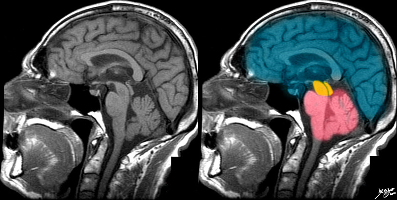
The T1 weighted MRI taken in sagittal projection reflects the 3 basic parts of the brain; forebrain (torquoise) midbrain (orange) and hind brain (salmon pink). Courtesy Ashley Davidoff MD copyright 2010 all rights reserved 49079b01b07.8s |
|
MRI of the Midbrain |
|
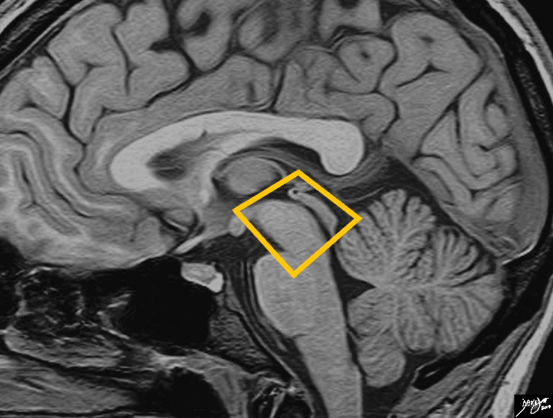
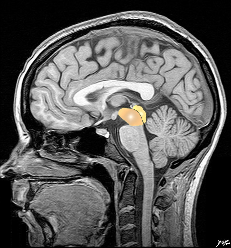
The midbrain is an almost rectangular structure that bridges the forebrain and hindbrain. It is the smallest of the 3 major components of the brain Davidoff art Image Courtesy Philips Medical System copyright 2010 92141.3kb03.81s |
|
Basic Divisions of the Midbrain Tegmentum and Tectum Cerebral Peduncles, Cerebral Crura and Colliculli
|
|
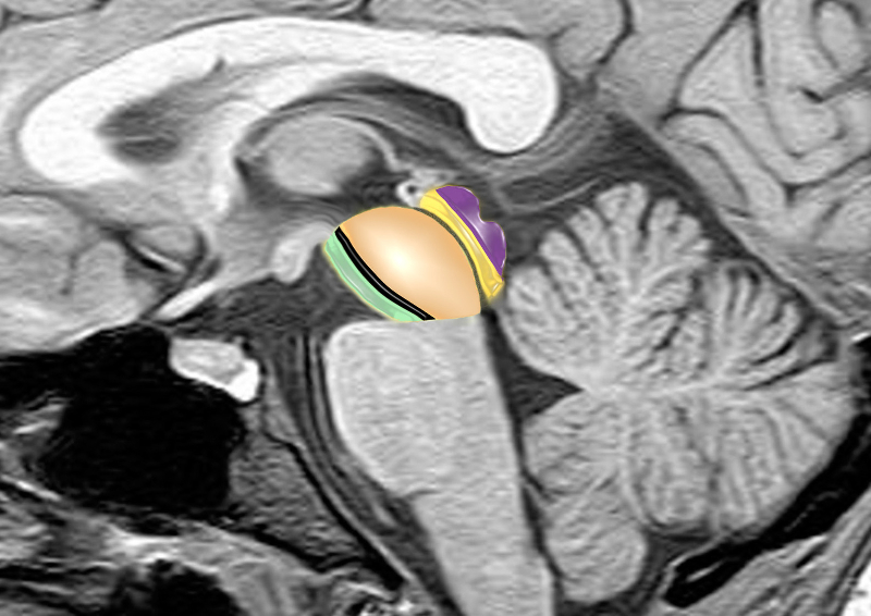
The midbrain consists of a larger anterior portion consisting of the cerebral peduncles, (green) the substantia nigra (black) and the tegmentum (light orange) that extends to the aqueduct (thin gray line) The posterior portion called tectum (orange) contains the colliculi (purple) which are the most posterior portions of the midbrain. brain anatomy neuroanatomy midbrain tegmentum colliculi tectum cerebral peduncle aqueduct of Sylvius conceptual diagram MRI principles Image provided by Philips Medical Systems Enhanced by Davidoff art Courtesy Ashley Davidoff MD 92141.3kb01ba02b02.8s |
DOMElement Object
(
    [schemaTypeInfo] =>
    [tagName] => table
    [firstElementChild] => (object value omitted)
    [lastElementChild] => (object value omitted)
    [childElementCount] => 1
    [previousElementSibling] => (object value omitted)
    [nextElementSibling] =>
    [nodeName] => table
    [nodeValue] =>


Basic Divisions of the Midbrain
Tegmentum and Tectum
Cerebral Peduncles, Cerebral Crura and Colliculli


The midbrain consists of a larger anterior portion consisting of the cerebral peduncles, (green) the substantia nigra (black) and the tegmentum (light orange) that extends to the aqueduct (thin gray line) The posterior portion called tectum (orange) contains the colliculi (purple) which are the most posterior portions of the midbrain. brain anatomy neuroanatomy midbrain tegmentum colliculi tectum cerebral peduncle aqueduct of Sylvius conceptual diagram MRI principles
Image provided by Philips Medical Systems Enhanced by Davidoff art Courtesy Ashley Davidoff MD 92141.3kb01ba02b02.8s


    [nodeType] => 1
    [parentNode] => (object value omitted)
    [childNodes] => (object value omitted)
    [firstChild] => (object value omitted)
    [lastChild] => (object value omitted)
    [previousSibling] => (object value omitted)
    [nextSibling] => (object value omitted)
    [attributes] => (object value omitted)
    [ownerDocument] => (object value omitted)
    [namespaceURI] =>
    [prefix] =>
    [localName] => table
    [baseURI] =>
    [textContent] =>


Basic Divisions of the Midbrain
Tegmentum and Tectum
Cerebral Peduncles, Cerebral Crura and Colliculli


The midbrain consists of a larger anterior portion consisting of the cerebral peduncles, (green) the substantia nigra (black) and the tegmentum (light orange) that extends to the aqueduct (thin gray line) The posterior portion called tectum (orange) contains the colliculi (purple) which are the most posterior portions of the midbrain. brain anatomy neuroanatomy midbrain tegmentum colliculi tectum cerebral peduncle aqueduct of Sylvius conceptual diagram MRI principles
Image provided by Philips Medical Systems Enhanced by Davidoff art Courtesy Ashley Davidoff MD 92141.3kb01ba02b02.8s


)
DOMElement Object
(
    [schemaTypeInfo] =>
    [tagName] => td
    [firstElementChild] => (object value omitted)
    [lastElementChild] => (object value omitted)
    [childElementCount] => 2
    [previousElementSibling] =>
    [nextElementSibling] =>
    [nodeName] => td
    [nodeValue] =>
The midbrain consists of a larger anterior portion consisting of the cerebral peduncles, (green) the substantia nigra (black) and the tegmentum (light orange) that extends to the aqueduct (thin gray line) The posterior portion called tectum (orange) contains the colliculi (purple) which are the most posterior portions of the midbrain. brain anatomy neuroanatomy midbrain tegmentum colliculi tectum cerebral peduncle aqueduct of Sylvius conceptual diagram MRI principles
Image provided by Philips Medical Systems Enhanced by Davidoff art Courtesy Ashley Davidoff MD 92141.3kb01ba02b02.8s

    [nodeType] => 1
    [parentNode] => (object value omitted)
    [childNodes] => (object value omitted)
    [firstChild] => (object value omitted)
    [lastChild] => (object value omitted)
    [previousSibling] => (object value omitted)
    [nextSibling] => (object value omitted)
    [attributes] => (object value omitted)
    [ownerDocument] => (object value omitted)
    [namespaceURI] =>
    [prefix] =>
    [localName] => td
    [baseURI] =>
    [textContent] =>
The midbrain consists of a larger anterior portion consisting of the cerebral peduncles, (green) the substantia nigra (black) and the tegmentum (light orange) that extends to the aqueduct (thin gray line) The posterior portion called tectum (orange) contains the colliculi (purple) which are the most posterior portions of the midbrain. brain anatomy neuroanatomy midbrain tegmentum colliculi tectum cerebral peduncle aqueduct of Sylvius conceptual diagram MRI principles
Image provided by Philips Medical Systems Enhanced by Davidoff art Courtesy Ashley Davidoff MD 92141.3kb01ba02b02.8s

)
DOMElement Object
(
    [schemaTypeInfo] =>
    [tagName] => td
    [firstElementChild] => (object value omitted)
    [lastElementChild] => (object value omitted)
    [childElementCount] => 5
    [previousElementSibling] =>
    [nextElementSibling] =>
    [nodeName] => td
    [nodeValue] =>

Basic Divisions of the Midbrain
Tegmentum and Tectum
Cerebral Peduncles, Cerebral Crura and Colliculli


    [nodeType] => 1
    [parentNode] => (object value omitted)
    [childNodes] => (object value omitted)
    [firstChild] => (object value omitted)
    [lastChild] => (object value omitted)
    [previousSibling] => (object value omitted)
    [nextSibling] => (object value omitted)
    [attributes] => (object value omitted)
    [ownerDocument] => (object value omitted)
    [namespaceURI] =>
    [prefix] =>
    [localName] => td
    [baseURI] =>
    [textContent] =>

Basic Divisions of the Midbrain
Tegmentum and Tectum
Cerebral Peduncles, Cerebral Crura and Colliculli


)
DOMElement Object
(
    [schemaTypeInfo] =>
    [tagName] => table
    [firstElementChild] => (object value omitted)
    [lastElementChild] => (object value omitted)
    [childElementCount] => 1
    [previousElementSibling] => (object value omitted)
    [nextElementSibling] => (object value omitted)
    [nodeName] => table
    [nodeValue] =>


Basic Parts of the Midbrain


The midbrain consists of a larger anterior portion called the cerebral peduncles incorporated into the tegmentum or floor of the midbrain (orange ).  The  posterior portion is called tectum or the roof  (yellow/orange) and are separated by the aqueduct of Sylvius (black)
Image provided by Philips Medical Systems Enhanced by Davidoff art Courtesy Ashley Davidoff MD 92141.3kb01bab02.8s


    [nodeType] => 1
    [parentNode] => (object value omitted)
    [childNodes] => (object value omitted)
    [firstChild] => (object value omitted)
    [lastChild] => (object value omitted)
    [previousSibling] => (object value omitted)
    [nextSibling] => (object value omitted)
    [attributes] => (object value omitted)
    [ownerDocument] => (object value omitted)
    [namespaceURI] =>
    [prefix] =>
    [localName] => table
    [baseURI] =>
    [textContent] =>


Basic Parts of the Midbrain


The midbrain consists of a larger anterior portion called the cerebral peduncles incorporated into the tegmentum or floor of the midbrain (orange ).  The  posterior portion is called tectum or the roof  (yellow/orange) and are separated by the aqueduct of Sylvius (black)
Image provided by Philips Medical Systems Enhanced by Davidoff art Courtesy Ashley Davidoff MD 92141.3kb01bab02.8s


)
DOMElement Object
(
    [schemaTypeInfo] =>
    [tagName] => td
    [firstElementChild] => (object value omitted)
    [lastElementChild] => (object value omitted)
    [childElementCount] => 2
    [previousElementSibling] =>
    [nextElementSibling] =>
    [nodeName] => td
    [nodeValue] =>
The midbrain consists of a larger anterior portion called the cerebral peduncles incorporated into the tegmentum or floor of the midbrain (orange ).  The  posterior portion is called tectum or the roof  (yellow/orange) and are separated by the aqueduct of Sylvius (black)
Image provided by Philips Medical Systems Enhanced by Davidoff art Courtesy Ashley Davidoff MD 92141.3kb01bab02.8s

    [nodeType] => 1
    [parentNode] => (object value omitted)
    [childNodes] => (object value omitted)
    [firstChild] => (object value omitted)
    [lastChild] => (object value omitted)
    [previousSibling] => (object value omitted)
    [nextSibling] => (object value omitted)
    [attributes] => (object value omitted)
    [ownerDocument] => (object value omitted)
    [namespaceURI] =>
    [prefix] =>
    [localName] => td
    [baseURI] =>
    [textContent] =>
The midbrain consists of a larger anterior portion called the cerebral peduncles incorporated into the tegmentum or floor of the midbrain (orange ).  The  posterior portion is called tectum or the roof  (yellow/orange) and are separated by the aqueduct of Sylvius (black)
Image provided by Philips Medical Systems Enhanced by Davidoff art Courtesy Ashley Davidoff MD 92141.3kb01bab02.8s

)
DOMElement Object
(
    [schemaTypeInfo] =>
    [tagName] => td
    [firstElementChild] => (object value omitted)
    [lastElementChild] => (object value omitted)
    [childElementCount] => 2
    [previousElementSibling] =>
    [nextElementSibling] =>
    [nodeName] => td
    [nodeValue] =>
Basic Parts of the Midbrain

    [nodeType] => 1
    [parentNode] => (object value omitted)
    [childNodes] => (object value omitted)
    [firstChild] => (object value omitted)
    [lastChild] => (object value omitted)
    [previousSibling] => (object value omitted)
    [nextSibling] => (object value omitted)
    [attributes] => (object value omitted)
    [ownerDocument] => (object value omitted)
    [namespaceURI] =>
    [prefix] =>
    [localName] => td
    [baseURI] =>
    [textContent] =>
Basic Parts of the Midbrain

)
DOMElement Object
(
    [schemaTypeInfo] =>
    [tagName] => table
    [firstElementChild] => (object value omitted)
    [lastElementChild] => (object value omitted)
    [childElementCount] => 1
    [previousElementSibling] => (object value omitted)
    [nextElementSibling] => (object value omitted)
    [nodeName] => table
    [nodeValue] =>


MRI of the Midbrain


The midbrain is an almost rectangular structure that bridges the forebrain and hindbrain. It is the smallest of the 3 major components of the brain
Davidoff art Image Courtesy Philips Medical System copyright 2010 92141.3kb03.81s


    [nodeType] => 1
    [parentNode] => (object value omitted)
    [childNodes] => (object value omitted)
    [firstChild] => (object value omitted)
    [lastChild] => (object value omitted)
    [previousSibling] => (object value omitted)
    [nextSibling] => (object value omitted)
    [attributes] => (object value omitted)
    [ownerDocument] => (object value omitted)
    [namespaceURI] =>
    [prefix] =>
    [localName] => table
    [baseURI] =>
    [textContent] =>


MRI of the Midbrain


The midbrain is an almost rectangular structure that bridges the forebrain and hindbrain. It is the smallest of the 3 major components of the brain
Davidoff art Image Courtesy Philips Medical System copyright 2010 92141.3kb03.81s


)
DOMElement Object
(
    [schemaTypeInfo] =>
    [tagName] => td
    [firstElementChild] => (object value omitted)
    [lastElementChild] => (object value omitted)
    [childElementCount] => 2
    [previousElementSibling] =>
    [nextElementSibling] =>
    [nodeName] => td
    [nodeValue] =>
The midbrain is an almost rectangular structure that bridges the forebrain and hindbrain. It is the smallest of the 3 major components of the brain
Davidoff art Image Courtesy Philips Medical System copyright 2010 92141.3kb03.81s

    [nodeType] => 1
    [parentNode] => (object value omitted)
    [childNodes] => (object value omitted)
    [firstChild] => (object value omitted)
    [lastChild] => (object value omitted)
    [previousSibling] => (object value omitted)
    [nextSibling] => (object value omitted)
    [attributes] => (object value omitted)
    [ownerDocument] => (object value omitted)
    [namespaceURI] =>
    [prefix] =>
    [localName] => td
    [baseURI] =>
    [textContent] =>
The midbrain is an almost rectangular structure that bridges the forebrain and hindbrain. It is the smallest of the 3 major components of the brain
Davidoff art Image Courtesy Philips Medical System copyright 2010 92141.3kb03.81s

)
DOMElement Object
(
    [schemaTypeInfo] =>
    [tagName] => td
    [firstElementChild] => (object value omitted)
    [lastElementChild] => (object value omitted)
    [childElementCount] => 2
    [previousElementSibling] =>
    [nextElementSibling] =>
    [nodeName] => td
    [nodeValue] =>

MRI of the Midbrain
    [nodeType] => 1
    [parentNode] => (object value omitted)
    [childNodes] => (object value omitted)
    [firstChild] => (object value omitted)
    [lastChild] => (object value omitted)
    [previousSibling] => (object value omitted)
    [nextSibling] => (object value omitted)
    [attributes] => (object value omitted)
    [ownerDocument] => (object value omitted)
    [namespaceURI] =>
    [prefix] =>
    [localName] => td
    [baseURI] =>
    [textContent] =>

MRI of the Midbrain
)
DOMElement Object
(
    [schemaTypeInfo] =>
    [tagName] => table
    [firstElementChild] => (object value omitted)
    [lastElementChild] => (object value omitted)
    [childElementCount] => 1
    [previousElementSibling] => (object value omitted)
    [nextElementSibling] => (object value omitted)
    [nodeName] => table
    [nodeValue] =>


Midbrain – bridge Between the Fore and Hindbrain


The T1 weighted MRI taken in sagittal projection reflects the 3 basic parts of the brain; forebrain (torquoise) midbrain (orange) and hind brain (salmon pink).
Courtesy Ashley Davidoff MD copyright 2010 all rights reserved 49079b01b07.8s


    [nodeType] => 1
    [parentNode] => (object value omitted)
    [childNodes] => (object value omitted)
    [firstChild] => (object value omitted)
    [lastChild] => (object value omitted)
    [previousSibling] => (object value omitted)
    [nextSibling] => (object value omitted)
    [attributes] => (object value omitted)
    [ownerDocument] => (object value omitted)
    [namespaceURI] =>
    [prefix] =>
    [localName] => table
    [baseURI] =>
    [textContent] =>


Midbrain – bridge Between the Fore and Hindbrain


The T1 weighted MRI taken in sagittal projection reflects the 3 basic parts of the brain; forebrain (torquoise) midbrain (orange) and hind brain (salmon pink).
Courtesy Ashley Davidoff MD copyright 2010 all rights reserved 49079b01b07.8s


)
DOMElement Object
(
    [schemaTypeInfo] =>
    [tagName] => td
    [firstElementChild] => (object value omitted)
    [lastElementChild] => (object value omitted)
    [childElementCount] => 2
    [previousElementSibling] =>
    [nextElementSibling] =>
    [nodeName] => td
    [nodeValue] =>
The T1 weighted MRI taken in sagittal projection reflects the 3 basic parts of the brain; forebrain (torquoise) midbrain (orange) and hind brain (salmon pink).
Courtesy Ashley Davidoff MD copyright 2010 all rights reserved 49079b01b07.8s

    [nodeType] => 1
    [parentNode] => (object value omitted)
    [childNodes] => (object value omitted)
    [firstChild] => (object value omitted)
    [lastChild] => (object value omitted)
    [previousSibling] => (object value omitted)
    [nextSibling] => (object value omitted)
    [attributes] => (object value omitted)
    [ownerDocument] => (object value omitted)
    [namespaceURI] =>
    [prefix] =>
    [localName] => td
    [baseURI] =>
    [textContent] =>
The T1 weighted MRI taken in sagittal projection reflects the 3 basic parts of the brain; forebrain (torquoise) midbrain (orange) and hind brain (salmon pink).
Courtesy Ashley Davidoff MD copyright 2010 all rights reserved 49079b01b07.8s

)
DOMElement Object
(
    [schemaTypeInfo] =>
    [tagName] => td
    [firstElementChild] => (object value omitted)
    [lastElementChild] => (object value omitted)
    [childElementCount] => 2
    [previousElementSibling] =>
    [nextElementSibling] =>
    [nodeName] => td
    [nodeValue] =>

Midbrain – bridge Between the Fore and Hindbrain

    [nodeType] => 1
    [parentNode] => (object value omitted)
    [childNodes] => (object value omitted)
    [firstChild] => (object value omitted)
    [lastChild] => (object value omitted)
    [previousSibling] => (object value omitted)
    [nextSibling] => (object value omitted)
    [attributes] => (object value omitted)
    [ownerDocument] => (object value omitted)
    [namespaceURI] =>
    [prefix] =>
    [localName] => td
    [baseURI] =>
    [textContent] =>

Midbrain – bridge Between the Fore and Hindbrain

)
DOMElement Object
(
    [schemaTypeInfo] =>
    [tagName] => table
    [firstElementChild] => (object value omitted)
    [lastElementChild] => (object value omitted)
    [childElementCount] => 1
    [previousElementSibling] => (object value omitted)
    [nextElementSibling] => (object value omitted)
    [nodeName] => table
    [nodeValue] =>


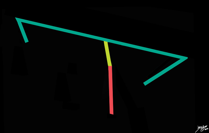
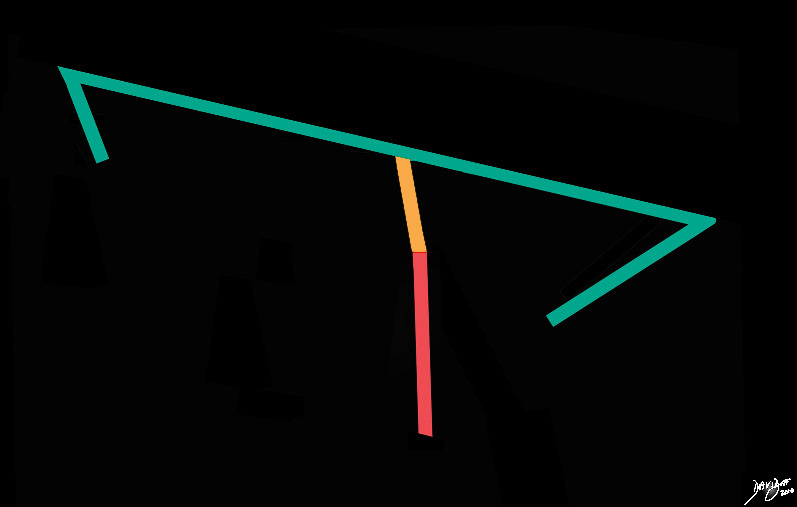
The Midbrain (orange) – A Bridge from the Forebrain (green)  to the Hindbrain (salmon)


The stick diagram starts to become more complex as the forebrain (green) demonstrates folds on its anterior and posterior extremes representing the fold of the frontal lobe anteriorly and temporal lobe posteriorly. The vertical limb also has a subtle angulation between the midbrain (yellow) and hind brain (salmon red).
Davidoff art Courtesy Ashley Davidoff MD copyright 2010 all rights reserved 93887b03a.81s


    [nodeType] => 1
    [parentNode] => (object value omitted)
    [childNodes] => (object value omitted)
    [firstChild] => (object value omitted)
    [lastChild] => (object value omitted)
    [previousSibling] => (object value omitted)
    [nextSibling] => (object value omitted)
    [attributes] => (object value omitted)
    [ownerDocument] => (object value omitted)
    [namespaceURI] =>
    [prefix] =>
    [localName] => table
    [baseURI] =>
    [textContent] =>


The Midbrain (orange) – A Bridge from the Forebrain (green)  to the Hindbrain (salmon)


The stick diagram starts to become more complex as the forebrain (green) demonstrates folds on its anterior and posterior extremes representing the fold of the frontal lobe anteriorly and temporal lobe posteriorly. The vertical limb also has a subtle angulation between the midbrain (yellow) and hind brain (salmon red).
Davidoff art Courtesy Ashley Davidoff MD copyright 2010 all rights reserved 93887b03a.81s


)
DOMElement Object
(
    [schemaTypeInfo] =>
    [tagName] => td
    [firstElementChild] => (object value omitted)
    [lastElementChild] => (object value omitted)
    [childElementCount] => 2
    [previousElementSibling] =>
    [nextElementSibling] =>
    [nodeName] => td
    [nodeValue] =>
The stick diagram starts to become more complex as the forebrain (green) demonstrates folds on its anterior and posterior extremes representing the fold of the frontal lobe anteriorly and temporal lobe posteriorly. The vertical limb also has a subtle angulation between the midbrain (yellow) and hind brain (salmon red).
Davidoff art Courtesy Ashley Davidoff MD copyright 2010 all rights reserved 93887b03a.81s

    [nodeType] => 1
    [parentNode] => (object value omitted)
    [childNodes] => (object value omitted)
    [firstChild] => (object value omitted)
    [lastChild] => (object value omitted)
    [previousSibling] => (object value omitted)
    [nextSibling] => (object value omitted)
    [attributes] => (object value omitted)
    [ownerDocument] => (object value omitted)
    [namespaceURI] =>
    [prefix] =>
    [localName] => td
    [baseURI] =>
    [textContent] =>
The stick diagram starts to become more complex as the forebrain (green) demonstrates folds on its anterior and posterior extremes representing the fold of the frontal lobe anteriorly and temporal lobe posteriorly. The vertical limb also has a subtle angulation between the midbrain (yellow) and hind brain (salmon red).
Davidoff art Courtesy Ashley Davidoff MD copyright 2010 all rights reserved 93887b03a.81s

)
DOMElement Object
(
    [schemaTypeInfo] =>
    [tagName] => td
    [firstElementChild] => (object value omitted)
    [lastElementChild] => (object value omitted)
    [childElementCount] => 2
    [previousElementSibling] =>
    [nextElementSibling] =>
    [nodeName] => td
    [nodeValue] =>
The Midbrain (orange) – A Bridge from the Forebrain (green)  to the Hindbrain (salmon)
    [nodeType] => 1
    [parentNode] => (object value omitted)
    [childNodes] => (object value omitted)
    [firstChild] => (object value omitted)
    [lastChild] => (object value omitted)
    [previousSibling] => (object value omitted)
    [nextSibling] => (object value omitted)
    [attributes] => (object value omitted)
    [ownerDocument] => (object value omitted)
    [namespaceURI] =>
    [prefix] =>
    [localName] => td
    [baseURI] =>
    [textContent] =>
The Midbrain (orange) – A Bridge from the Forebrain (green)  to the Hindbrain (salmon)
)

