DOMElement Object
(
    [schemaTypeInfo] =>
    [tagName] => table
    [firstElementChild] => (object value omitted)
    [lastElementChild] => (object value omitted)
    [childElementCount] => 1
    [previousElementSibling] => (object value omitted)
    [nextElementSibling] =>
    [nodeName] => table
    [nodeValue] =>


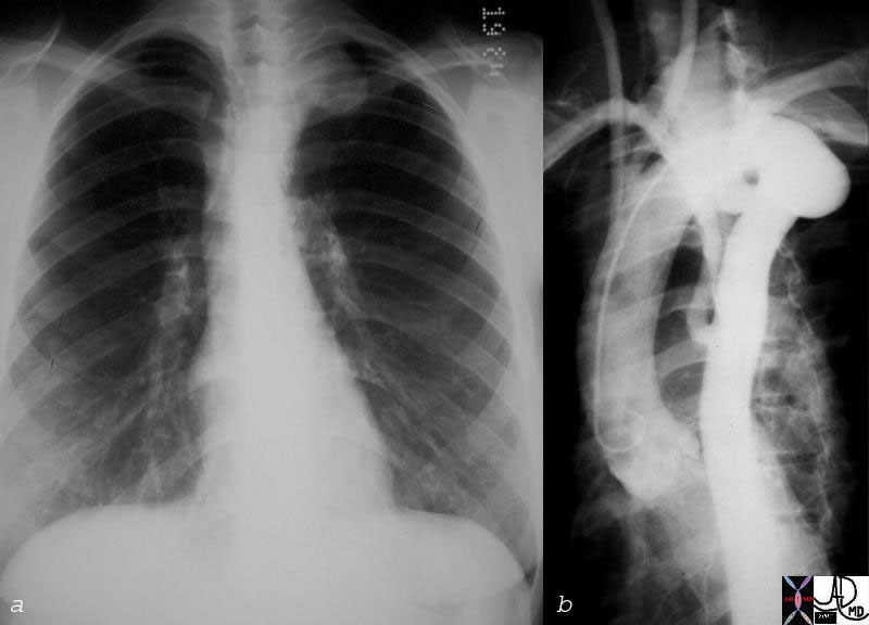
Cervical Left Sided Aortic Arch with Severe Kinking and Aberrant Right Subclavian Artery


35361c01 aorta arch cervical arch pseudocoarctation angiogram angiography Courtesy Laura Feldman MD


    [nodeType] => 1
    [parentNode] => (object value omitted)
    [childNodes] => (object value omitted)
    [firstChild] => (object value omitted)
    [lastChild] => (object value omitted)
    [previousSibling] => (object value omitted)
    [nextSibling] => (object value omitted)
    [attributes] => (object value omitted)
    [ownerDocument] => (object value omitted)
    [namespaceURI] =>
    [prefix] =>
    [localName] => table
    [baseURI] =>
    [textContent] =>


Cervical Left Sided Aortic Arch with Severe Kinking and Aberrant Right Subclavian Artery


35361c01 aorta arch cervical arch pseudocoarctation angiogram angiography Courtesy Laura Feldman MD


)
DOMElement Object
(
    [schemaTypeInfo] =>
    [tagName] => td
    [firstElementChild] => (object value omitted)
    [lastElementChild] => (object value omitted)
    [childElementCount] => 1
    [previousElementSibling] =>
    [nextElementSibling] =>
    [nodeName] => td
    [nodeValue] => 35361c01 aorta arch cervical arch pseudocoarctation angiogram angiography Courtesy Laura Feldman MD
    [nodeType] => 1
    [parentNode] => (object value omitted)
    [childNodes] => (object value omitted)
    [firstChild] => (object value omitted)
    [lastChild] => (object value omitted)
    [previousSibling] => (object value omitted)
    [nextSibling] => (object value omitted)
    [attributes] => (object value omitted)
    [ownerDocument] => (object value omitted)
    [namespaceURI] =>
    [prefix] =>
    [localName] => td
    [baseURI] =>
    [textContent] => 35361c01 aorta arch cervical arch pseudocoarctation angiogram angiography Courtesy Laura Feldman MD
)
DOMElement Object
(
    [schemaTypeInfo] =>
    [tagName] => td
    [firstElementChild] => (object value omitted)
    [lastElementChild] => (object value omitted)
    [childElementCount] => 2
    [previousElementSibling] =>
    [nextElementSibling] =>
    [nodeName] => td
    [nodeValue] =>
Cervical Left Sided Aortic Arch with Severe Kinking and Aberrant Right Subclavian Artery

    [nodeType] => 1
    [parentNode] => (object value omitted)
    [childNodes] => (object value omitted)
    [firstChild] => (object value omitted)
    [lastChild] => (object value omitted)
    [previousSibling] => (object value omitted)
    [nextSibling] => (object value omitted)
    [attributes] => (object value omitted)
    [ownerDocument] => (object value omitted)
    [namespaceURI] =>
    [prefix] =>
    [localName] => td
    [baseURI] =>
    [textContent] =>
Cervical Left Sided Aortic Arch with Severe Kinking and Aberrant Right Subclavian Artery

)
DOMElement Object
(
    [schemaTypeInfo] =>
    [tagName] => table
    [firstElementChild] => (object value omitted)
    [lastElementChild] => (object value omitted)
    [childElementCount] => 1
    [previousElementSibling] => (object value omitted)
    [nextElementSibling] => (object value omitted)
    [nodeName] => table
    [nodeValue] =>


Cervical Arch and Pseudocoarctation


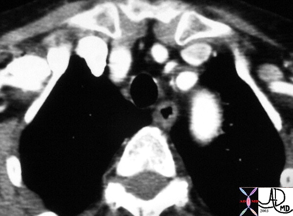
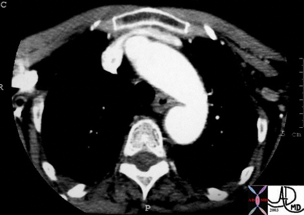
This cross sectional view of the chest at the apex of the lungs at the level of the manubrial notch shows the top of the aortic ach which is more superior than usual and is known as a cervical arch. The aorta more inferiorly is also redundant and tends to kink eventually appearing as a pseudocoarctation. In this case only a kink is present.
Courtesy Ashley Davidoff MD 33539 see 33535 code aorta arch cervical kink pseudocoarctation imaging radiology CTscan


    [nodeType] => 1
    [parentNode] => (object value omitted)
    [childNodes] => (object value omitted)
    [firstChild] => (object value omitted)
    [lastChild] => (object value omitted)
    [previousSibling] => (object value omitted)
    [nextSibling] => (object value omitted)
    [attributes] => (object value omitted)
    [ownerDocument] => (object value omitted)
    [namespaceURI] =>
    [prefix] =>
    [localName] => table
    [baseURI] =>
    [textContent] =>


Cervical Arch and Pseudocoarctation


This cross sectional view of the chest at the apex of the lungs at the level of the manubrial notch shows the top of the aortic ach which is more superior than usual and is known as a cervical arch. The aorta more inferiorly is also redundant and tends to kink eventually appearing as a pseudocoarctation. In this case only a kink is present.
Courtesy Ashley Davidoff MD 33539 see 33535 code aorta arch cervical kink pseudocoarctation imaging radiology CTscan


)
DOMElement Object
(
    [schemaTypeInfo] =>
    [tagName] => td
    [firstElementChild] => (object value omitted)
    [lastElementChild] => (object value omitted)
    [childElementCount] => 2
    [previousElementSibling] =>
    [nextElementSibling] =>
    [nodeName] => td
    [nodeValue] => This cross sectional view of the chest at the apex of the lungs at the level of the manubrial notch shows the top of the aortic ach which is more superior than usual and is known as a cervical arch. The aorta more inferiorly is also redundant and tends to kink eventually appearing as a pseudocoarctation. In this case only a kink is present.
Courtesy Ashley Davidoff MD 33539 see 33535 code aorta arch cervical kink pseudocoarctation imaging radiology CTscan
    [nodeType] => 1
    [parentNode] => (object value omitted)
    [childNodes] => (object value omitted)
    [firstChild] => (object value omitted)
    [lastChild] => (object value omitted)
    [previousSibling] => (object value omitted)
    [nextSibling] => (object value omitted)
    [attributes] => (object value omitted)
    [ownerDocument] => (object value omitted)
    [namespaceURI] =>
    [prefix] =>
    [localName] => td
    [baseURI] =>
    [textContent] => This cross sectional view of the chest at the apex of the lungs at the level of the manubrial notch shows the top of the aortic ach which is more superior than usual and is known as a cervical arch. The aorta more inferiorly is also redundant and tends to kink eventually appearing as a pseudocoarctation. In this case only a kink is present.
Courtesy Ashley Davidoff MD 33539 see 33535 code aorta arch cervical kink pseudocoarctation imaging radiology CTscan
)
DOMElement Object
(
    [schemaTypeInfo] =>
    [tagName] => td
    [firstElementChild] => (object value omitted)
    [lastElementChild] => (object value omitted)
    [childElementCount] => 3
    [previousElementSibling] =>
    [nextElementSibling] =>
    [nodeName] => td
    [nodeValue] =>
Cervical Arch and Pseudocoarctation

    [nodeType] => 1
    [parentNode] => (object value omitted)
    [childNodes] => (object value omitted)
    [firstChild] => (object value omitted)
    [lastChild] => (object value omitted)
    [previousSibling] => (object value omitted)
    [nextSibling] => (object value omitted)
    [attributes] => (object value omitted)
    [ownerDocument] => (object value omitted)
    [namespaceURI] =>
    [prefix] =>
    [localName] => td
    [baseURI] =>
    [textContent] =>
Cervical Arch and Pseudocoarctation

)
DOMElement Object
(
    [schemaTypeInfo] =>
    [tagName] => table
    [firstElementChild] => (object value omitted)
    [lastElementChild] => (object value omitted)
    [childElementCount] => 1
    [previousElementSibling] =>
    [nextElementSibling] =>
    [nodeName] => table
    [nodeValue] =>


Pseudocoarctation of the Aorta
The Common Vein Copyright 2008
Definition
Pseudocoarctation of the aorta is a narrowing of the aorta that has both congenital and acquired forms.
Its is caused by kinking or buckling of an elongated thoracic aortic arch distal to the origin of the left subclavian artery without meausarable gradient. Asociated diseases include a cervical arch, bicuspid aortic valve, VSD, PDA, and it is also seen in middle aged men with hypertension.
Diagnosis is made by angiography, CT or MRI where the kinking of the proximal thoracic aorta, in the absence of the a measurable gradient is noted. Plain film may suggest the diagnosis where a “three” sign reminscent of true coarctation in both the P-A and lateral can be seen. Rib notching is absent. There is no therapy needed since there is no functional abnormality.


Cervical Arch and Pseudocoarctation


This cross sectional view of the chest at the apex of the lungs at the level of the manubrial notch shows the top of the aortic ach which is more superior than usual and is known as a cervical arch. The aorta more inferiorly is also redundant and tends to kink eventually appearing as a pseudocoarctation. In this case only a kink is present.
Courtesy Ashley Davidoff MD 33539 see 33535 code aorta arch cervical kink pseudocoarctation imaging radiology CTscan


Cervical Left Sided Aortic Arch with Severe Kinking and Aberrant Right Subclavian Artery


35361c01 aorta arch cervical arch pseudocoarctation angiogram angiography Courtesy Laura Feldman MD


    [nodeType] => 1
    [parentNode] => (object value omitted)
    [childNodes] => (object value omitted)
    [firstChild] => (object value omitted)
    [lastChild] => (object value omitted)
    [previousSibling] =>
    [nextSibling] => (object value omitted)
    [attributes] => (object value omitted)
    [ownerDocument] => (object value omitted)
    [namespaceURI] =>
    [prefix] =>
    [localName] => table
    [baseURI] =>
    [textContent] =>


Pseudocoarctation of the Aorta
The Common Vein Copyright 2008
Definition
Pseudocoarctation of the aorta is a narrowing of the aorta that has both congenital and acquired forms.
Its is caused by kinking or buckling of an elongated thoracic aortic arch distal to the origin of the left subclavian artery without meausarable gradient. Asociated diseases include a cervical arch, bicuspid aortic valve, VSD, PDA, and it is also seen in middle aged men with hypertension.
Diagnosis is made by angiography, CT or MRI where the kinking of the proximal thoracic aorta, in the absence of the a measurable gradient is noted. Plain film may suggest the diagnosis where a “three” sign reminscent of true coarctation in both the P-A and lateral can be seen. Rib notching is absent. There is no therapy needed since there is no functional abnormality.


Cervical Arch and Pseudocoarctation


This cross sectional view of the chest at the apex of the lungs at the level of the manubrial notch shows the top of the aortic ach which is more superior than usual and is known as a cervical arch. The aorta more inferiorly is also redundant and tends to kink eventually appearing as a pseudocoarctation. In this case only a kink is present.
Courtesy Ashley Davidoff MD 33539 see 33535 code aorta arch cervical kink pseudocoarctation imaging radiology CTscan


Cervical Left Sided Aortic Arch with Severe Kinking and Aberrant Right Subclavian Artery


35361c01 aorta arch cervical arch pseudocoarctation angiogram angiography Courtesy Laura Feldman MD


)
DOMElement Object
(
    [schemaTypeInfo] =>
    [tagName] => td
    [firstElementChild] => (object value omitted)
    [lastElementChild] => (object value omitted)
    [childElementCount] => 1
    [previousElementSibling] =>
    [nextElementSibling] =>
    [nodeName] => td
    [nodeValue] => 35361c01 aorta arch cervical arch pseudocoarctation angiogram angiography Courtesy Laura Feldman MD
    [nodeType] => 1
    [parentNode] => (object value omitted)
    [childNodes] => (object value omitted)
    [firstChild] => (object value omitted)
    [lastChild] => (object value omitted)
    [previousSibling] => (object value omitted)
    [nextSibling] => (object value omitted)
    [attributes] => (object value omitted)
    [ownerDocument] => (object value omitted)
    [namespaceURI] =>
    [prefix] =>
    [localName] => td
    [baseURI] =>
    [textContent] => 35361c01 aorta arch cervical arch pseudocoarctation angiogram angiography Courtesy Laura Feldman MD
)
DOMElement Object
(
    [schemaTypeInfo] =>
    [tagName] => td
    [firstElementChild] => (object value omitted)
    [lastElementChild] => (object value omitted)
    [childElementCount] => 2
    [previousElementSibling] =>
    [nextElementSibling] =>
    [nodeName] => td
    [nodeValue] =>
Cervical Left Sided Aortic Arch with Severe Kinking and Aberrant Right Subclavian Artery

    [nodeType] => 1
    [parentNode] => (object value omitted)
    [childNodes] => (object value omitted)
    [firstChild] => (object value omitted)
    [lastChild] => (object value omitted)
    [previousSibling] => (object value omitted)
    [nextSibling] => (object value omitted)
    [attributes] => (object value omitted)
    [ownerDocument] => (object value omitted)
    [namespaceURI] =>
    [prefix] =>
    [localName] => td
    [baseURI] =>
    [textContent] =>
Cervical Left Sided Aortic Arch with Severe Kinking and Aberrant Right Subclavian Artery

)
DOMElement Object
(
    [schemaTypeInfo] =>
    [tagName] => td
    [firstElementChild] => (object value omitted)
    [lastElementChild] => (object value omitted)
    [childElementCount] => 2
    [previousElementSibling] =>
    [nextElementSibling] =>
    [nodeName] => td
    [nodeValue] => This cross sectional view of the chest at the apex of the lungs at the level of the manubrial notch shows the top of the aortic ach which is more superior than usual and is known as a cervical arch. The aorta more inferiorly is also redundant and tends to kink eventually appearing as a pseudocoarctation. In this case only a kink is present.
Courtesy Ashley Davidoff MD 33539 see 33535 code aorta arch cervical kink pseudocoarctation imaging radiology CTscan
    [nodeType] => 1
    [parentNode] => (object value omitted)
    [childNodes] => (object value omitted)
    [firstChild] => (object value omitted)
    [lastChild] => (object value omitted)
    [previousSibling] => (object value omitted)
    [nextSibling] => (object value omitted)
    [attributes] => (object value omitted)
    [ownerDocument] => (object value omitted)
    [namespaceURI] =>
    [prefix] =>
    [localName] => td
    [baseURI] =>
    [textContent] => This cross sectional view of the chest at the apex of the lungs at the level of the manubrial notch shows the top of the aortic ach which is more superior than usual and is known as a cervical arch. The aorta more inferiorly is also redundant and tends to kink eventually appearing as a pseudocoarctation. In this case only a kink is present.
Courtesy Ashley Davidoff MD 33539 see 33535 code aorta arch cervical kink pseudocoarctation imaging radiology CTscan
)
https://beta.thecommonvein.net/wp-content/uploads/2023/05/35361c01.jpg
DOMElement Object
(
    [schemaTypeInfo] =>
    [tagName] => td
    [firstElementChild] => (object value omitted)
    [lastElementChild] => (object value omitted)
    [childElementCount] => 3
    [previousElementSibling] =>
    [nextElementSibling] =>
    [nodeName] => td
    [nodeValue] =>
Cervical Arch and Pseudocoarctation

    [nodeType] => 1
    [parentNode] => (object value omitted)
    [childNodes] => (object value omitted)
    [firstChild] => (object value omitted)
    [lastChild] => (object value omitted)
    [previousSibling] => (object value omitted)
    [nextSibling] => (object value omitted)
    [attributes] => (object value omitted)
    [ownerDocument] => (object value omitted)
    [namespaceURI] =>
    [prefix] =>
    [localName] => td
    [baseURI] =>
    [textContent] =>
Cervical Arch and Pseudocoarctation

)
https://beta.thecommonvein.net/wp-content/uploads/2023/05/35361c01.jpg
http://thecommonvein.net/media/33535.jpg http://thecommonvein.net/media/33539.jpg
DOMElement Object
(
    [schemaTypeInfo] =>
    [tagName] => td
    [firstElementChild] => (object value omitted)
    [lastElementChild] => (object value omitted)
    [childElementCount] => 8
    [previousElementSibling] =>
    [nextElementSibling] =>
    [nodeName] => td
    [nodeValue] =>
Pseudocoarctation of the Aorta
The Common Vein Copyright 2008
Definition
Pseudocoarctation of the aorta is a narrowing of the aorta that has both congenital and acquired forms.
Its is caused by kinking or buckling of an elongated thoracic aortic arch distal to the origin of the left subclavian artery without meausarable gradient. Asociated diseases include a cervical arch, bicuspid aortic valve, VSD, PDA, and it is also seen in middle aged men with hypertension.
Diagnosis is made by angiography, CT or MRI where the kinking of the proximal thoracic aorta, in the absence of the a measurable gradient is noted. Plain film may suggest the diagnosis where a “three” sign reminscent of true coarctation in both the P-A and lateral can be seen. Rib notching is absent. There is no therapy needed since there is no functional abnormality.


Cervical Arch and Pseudocoarctation


This cross sectional view of the chest at the apex of the lungs at the level of the manubrial notch shows the top of the aortic ach which is more superior than usual and is known as a cervical arch. The aorta more inferiorly is also redundant and tends to kink eventually appearing as a pseudocoarctation. In this case only a kink is present.
Courtesy Ashley Davidoff MD 33539 see 33535 code aorta arch cervical kink pseudocoarctation imaging radiology CTscan


Cervical Left Sided Aortic Arch with Severe Kinking and Aberrant Right Subclavian Artery


35361c01 aorta arch cervical arch pseudocoarctation angiogram angiography Courtesy Laura Feldman MD


    [nodeType] => 1
    [parentNode] => (object value omitted)
    [childNodes] => (object value omitted)
    [firstChild] => (object value omitted)
    [lastChild] => (object value omitted)
    [previousSibling] => (object value omitted)
    [nextSibling] => (object value omitted)
    [attributes] => (object value omitted)
    [ownerDocument] => (object value omitted)
    [namespaceURI] =>
    [prefix] =>
    [localName] => td
    [baseURI] =>
    [textContent] =>
Pseudocoarctation of the Aorta
The Common Vein Copyright 2008
Definition
Pseudocoarctation of the aorta is a narrowing of the aorta that has both congenital and acquired forms.
Its is caused by kinking or buckling of an elongated thoracic aortic arch distal to the origin of the left subclavian artery without meausarable gradient. Asociated diseases include a cervical arch, bicuspid aortic valve, VSD, PDA, and it is also seen in middle aged men with hypertension.
Diagnosis is made by angiography, CT or MRI where the kinking of the proximal thoracic aorta, in the absence of the a measurable gradient is noted. Plain film may suggest the diagnosis where a “three” sign reminscent of true coarctation in both the P-A and lateral can be seen. Rib notching is absent. There is no therapy needed since there is no functional abnormality.


Cervical Arch and Pseudocoarctation


This cross sectional view of the chest at the apex of the lungs at the level of the manubrial notch shows the top of the aortic ach which is more superior than usual and is known as a cervical arch. The aorta more inferiorly is also redundant and tends to kink eventually appearing as a pseudocoarctation. In this case only a kink is present.
Courtesy Ashley Davidoff MD 33539 see 33535 code aorta arch cervical kink pseudocoarctation imaging radiology CTscan


Cervical Left Sided Aortic Arch with Severe Kinking and Aberrant Right Subclavian Artery


35361c01 aorta arch cervical arch pseudocoarctation angiogram angiography Courtesy Laura Feldman MD


)


