Author
Assistant
Introduction
In the Saggital Plane
|
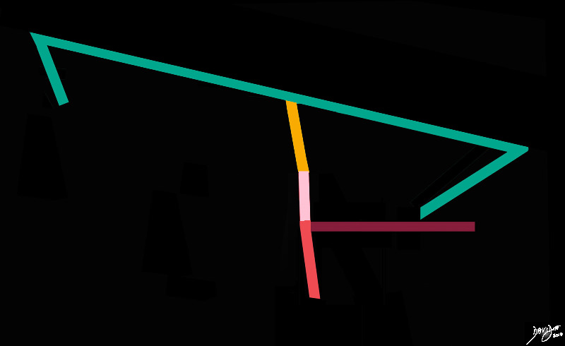
The Cerebellum (maroon) ? Part of the Hindbrain Stick Diagram |
|
The hindbrain consists of 3 major components;the pons (light pink, the medulla, (salmon) and the cerebellum (maroon) Courtesy Ashley DAvidoff MD copyright 2010 93887b03ab03.8s |
|
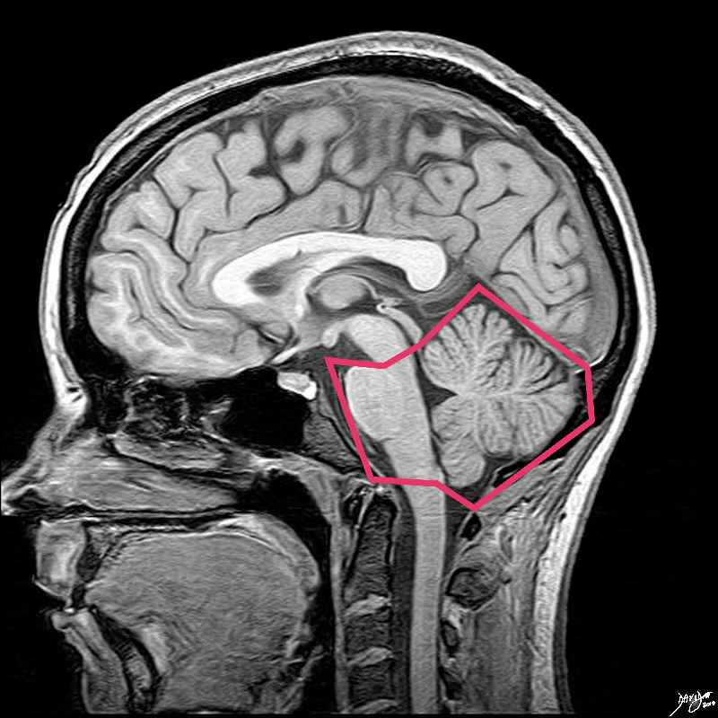
The Hindbrain Posterior and Largest Part of the Hindbrain |
|
The hindbrain in the sagital plane is bordered in salmon pink and consists of the pons, the medulla, and the cerebellum Image Courtesy Philips Medical Systems Rendered by Ashley Davidoff MD Copyright 2010 92141.3ka.8s |
|
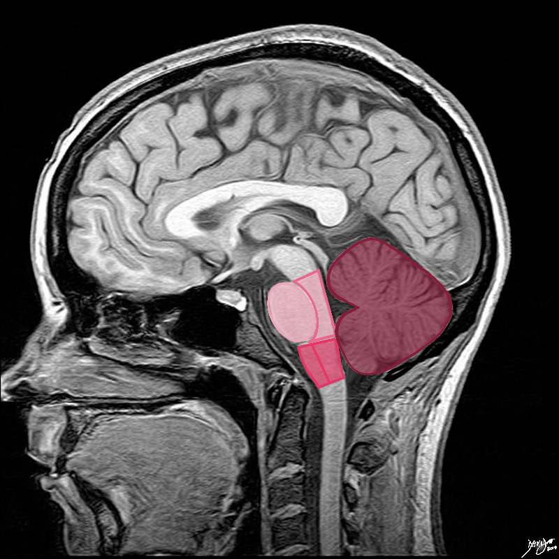
Advancing the Parts of the Hindbrain |
|
The shapes in fact are not quite ovoid. The pons (lightest pink) has shapes reminiscent of an oversize egg lying in a small bed The anterior eggshaped portion is called the ventral or anterior pons and the posterior portion called the tegmentum which is a continuation of the tegmentum of the midbrain. The medulla oblongata is almost rectangular but has a subtle forward bulging to its anterior surface. The anterior portion is called the ventral or anterior medulla and the posterior portion is called the tegmentum,similar in name a nad a continuation of th etegmentum of the midbrain and pons The cerebellum is situated posteriorly, is the largest of the three major components and it consists of two hemispheres and a central vermis better appreciated in the axial plane. Courtesy Ashley Davidoff MD Copyright 2010 all rights reserved 92141.3kd03b04b.8s |
In the Coronal Plane
|
Space Position and the Tentorium in the Coronal Plane Lying Under the Tent |
|
In this conceptual coronal drawing the distinction between the supratentorial and infratentorial space is made apparent by the bright pink tentorium that acts as a roof of the posterior fossa. The forebrain and the upper part of the midbrain lie above the tentorium, and the lower midbrain and hindbrain lie below. All the structures above the pink line are classified as supratentorial structures , and those below are infratentorial. Courtesy Ashley Davidoff MD copyright 2010 all rights reserved 93914b07g02.8s |
|
Supratentorial and Infratentorial Structures |
|
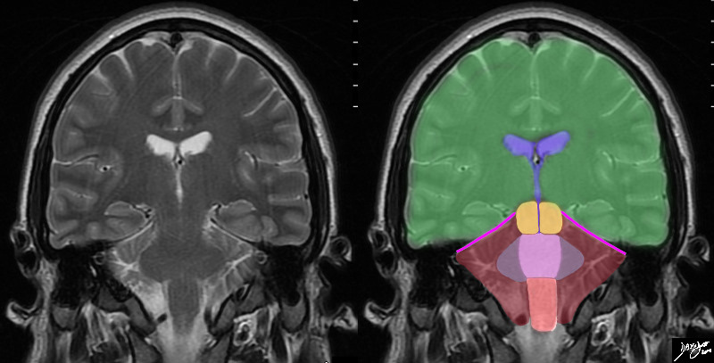
In this T2 weighted coronal MRI image the distinction between the supratentorial and infratentorial structures is made apparent by the bright pink tentorium that acts as a roof of the posterior fossa. The forebrain (green) midbrain (orange) and hindbrain (pink salmon and maroon) and the cerebellum (maroon), with other parts of the hindbrain filled in including the pons (light pink) middle cerebellar peduncles (mauve) and medulla (salmon) All the structures above the pink line are supratentorial, and those below are infratentorial. Part of the midbrain is supratentorial and part is infratentorial. The ventricular system is outlined in blue Courtesy Ashley Davidoff MD copyright 2010 all rights reserved 89721c06b.8sg01 |
Axial
|
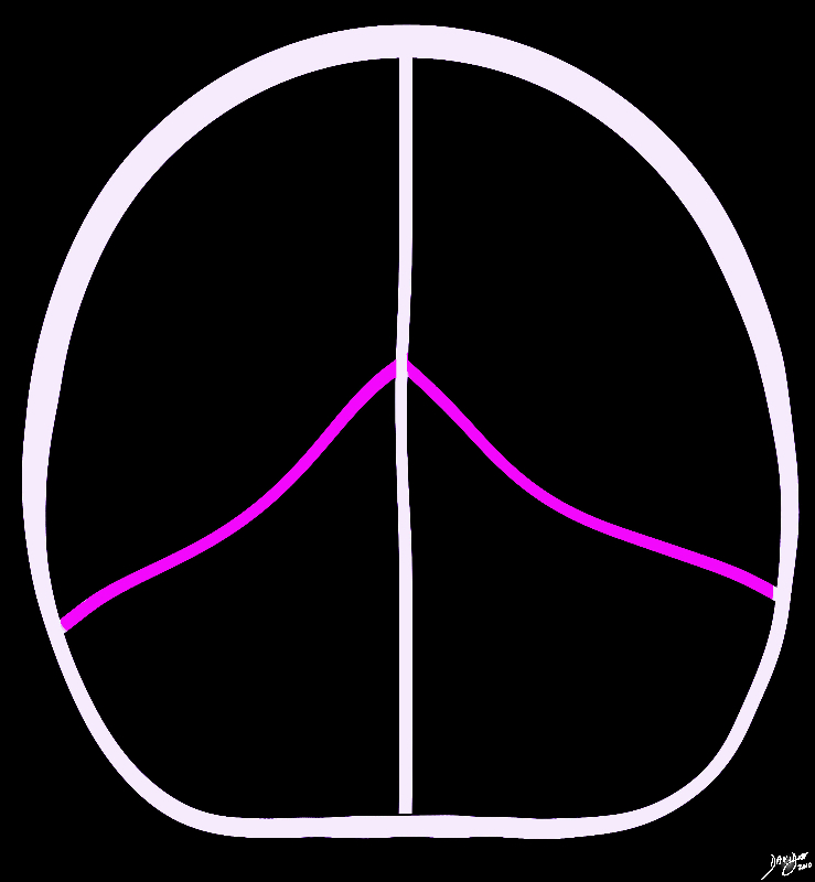
The Hemispheres and the Vermis |
|
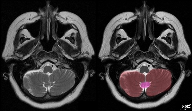
The T2 weighted MRI shows the 2 major components of the cerebellum ? the central vermis (pink), and the two cerebellar hemispheres. The cerebellum consists of two cerebellar hemispheres and a central worm like vermis Courtesy Ashley Davidoff MD copyright 2010 all rights reserved 49037c.8s |
DOMElement Object
(
    [schemaTypeInfo] =>
    [tagName] => table
    [firstElementChild] => (object value omitted)
    [lastElementChild] => (object value omitted)
    [childElementCount] => 1
    [previousElementSibling] => (object value omitted)
    [nextElementSibling] =>
    [nodeName] => table
    [nodeValue] =>


Winking Tabby Cat


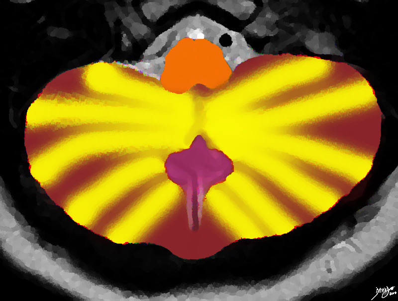
The Winking Cat The MRI of the posterior fossa and cerebellum was rendered and reminded the author of a winking tabby cat
Davidoff Art Copyright 2010 all rights reserved 49037.3kb03.81s


    [nodeType] => 1
    [parentNode] => (object value omitted)
    [childNodes] => (object value omitted)
    [firstChild] => (object value omitted)
    [lastChild] => (object value omitted)
    [previousSibling] => (object value omitted)
    [nextSibling] => (object value omitted)
    [attributes] => (object value omitted)
    [ownerDocument] => (object value omitted)
    [namespaceURI] =>
    [prefix] =>
    [localName] => table
    [baseURI] =>
    [textContent] =>


Winking Tabby Cat


The Winking Cat The MRI of the posterior fossa and cerebellum was rendered and reminded the author of a winking tabby cat
Davidoff Art Copyright 2010 all rights reserved 49037.3kb03.81s


)
DOMElement Object
(
    [schemaTypeInfo] =>
    [tagName] => td
    [firstElementChild] => (object value omitted)
    [lastElementChild] => (object value omitted)
    [childElementCount] => 2
    [previousElementSibling] =>
    [nextElementSibling] =>
    [nodeName] => td
    [nodeValue] =>
The Winking Cat The MRI of the posterior fossa and cerebellum was rendered and reminded the author of a winking tabby cat
Davidoff Art Copyright 2010 all rights reserved 49037.3kb03.81s

    [nodeType] => 1
    [parentNode] => (object value omitted)
    [childNodes] => (object value omitted)
    [firstChild] => (object value omitted)
    [lastChild] => (object value omitted)
    [previousSibling] => (object value omitted)
    [nextSibling] => (object value omitted)
    [attributes] => (object value omitted)
    [ownerDocument] => (object value omitted)
    [namespaceURI] =>
    [prefix] =>
    [localName] => td
    [baseURI] =>
    [textContent] =>
The Winking Cat The MRI of the posterior fossa and cerebellum was rendered and reminded the author of a winking tabby cat
Davidoff Art Copyright 2010 all rights reserved 49037.3kb03.81s

)
DOMElement Object
(
    [schemaTypeInfo] =>
    [tagName] => td
    [firstElementChild] => (object value omitted)
    [lastElementChild] => (object value omitted)
    [childElementCount] => 2
    [previousElementSibling] =>
    [nextElementSibling] =>
    [nodeName] => td
    [nodeValue] => Winking Tabby Cat
    [nodeType] => 1
    [parentNode] => (object value omitted)
    [childNodes] => (object value omitted)
    [firstChild] => (object value omitted)
    [lastChild] => (object value omitted)
    [previousSibling] => (object value omitted)
    [nextSibling] => (object value omitted)
    [attributes] => (object value omitted)
    [ownerDocument] => (object value omitted)
    [namespaceURI] =>
    [prefix] =>
    [localName] => td
    [baseURI] =>
    [textContent] => Winking Tabby Cat
)
DOMElement Object
(
    [schemaTypeInfo] =>
    [tagName] => table
    [firstElementChild] => (object value omitted)
    [lastElementChild] => (object value omitted)
    [childElementCount] => 1
    [previousElementSibling] => (object value omitted)
    [nextElementSibling] => (object value omitted)
    [nodeName] => table
    [nodeValue] =>


The Hemispheres and the Vermis


The T2 weighted MRI shows the 2 major components of the cerebellum ? the central vermis (pink), and the two cerebellar hemispheres. The cerebellum consists of two cerebellar hemispheres and a central worm like vermis
Courtesy Ashley Davidoff MD copyright 2010 all rights reserved 49037c.8s


    [nodeType] => 1
    [parentNode] => (object value omitted)
    [childNodes] => (object value omitted)
    [firstChild] => (object value omitted)
    [lastChild] => (object value omitted)
    [previousSibling] => (object value omitted)
    [nextSibling] => (object value omitted)
    [attributes] => (object value omitted)
    [ownerDocument] => (object value omitted)
    [namespaceURI] =>
    [prefix] =>
    [localName] => table
    [baseURI] =>
    [textContent] =>


The Hemispheres and the Vermis


The T2 weighted MRI shows the 2 major components of the cerebellum ? the central vermis (pink), and the two cerebellar hemispheres. The cerebellum consists of two cerebellar hemispheres and a central worm like vermis
Courtesy Ashley Davidoff MD copyright 2010 all rights reserved 49037c.8s


)
DOMElement Object
(
    [schemaTypeInfo] =>
    [tagName] => td
    [firstElementChild] => (object value omitted)
    [lastElementChild] => (object value omitted)
    [childElementCount] => 2
    [previousElementSibling] =>
    [nextElementSibling] =>
    [nodeName] => td
    [nodeValue] =>
The T2 weighted MRI shows the 2 major components of the cerebellum ? the central vermis (pink), and the two cerebellar hemispheres. The cerebellum consists of two cerebellar hemispheres and a central worm like vermis
Courtesy Ashley Davidoff MD copyright 2010 all rights reserved 49037c.8s

    [nodeType] => 1
    [parentNode] => (object value omitted)
    [childNodes] => (object value omitted)
    [firstChild] => (object value omitted)
    [lastChild] => (object value omitted)
    [previousSibling] => (object value omitted)
    [nextSibling] => (object value omitted)
    [attributes] => (object value omitted)
    [ownerDocument] => (object value omitted)
    [namespaceURI] =>
    [prefix] =>
    [localName] => td
    [baseURI] =>
    [textContent] =>
The T2 weighted MRI shows the 2 major components of the cerebellum ? the central vermis (pink), and the two cerebellar hemispheres. The cerebellum consists of two cerebellar hemispheres and a central worm like vermis
Courtesy Ashley Davidoff MD copyright 2010 all rights reserved 49037c.8s

)
DOMElement Object
(
    [schemaTypeInfo] =>
    [tagName] => td
    [firstElementChild] => (object value omitted)
    [lastElementChild] => (object value omitted)
    [childElementCount] => 2
    [previousElementSibling] =>
    [nextElementSibling] =>
    [nodeName] => td
    [nodeValue] =>

The Hemispheres and the Vermis
    [nodeType] => 1
    [parentNode] => (object value omitted)
    [childNodes] => (object value omitted)
    [firstChild] => (object value omitted)
    [lastChild] => (object value omitted)
    [previousSibling] => (object value omitted)
    [nextSibling] => (object value omitted)
    [attributes] => (object value omitted)
    [ownerDocument] => (object value omitted)
    [namespaceURI] =>
    [prefix] =>
    [localName] => td
    [baseURI] =>
    [textContent] =>

The Hemispheres and the Vermis
)
DOMElement Object
(
    [schemaTypeInfo] =>
    [tagName] => table
    [firstElementChild] => (object value omitted)
    [lastElementChild] => (object value omitted)
    [childElementCount] => 1
    [previousElementSibling] => (object value omitted)
    [nextElementSibling] => (object value omitted)
    [nodeName] => table
    [nodeValue] =>


Supratentorial and Infratentorial Structures


In this T2 weighted coronal MRI image the distinction between the supratentorial and infratentorial structures is made apparent by the bright pink tentorium that acts as a roof of the posterior fossa. The forebrain (green) midbrain (orange) and hindbrain (pink salmon and maroon) and the cerebellum (maroon), with other parts of the hindbrain filled in including the pons (light pink) middle cerebellar peduncles (mauve) and medulla (salmon) All the structures above the pink line are supratentorial, and those below are infratentorial. Part of the midbrain is supratentorial and part is infratentorial. The ventricular system is outlined in blue
Courtesy Ashley Davidoff MD copyright 2010 all rights reserved 89721c06b.8sg01


    [nodeType] => 1
    [parentNode] => (object value omitted)
    [childNodes] => (object value omitted)
    [firstChild] => (object value omitted)
    [lastChild] => (object value omitted)
    [previousSibling] => (object value omitted)
    [nextSibling] => (object value omitted)
    [attributes] => (object value omitted)
    [ownerDocument] => (object value omitted)
    [namespaceURI] =>
    [prefix] =>
    [localName] => table
    [baseURI] =>
    [textContent] =>


Supratentorial and Infratentorial Structures


In this T2 weighted coronal MRI image the distinction between the supratentorial and infratentorial structures is made apparent by the bright pink tentorium that acts as a roof of the posterior fossa. The forebrain (green) midbrain (orange) and hindbrain (pink salmon and maroon) and the cerebellum (maroon), with other parts of the hindbrain filled in including the pons (light pink) middle cerebellar peduncles (mauve) and medulla (salmon) All the structures above the pink line are supratentorial, and those below are infratentorial. Part of the midbrain is supratentorial and part is infratentorial. The ventricular system is outlined in blue
Courtesy Ashley Davidoff MD copyright 2010 all rights reserved 89721c06b.8sg01


)
DOMElement Object
(
    [schemaTypeInfo] =>
    [tagName] => td
    [firstElementChild] => (object value omitted)
    [lastElementChild] => (object value omitted)
    [childElementCount] => 2
    [previousElementSibling] =>
    [nextElementSibling] =>
    [nodeName] => td
    [nodeValue] =>
In this T2 weighted coronal MRI image the distinction between the supratentorial and infratentorial structures is made apparent by the bright pink tentorium that acts as a roof of the posterior fossa. The forebrain (green) midbrain (orange) and hindbrain (pink salmon and maroon) and the cerebellum (maroon), with other parts of the hindbrain filled in including the pons (light pink) middle cerebellar peduncles (mauve) and medulla (salmon) All the structures above the pink line are supratentorial, and those below are infratentorial. Part of the midbrain is supratentorial and part is infratentorial. The ventricular system is outlined in blue
Courtesy Ashley Davidoff MD copyright 2010 all rights reserved 89721c06b.8sg01

    [nodeType] => 1
    [parentNode] => (object value omitted)
    [childNodes] => (object value omitted)
    [firstChild] => (object value omitted)
    [lastChild] => (object value omitted)
    [previousSibling] => (object value omitted)
    [nextSibling] => (object value omitted)
    [attributes] => (object value omitted)
    [ownerDocument] => (object value omitted)
    [namespaceURI] =>
    [prefix] =>
    [localName] => td
    [baseURI] =>
    [textContent] =>
In this T2 weighted coronal MRI image the distinction between the supratentorial and infratentorial structures is made apparent by the bright pink tentorium that acts as a roof of the posterior fossa. The forebrain (green) midbrain (orange) and hindbrain (pink salmon and maroon) and the cerebellum (maroon), with other parts of the hindbrain filled in including the pons (light pink) middle cerebellar peduncles (mauve) and medulla (salmon) All the structures above the pink line are supratentorial, and those below are infratentorial. Part of the midbrain is supratentorial and part is infratentorial. The ventricular system is outlined in blue
Courtesy Ashley Davidoff MD copyright 2010 all rights reserved 89721c06b.8sg01

)
DOMElement Object
(
    [schemaTypeInfo] =>
    [tagName] => td
    [firstElementChild] => (object value omitted)
    [lastElementChild] => (object value omitted)
    [childElementCount] => 2
    [previousElementSibling] =>
    [nextElementSibling] =>
    [nodeName] => td
    [nodeValue] =>

Supratentorial and Infratentorial Structures
    [nodeType] => 1
    [parentNode] => (object value omitted)
    [childNodes] => (object value omitted)
    [firstChild] => (object value omitted)
    [lastChild] => (object value omitted)
    [previousSibling] => (object value omitted)
    [nextSibling] => (object value omitted)
    [attributes] => (object value omitted)
    [ownerDocument] => (object value omitted)
    [namespaceURI] =>
    [prefix] =>
    [localName] => td
    [baseURI] =>
    [textContent] =>

Supratentorial and Infratentorial Structures
)
DOMElement Object
(
    [schemaTypeInfo] =>
    [tagName] => table
    [firstElementChild] => (object value omitted)
    [lastElementChild] => (object value omitted)
    [childElementCount] => 1
    [previousElementSibling] => (object value omitted)
    [nextElementSibling] => (object value omitted)
    [nodeName] => table
    [nodeValue] =>


Space Position and the Tentorium in the Coronal Plane
Lying Under the Tent


In this conceptual coronal drawing the distinction between the supratentorial and infratentorial space is made apparent by the bright pink tentorium that acts as a roof of the posterior fossa. The forebrain and the upper part of the midbrain lie above the tentorium, and the lower midbrain and hindbrain lie below. All the structures above the pink line are classified as supratentorial structures , and those below are infratentorial.
Courtesy Ashley Davidoff MD copyright 2010 all rights reserved 93914b07g02.8s


    [nodeType] => 1
    [parentNode] => (object value omitted)
    [childNodes] => (object value omitted)
    [firstChild] => (object value omitted)
    [lastChild] => (object value omitted)
    [previousSibling] => (object value omitted)
    [nextSibling] => (object value omitted)
    [attributes] => (object value omitted)
    [ownerDocument] => (object value omitted)
    [namespaceURI] =>
    [prefix] =>
    [localName] => table
    [baseURI] =>
    [textContent] =>


Space Position and the Tentorium in the Coronal Plane
Lying Under the Tent


In this conceptual coronal drawing the distinction between the supratentorial and infratentorial space is made apparent by the bright pink tentorium that acts as a roof of the posterior fossa. The forebrain and the upper part of the midbrain lie above the tentorium, and the lower midbrain and hindbrain lie below. All the structures above the pink line are classified as supratentorial structures , and those below are infratentorial.
Courtesy Ashley Davidoff MD copyright 2010 all rights reserved 93914b07g02.8s


)
DOMElement Object
(
    [schemaTypeInfo] =>
    [tagName] => td
    [firstElementChild] => (object value omitted)
    [lastElementChild] => (object value omitted)
    [childElementCount] => 2
    [previousElementSibling] =>
    [nextElementSibling] =>
    [nodeName] => td
    [nodeValue] =>
In this conceptual coronal drawing the distinction between the supratentorial and infratentorial space is made apparent by the bright pink tentorium that acts as a roof of the posterior fossa. The forebrain and the upper part of the midbrain lie above the tentorium, and the lower midbrain and hindbrain lie below. All the structures above the pink line are classified as supratentorial structures , and those below are infratentorial.
Courtesy Ashley Davidoff MD copyright 2010 all rights reserved 93914b07g02.8s

    [nodeType] => 1
    [parentNode] => (object value omitted)
    [childNodes] => (object value omitted)
    [firstChild] => (object value omitted)
    [lastChild] => (object value omitted)
    [previousSibling] => (object value omitted)
    [nextSibling] => (object value omitted)
    [attributes] => (object value omitted)
    [ownerDocument] => (object value omitted)
    [namespaceURI] =>
    [prefix] =>
    [localName] => td
    [baseURI] =>
    [textContent] =>
In this conceptual coronal drawing the distinction between the supratentorial and infratentorial space is made apparent by the bright pink tentorium that acts as a roof of the posterior fossa. The forebrain and the upper part of the midbrain lie above the tentorium, and the lower midbrain and hindbrain lie below. All the structures above the pink line are classified as supratentorial structures , and those below are infratentorial.
Courtesy Ashley Davidoff MD copyright 2010 all rights reserved 93914b07g02.8s

)
DOMElement Object
(
    [schemaTypeInfo] =>
    [tagName] => td
    [firstElementChild] => (object value omitted)
    [lastElementChild] => (object value omitted)
    [childElementCount] => 3
    [previousElementSibling] =>
    [nextElementSibling] =>
    [nodeName] => td
    [nodeValue] =>

Space Position and the Tentorium in the Coronal Plane
Lying Under the Tent
    [nodeType] => 1
    [parentNode] => (object value omitted)
    [childNodes] => (object value omitted)
    [firstChild] => (object value omitted)
    [lastChild] => (object value omitted)
    [previousSibling] => (object value omitted)
    [nextSibling] => (object value omitted)
    [attributes] => (object value omitted)
    [ownerDocument] => (object value omitted)
    [namespaceURI] =>
    [prefix] =>
    [localName] => td
    [baseURI] =>
    [textContent] =>

Space Position and the Tentorium in the Coronal Plane
Lying Under the Tent
)
DOMElement Object
(
    [schemaTypeInfo] =>
    [tagName] => table
    [firstElementChild] => (object value omitted)
    [lastElementChild] => (object value omitted)
    [childElementCount] => 1
    [previousElementSibling] => (object value omitted)
    [nextElementSibling] => (object value omitted)
    [nodeName] => table
    [nodeValue] =>


Advancing the Parts of the Hindbrain


The shapes in fact are not quite ovoid. The pons (lightest pink) has shapes reminiscent of an oversize egg lying in a small bed The anterior eggshaped portion is called the ventral or anterior pons and the posterior portion called the tegmentum which is a continuation of the tegmentum of the midbrain. The medulla oblongata is almost rectangular but has a subtle forward bulging to its anterior surface. The anterior portion is called the ventral or anterior medulla and the posterior portion is called the tegmentum,similar in name a nad a continuation of th etegmentum of the midbrain and pons The cerebellum is situated posteriorly, is the largest of the three major components and it consists of two hemispheres and a central vermis better appreciated in the axial plane.
Courtesy Ashley Davidoff MD Copyright 2010 all rights reserved 92141.3kd03b04b.8s


    [nodeType] => 1
    [parentNode] => (object value omitted)
    [childNodes] => (object value omitted)
    [firstChild] => (object value omitted)
    [lastChild] => (object value omitted)
    [previousSibling] => (object value omitted)
    [nextSibling] => (object value omitted)
    [attributes] => (object value omitted)
    [ownerDocument] => (object value omitted)
    [namespaceURI] =>
    [prefix] =>
    [localName] => table
    [baseURI] =>
    [textContent] =>


Advancing the Parts of the Hindbrain


The shapes in fact are not quite ovoid. The pons (lightest pink) has shapes reminiscent of an oversize egg lying in a small bed The anterior eggshaped portion is called the ventral or anterior pons and the posterior portion called the tegmentum which is a continuation of the tegmentum of the midbrain. The medulla oblongata is almost rectangular but has a subtle forward bulging to its anterior surface. The anterior portion is called the ventral or anterior medulla and the posterior portion is called the tegmentum,similar in name a nad a continuation of th etegmentum of the midbrain and pons The cerebellum is situated posteriorly, is the largest of the three major components and it consists of two hemispheres and a central vermis better appreciated in the axial plane.
Courtesy Ashley Davidoff MD Copyright 2010 all rights reserved 92141.3kd03b04b.8s


)
DOMElement Object
(
    [schemaTypeInfo] =>
    [tagName] => td
    [firstElementChild] => (object value omitted)
    [lastElementChild] => (object value omitted)
    [childElementCount] => 2
    [previousElementSibling] =>
    [nextElementSibling] =>
    [nodeName] => td
    [nodeValue] =>
The shapes in fact are not quite ovoid. The pons (lightest pink) has shapes reminiscent of an oversize egg lying in a small bed The anterior eggshaped portion is called the ventral or anterior pons and the posterior portion called the tegmentum which is a continuation of the tegmentum of the midbrain. The medulla oblongata is almost rectangular but has a subtle forward bulging to its anterior surface. The anterior portion is called the ventral or anterior medulla and the posterior portion is called the tegmentum,similar in name a nad a continuation of th etegmentum of the midbrain and pons The cerebellum is situated posteriorly, is the largest of the three major components and it consists of two hemispheres and a central vermis better appreciated in the axial plane.
Courtesy Ashley Davidoff MD Copyright 2010 all rights reserved 92141.3kd03b04b.8s

    [nodeType] => 1
    [parentNode] => (object value omitted)
    [childNodes] => (object value omitted)
    [firstChild] => (object value omitted)
    [lastChild] => (object value omitted)
    [previousSibling] => (object value omitted)
    [nextSibling] => (object value omitted)
    [attributes] => (object value omitted)
    [ownerDocument] => (object value omitted)
    [namespaceURI] =>
    [prefix] =>
    [localName] => td
    [baseURI] =>
    [textContent] =>
The shapes in fact are not quite ovoid. The pons (lightest pink) has shapes reminiscent of an oversize egg lying in a small bed The anterior eggshaped portion is called the ventral or anterior pons and the posterior portion called the tegmentum which is a continuation of the tegmentum of the midbrain. The medulla oblongata is almost rectangular but has a subtle forward bulging to its anterior surface. The anterior portion is called the ventral or anterior medulla and the posterior portion is called the tegmentum,similar in name a nad a continuation of th etegmentum of the midbrain and pons The cerebellum is situated posteriorly, is the largest of the three major components and it consists of two hemispheres and a central vermis better appreciated in the axial plane.
Courtesy Ashley Davidoff MD Copyright 2010 all rights reserved 92141.3kd03b04b.8s

)
DOMElement Object
(
    [schemaTypeInfo] =>
    [tagName] => td
    [firstElementChild] => (object value omitted)
    [lastElementChild] => (object value omitted)
    [childElementCount] => 2
    [previousElementSibling] =>
    [nextElementSibling] =>
    [nodeName] => td
    [nodeValue] =>

Advancing the Parts of the Hindbrain
    [nodeType] => 1
    [parentNode] => (object value omitted)
    [childNodes] => (object value omitted)
    [firstChild] => (object value omitted)
    [lastChild] => (object value omitted)
    [previousSibling] => (object value omitted)
    [nextSibling] => (object value omitted)
    [attributes] => (object value omitted)
    [ownerDocument] => (object value omitted)
    [namespaceURI] =>
    [prefix] =>
    [localName] => td
    [baseURI] =>
    [textContent] =>

Advancing the Parts of the Hindbrain
)
DOMElement Object
(
    [schemaTypeInfo] =>
    [tagName] => table
    [firstElementChild] => (object value omitted)
    [lastElementChild] => (object value omitted)
    [childElementCount] => 1
    [previousElementSibling] => (object value omitted)
    [nextElementSibling] => (object value omitted)
    [nodeName] => table
    [nodeValue] =>


The Hindbrain
Posterior and Largest Part of the Hindbrain


The hindbrain in the sagital plane is bordered in salmon pink and consists of the pons, the medulla, and the cerebellum
Image Courtesy Philips Medical Systems Rendered by Ashley Davidoff MD Copyright 2010 92141.3ka.8s


    [nodeType] => 1
    [parentNode] => (object value omitted)
    [childNodes] => (object value omitted)
    [firstChild] => (object value omitted)
    [lastChild] => (object value omitted)
    [previousSibling] => (object value omitted)
    [nextSibling] => (object value omitted)
    [attributes] => (object value omitted)
    [ownerDocument] => (object value omitted)
    [namespaceURI] =>
    [prefix] =>
    [localName] => table
    [baseURI] =>
    [textContent] =>


The Hindbrain
Posterior and Largest Part of the Hindbrain


The hindbrain in the sagital plane is bordered in salmon pink and consists of the pons, the medulla, and the cerebellum
Image Courtesy Philips Medical Systems Rendered by Ashley Davidoff MD Copyright 2010 92141.3ka.8s


)
DOMElement Object
(
    [schemaTypeInfo] =>
    [tagName] => td
    [firstElementChild] => (object value omitted)
    [lastElementChild] => (object value omitted)
    [childElementCount] => 2
    [previousElementSibling] =>
    [nextElementSibling] =>
    [nodeName] => td
    [nodeValue] =>
The hindbrain in the sagital plane is bordered in salmon pink and consists of the pons, the medulla, and the cerebellum
Image Courtesy Philips Medical Systems Rendered by Ashley Davidoff MD Copyright 2010 92141.3ka.8s

    [nodeType] => 1
    [parentNode] => (object value omitted)
    [childNodes] => (object value omitted)
    [firstChild] => (object value omitted)
    [lastChild] => (object value omitted)
    [previousSibling] => (object value omitted)
    [nextSibling] => (object value omitted)
    [attributes] => (object value omitted)
    [ownerDocument] => (object value omitted)
    [namespaceURI] =>
    [prefix] =>
    [localName] => td
    [baseURI] =>
    [textContent] =>
The hindbrain in the sagital plane is bordered in salmon pink and consists of the pons, the medulla, and the cerebellum
Image Courtesy Philips Medical Systems Rendered by Ashley Davidoff MD Copyright 2010 92141.3ka.8s

)
DOMElement Object
(
    [schemaTypeInfo] =>
    [tagName] => td
    [firstElementChild] => (object value omitted)
    [lastElementChild] => (object value omitted)
    [childElementCount] => 3
    [previousElementSibling] =>
    [nextElementSibling] =>
    [nodeName] => td
    [nodeValue] =>

The Hindbrain
Posterior and Largest Part of the Hindbrain
    [nodeType] => 1
    [parentNode] => (object value omitted)
    [childNodes] => (object value omitted)
    [firstChild] => (object value omitted)
    [lastChild] => (object value omitted)
    [previousSibling] => (object value omitted)
    [nextSibling] => (object value omitted)
    [attributes] => (object value omitted)
    [ownerDocument] => (object value omitted)
    [namespaceURI] =>
    [prefix] =>
    [localName] => td
    [baseURI] =>
    [textContent] =>

The Hindbrain
Posterior and Largest Part of the Hindbrain
)
DOMElement Object
(
    [schemaTypeInfo] =>
    [tagName] => table
    [firstElementChild] => (object value omitted)
    [lastElementChild] => (object value omitted)
    [childElementCount] => 1
    [previousElementSibling] => (object value omitted)
    [nextElementSibling] => (object value omitted)
    [nodeName] => table
    [nodeValue] =>


The Cerebellum (maroon) ? Part of the Hindbrain
Stick Diagram


The hindbrain consists of 3 major components;the pons (light pink, the medulla, (salmon) and the cerebellum (maroon)
Courtesy Ashley DAvidoff MD copyright 2010  93887b03ab03.8s


    [nodeType] => 1
    [parentNode] => (object value omitted)
    [childNodes] => (object value omitted)
    [firstChild] => (object value omitted)
    [lastChild] => (object value omitted)
    [previousSibling] => (object value omitted)
    [nextSibling] => (object value omitted)
    [attributes] => (object value omitted)
    [ownerDocument] => (object value omitted)
    [namespaceURI] =>
    [prefix] =>
    [localName] => table
    [baseURI] =>
    [textContent] =>


The Cerebellum (maroon) ? Part of the Hindbrain
Stick Diagram


The hindbrain consists of 3 major components;the pons (light pink, the medulla, (salmon) and the cerebellum (maroon)
Courtesy Ashley DAvidoff MD copyright 2010  93887b03ab03.8s


)
DOMElement Object
(
    [schemaTypeInfo] =>
    [tagName] => td
    [firstElementChild] => (object value omitted)
    [lastElementChild] => (object value omitted)
    [childElementCount] => 2
    [previousElementSibling] =>
    [nextElementSibling] =>
    [nodeName] => td
    [nodeValue] =>
The hindbrain consists of 3 major components;the pons (light pink, the medulla, (salmon) and the cerebellum (maroon)
Courtesy Ashley DAvidoff MD copyright 2010  93887b03ab03.8s

    [nodeType] => 1
    [parentNode] => (object value omitted)
    [childNodes] => (object value omitted)
    [firstChild] => (object value omitted)
    [lastChild] => (object value omitted)
    [previousSibling] => (object value omitted)
    [nextSibling] => (object value omitted)
    [attributes] => (object value omitted)
    [ownerDocument] => (object value omitted)
    [namespaceURI] =>
    [prefix] =>
    [localName] => td
    [baseURI] =>
    [textContent] =>
The hindbrain consists of 3 major components;the pons (light pink, the medulla, (salmon) and the cerebellum (maroon)
Courtesy Ashley DAvidoff MD copyright 2010  93887b03ab03.8s

)
DOMElement Object
(
    [schemaTypeInfo] =>
    [tagName] => td
    [firstElementChild] => (object value omitted)
    [lastElementChild] => (object value omitted)
    [childElementCount] => 3
    [previousElementSibling] =>
    [nextElementSibling] =>
    [nodeName] => td
    [nodeValue] =>

The Cerebellum (maroon) ? Part of the Hindbrain
Stick Diagram
    [nodeType] => 1
    [parentNode] => (object value omitted)
    [childNodes] => (object value omitted)
    [firstChild] => (object value omitted)
    [lastChild] => (object value omitted)
    [previousSibling] => (object value omitted)
    [nextSibling] => (object value omitted)
    [attributes] => (object value omitted)
    [ownerDocument] => (object value omitted)
    [namespaceURI] =>
    [prefix] =>
    [localName] => td
    [baseURI] =>
    [textContent] =>

The Cerebellum (maroon) ? Part of the Hindbrain
Stick Diagram
)

