

Hi Barry
- The points I want to try and get across
- How to diagnose constrictive pericarditis on CT
- Questions and Answers
-
- What are the signs of constrictive pericarditis on CT
- pericardial thickening
- diffuse or localized(2mm – 3mm equivocal)
- signs of impaired diastolic filling of the right ventricle
- ie signs of right heart failure including
- dilatation of the
- RA
- IVC and hepatic veins
- Coronary Sinus
- Azygos vein
- Hepatomegaly
- dilatation of the
- What is the role of the CXR and LA in Dx
- increase in left atrial because it cannot fill therefore
- increase in pulmonary venous pressure
- Usually normal size LA
- However since LA hmay be only partly covered by pericardium, it may in fact enlar
- pericardial thickening
- What are the signs of constrictive pericarditis on CT
-
- Questions and Answers
-
In our patient there is
- no pericardial thickening
- minor pericardial calcification
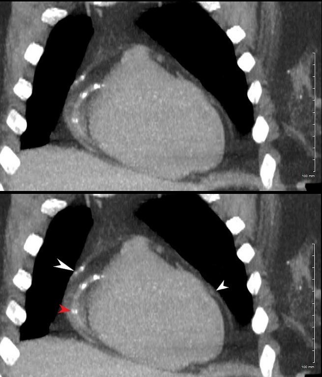
- There are
-
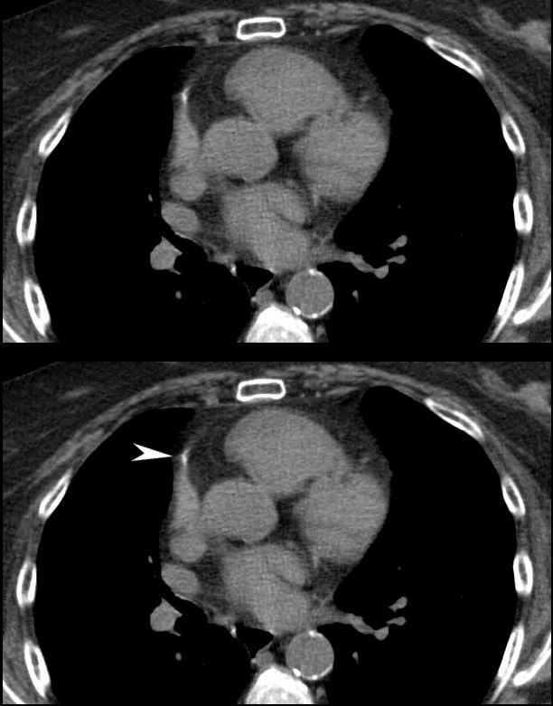
- focal adhesions near the LAD
-
Are There Signs of Constriction?
Consider
Pericardium Thickness
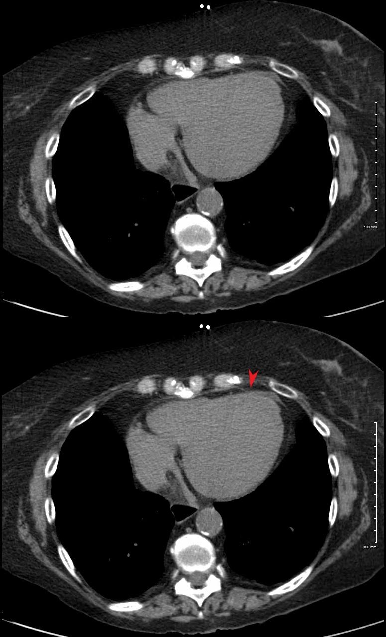
Size of the Right Atrium
The axial image through the region of the AV valves shows normal sized atria. Constriction would be unlikely in the face of a normal sized right atrium. Note the flattened surface of the right atrium
Ashley Davidoff MD
? Size of the Coronary Sinus
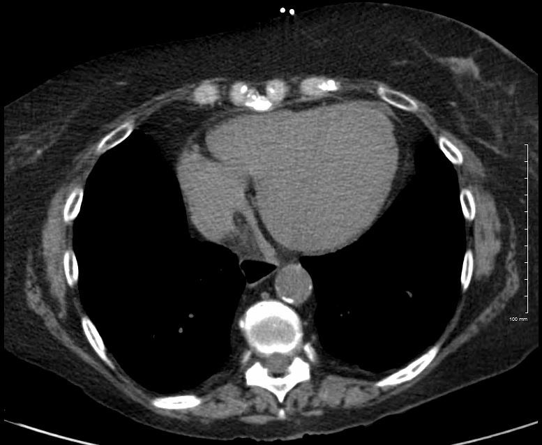
The axial image through the region of the normal sized coronary sinus. Constriction would be unlikely in the face of a normal sized coronary sinus. Note the flattened surface of the right atrium.
Ashley Davidoff MD
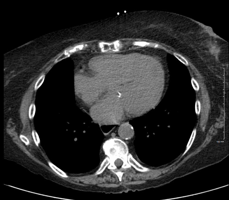
? Size of the Azygos Vein and IVC
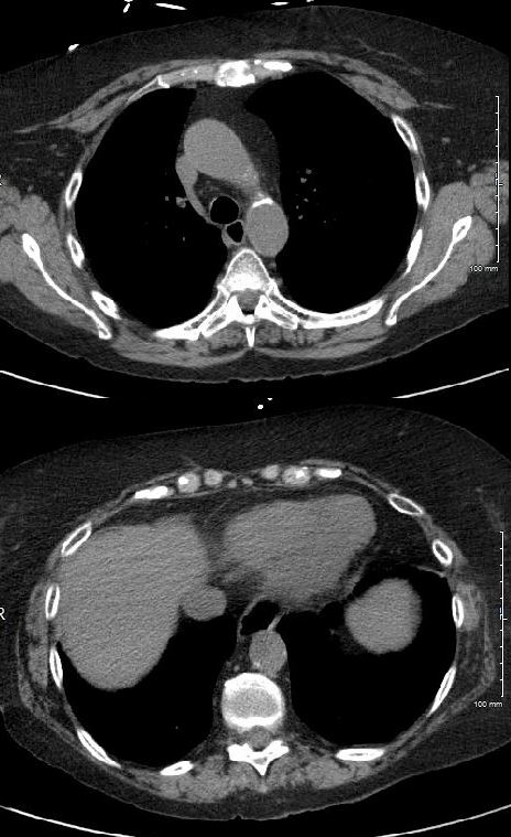
The axial image is through the region of the normal azygos vein (above) and the normal sized IVC (below). Constrictive pericarditis would be unlikely in the face of a normal sized azygos vein and IVC.
Thus constrictive pericarditis is not present
ABR
Links and References
Napolitano G et al Imaging Features of Constrictive Pericarditis: Beyond Pericardial Thickening Canadian Association of Radiologists Journal Volume 60, Issue 1, February 2009, (good review)
Senapati A et al, Disparity in spatial distribution of pericardial calcifications in constrictive pericarditis Openheart BMJmj. Volume 5, Issue 2
Khalid, N et al Pericardial Calcification StatPearls