the Common Vein Copyright 2010
Definition
end
DOMElement Object
(
    [schemaTypeInfo] =>
    [tagName] => table
    [firstElementChild] => (object value omitted)
    [lastElementChild] => (object value omitted)
    [childElementCount] => 1
    [previousElementSibling] => (object value omitted)
    [nextElementSibling] =>
    [nodeName] => table
    [nodeValue] =>


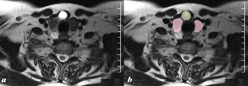
MRI T2 Weighted Thyroglossal Duct Cyst


The MRI T2 weighted image through the inferior aspect of the thyroid gland (pink)shows a cystic structure (yellow), intensely T2 bright, in the region of the isthmus in the ventral aspect of the gland. Findings are consistent with a thyroglossal cyst
Courtesy Ashley Davidoff MD Copyright 2010 97310cL.8


    [nodeType] => 1
    [parentNode] => (object value omitted)
    [childNodes] => (object value omitted)
    [firstChild] => (object value omitted)
    [lastChild] => (object value omitted)
    [previousSibling] => (object value omitted)
    [nextSibling] => (object value omitted)
    [attributes] => (object value omitted)
    [ownerDocument] => (object value omitted)
    [namespaceURI] =>
    [prefix] =>
    [localName] => table
    [baseURI] =>
    [textContent] =>


MRI T2 Weighted Thyroglossal Duct Cyst


The MRI T2 weighted image through the inferior aspect of the thyroid gland (pink)shows a cystic structure (yellow), intensely T2 bright, in the region of the isthmus in the ventral aspect of the gland. Findings are consistent with a thyroglossal cyst
Courtesy Ashley Davidoff MD Copyright 2010 97310cL.8


)
DOMElement Object
(
    [schemaTypeInfo] =>
    [tagName] => td
    [firstElementChild] => (object value omitted)
    [lastElementChild] => (object value omitted)
    [childElementCount] => 2
    [previousElementSibling] =>
    [nextElementSibling] =>
    [nodeName] => td
    [nodeValue] =>
The MRI T2 weighted image through the inferior aspect of the thyroid gland (pink)shows a cystic structure (yellow), intensely T2 bright, in the region of the isthmus in the ventral aspect of the gland. Findings are consistent with a thyroglossal cyst
Courtesy Ashley Davidoff MD Copyright 2010 97310cL.8

    [nodeType] => 1
    [parentNode] => (object value omitted)
    [childNodes] => (object value omitted)
    [firstChild] => (object value omitted)
    [lastChild] => (object value omitted)
    [previousSibling] => (object value omitted)
    [nextSibling] => (object value omitted)
    [attributes] => (object value omitted)
    [ownerDocument] => (object value omitted)
    [namespaceURI] =>
    [prefix] =>
    [localName] => td
    [baseURI] =>
    [textContent] =>
The MRI T2 weighted image through the inferior aspect of the thyroid gland (pink)shows a cystic structure (yellow), intensely T2 bright, in the region of the isthmus in the ventral aspect of the gland. Findings are consistent with a thyroglossal cyst
Courtesy Ashley Davidoff MD Copyright 2010 97310cL.8

)
DOMElement Object
(
    [schemaTypeInfo] =>
    [tagName] => td
    [firstElementChild] => (object value omitted)
    [lastElementChild] => (object value omitted)
    [childElementCount] => 2
    [previousElementSibling] =>
    [nextElementSibling] =>
    [nodeName] => td
    [nodeValue] =>
MRI T2 Weighted Thyroglossal Duct Cyst
    [nodeType] => 1
    [parentNode] => (object value omitted)
    [childNodes] => (object value omitted)
    [firstChild] => (object value omitted)
    [lastChild] => (object value omitted)
    [previousSibling] => (object value omitted)
    [nextSibling] => (object value omitted)
    [attributes] => (object value omitted)
    [ownerDocument] => (object value omitted)
    [namespaceURI] =>
    [prefix] =>
    [localName] => td
    [baseURI] =>
    [textContent] =>
MRI T2 Weighted Thyroglossal Duct Cyst
)

