Function
The Common Vein Copyright 2007
There are several phases of the swallowing mechanism. It originates with the cerebral aspect of thinking about food, followed by mastication and bolus propulsion into the posterior oro-pharynx. What follows is the esophageal component of swallowing:
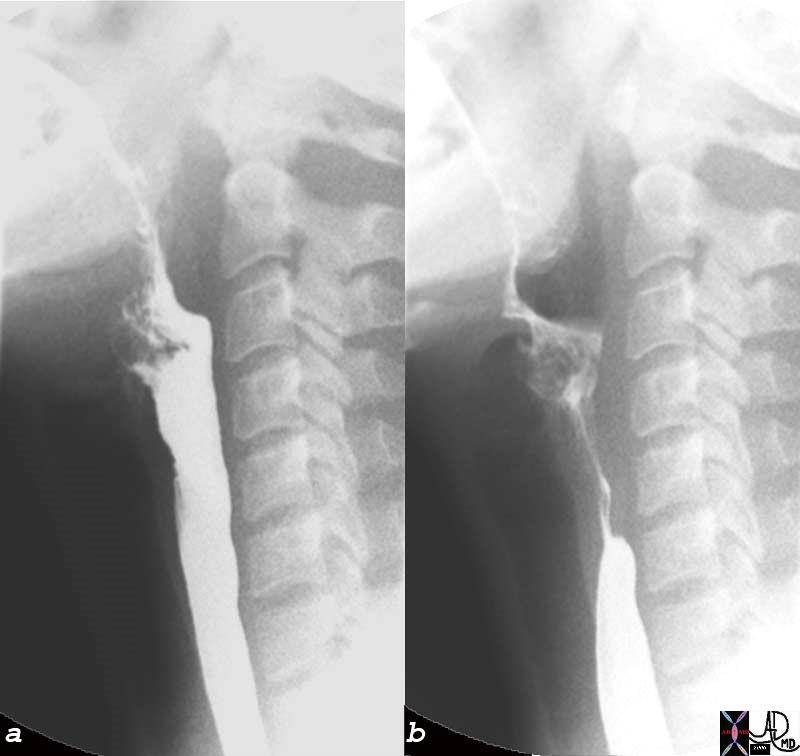
At the Upper Esophageal Sphincter, the visceral striated muscle keeps the UES closed until there is the stimulation of a food bolus. The UES then relaxes with a swallow and is integrated with the tongue and posterior oro-pharynx. Regardless of the size of the bolus, the degree of UES relaxation is the same, however, the amount the UES opens is based on size of the bolus. Relaxation of the UES preceeds the sphincter opening by 0.1 seconds. Once the food enters the esophagus proper, the UES again regains its tonicity.
Within the esophagus the longitudinal muscles begin to contract causing an overall shortening of the esophageal body. The esophagus can shorten approximately 2-2.5 cm in total length. This is followed by the contraction of the circular muscles that propel the bolus down the esophagus. The bolus typically travels 2-4cm/second which means that a normal esophagus (length 20cm) should empty its bolus in 10 seconds.
The movement of the bolus thru the esophagus should be one directional and somewhat uniform. There are two types of peristaltic waves that empty the esophagus.
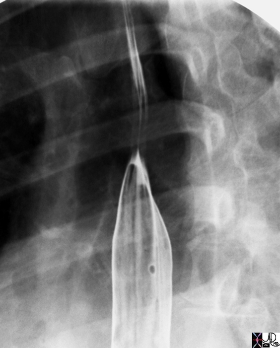
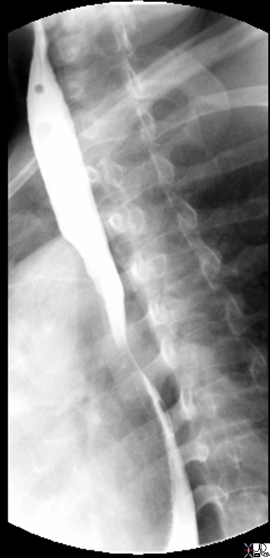
Primary Peristalsis is initiated by the pharyngeal phase of swallowing and thus is a voluntary process. A single wave caused by contraction of the circular muscles essentially strips the esophagus of its content and deposits it into the stomach. This may be followed by secondary peristalsis which is an involuntary process initiated by the neurologic properties of the esophagus itself. In response to distension from the lack of complete emptying with primary peristalsis or reflux of debris back into the esophagus.
Therefore, the competence of the LES is quite important to maintaining proper esophageal peristalsis. The LES relaxes about 2 seconds after a swallow is initiated to allow for bolus passage. The normal resting pressure is thought to be 15-25mmHg and relaxes to near 0 with swallowing and a rebound higher value once the muscles tense.
 Primary Stripping Wave – Pressure Changes Primary Stripping Wave – Pressure Changes |
| The normal propagation can be seen in the above diagram of a motility study that follows the pressure gradient within the esophagus during a wet swallow. The initial dip on the diagram at the upper sphincter indicates a relaxation of this muscle to allow for passage of food and then the rapid return to the closed state indicated by the rapid upswing. Similarly, there is a drop in muscle pressure at the lower sphincter approximately 2 seconds (2 boxes) after the swallow indicating relaxation of the muscle to allow food to enter the stomach.
73368b05.800 Davidoff Art Courtesy Ashley Davidoff MD
Image modified from Sleisenger and Fordtran?s Gastrointestinal and Liver Disease. Saunders. 2002. |
 Upper Esophageal Sphincter Open, (a) Closed (b) and beginning of Primary Stripping Wave Upper Esophageal Sphincter Open, (a) Closed (b) and beginning of Primary Stripping Wave |
| 76204c esophagus cervical esophagus UES upper esophageal sphincter beginning of primary stripping wave normal physiology swallowing mechanism function single contrast barium swallow Courtesy Ashley Davidoff MD |
 Primary Stripping Wave Primary Stripping Wave |
| 76159 esophagus proximal thoracic esophagus primary stripping wave normal anatomy physiology double contrast barium swallow Courtesy Ashley Davidoff MD |
 Primary Stripping Wave in Mid Esophagus Primary Stripping Wave in Mid Esophagus |
| 76199 esophagus swallowing normal anatomy physiology function shape muscle contraction relaxation single contrast barium swallow Courtesy Ashley Davidoff MD |
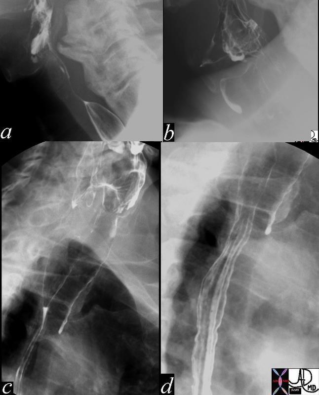
 Aspiration Aspiration |
| 46508c01.800 esophagus dysphagia fx large osteophyte causing difficulty with passing a scope fx aspiration barium swallow x-ray contrast Davidoff MD |
References
Web
The Longitudinal Muscle in Esophageal Disease Arthur Stiennon MD
Anatomy of the SwallowPatrick McCaffrey, Ph.D The neuroscience on the Web Series CSUCHICO 5star
DOMElement Object
(
    [schemaTypeInfo] =>
    [tagName] => table
    [firstElementChild] => (object value omitted)
    [lastElementChild] => (object value omitted)
    [childElementCount] => 1
    [previousElementSibling] => (object value omitted)
    [nextElementSibling] => (object value omitted)
    [nodeName] => table
    [nodeValue] =>


Aspiration


46508c01.800 esophagus dysphagia fx large osteophyte causing difficulty with passing a scope fx aspiration barium swallow x-ray contrast Davidoff MD


    [nodeType] => 1
    [parentNode] => (object value omitted)
    [childNodes] => (object value omitted)
    [firstChild] => (object value omitted)
    [lastChild] => (object value omitted)
    [previousSibling] => (object value omitted)
    [nextSibling] => (object value omitted)
    [attributes] => (object value omitted)
    [ownerDocument] => (object value omitted)
    [namespaceURI] =>
    [prefix] =>
    [localName] => table
    [baseURI] =>
    [textContent] =>


Aspiration


46508c01.800 esophagus dysphagia fx large osteophyte causing difficulty with passing a scope fx aspiration barium swallow x-ray contrast Davidoff MD


)
DOMElement Object
(
    [schemaTypeInfo] =>
    [tagName] => td
    [firstElementChild] =>
    [lastElementChild] =>
    [childElementCount] => 0
    [previousElementSibling] =>
    [nextElementSibling] =>
    [nodeName] => td
    [nodeValue] => 46508c01.800 esophagus dysphagia fx large osteophyte causing difficulty with passing a scope fx aspiration barium swallow x-ray contrast Davidoff MD
    [nodeType] => 1
    [parentNode] => (object value omitted)
    [childNodes] => (object value omitted)
    [firstChild] => (object value omitted)
    [lastChild] => (object value omitted)
    [previousSibling] => (object value omitted)
    [nextSibling] => (object value omitted)
    [attributes] => (object value omitted)
    [ownerDocument] => (object value omitted)
    [namespaceURI] =>
    [prefix] =>
    [localName] => td
    [baseURI] =>
    [textContent] => 46508c01.800 esophagus dysphagia fx large osteophyte causing difficulty with passing a scope fx aspiration barium swallow x-ray contrast Davidoff MD
)
DOMElement Object
(
    [schemaTypeInfo] =>
    [tagName] => td
    [firstElementChild] => (object value omitted)
    [lastElementChild] => (object value omitted)
    [childElementCount] => 1
    [previousElementSibling] =>
    [nextElementSibling] =>
    [nodeName] => td
    [nodeValue] => Aspiration
    [nodeType] => 1
    [parentNode] => (object value omitted)
    [childNodes] => (object value omitted)
    [firstChild] => (object value omitted)
    [lastChild] => (object value omitted)
    [previousSibling] => (object value omitted)
    [nextSibling] => (object value omitted)
    [attributes] => (object value omitted)
    [ownerDocument] => (object value omitted)
    [namespaceURI] =>
    [prefix] =>
    [localName] => td
    [baseURI] =>
    [textContent] => Aspiration
)
DOMElement Object
(
    [schemaTypeInfo] =>
    [tagName] => table
    [firstElementChild] => (object value omitted)
    [lastElementChild] => (object value omitted)
    [childElementCount] => 1
    [previousElementSibling] => (object value omitted)
    [nextElementSibling] => (object value omitted)
    [nodeName] => table
    [nodeValue] =>


Primary Stripping Wave in Mid Esophagus


76199 esophagus swallowing normal anatomy physiology function shape muscle contraction relaxation single contrast barium swallow Courtesy Ashley Davidoff MD


    [nodeType] => 1
    [parentNode] => (object value omitted)
    [childNodes] => (object value omitted)
    [firstChild] => (object value omitted)
    [lastChild] => (object value omitted)
    [previousSibling] => (object value omitted)
    [nextSibling] => (object value omitted)
    [attributes] => (object value omitted)
    [ownerDocument] => (object value omitted)
    [namespaceURI] =>
    [prefix] =>
    [localName] => table
    [baseURI] =>
    [textContent] =>


Primary Stripping Wave in Mid Esophagus


76199 esophagus swallowing normal anatomy physiology function shape muscle contraction relaxation single contrast barium swallow Courtesy Ashley Davidoff MD


)
DOMElement Object
(
    [schemaTypeInfo] =>
    [tagName] => td
    [firstElementChild] =>
    [lastElementChild] =>
    [childElementCount] => 0
    [previousElementSibling] =>
    [nextElementSibling] =>
    [nodeName] => td
    [nodeValue] => 76199 esophagus swallowing normal anatomy physiology function shape muscle contraction relaxation single contrast barium swallow Courtesy Ashley Davidoff MD
    [nodeType] => 1
    [parentNode] => (object value omitted)
    [childNodes] => (object value omitted)
    [firstChild] => (object value omitted)
    [lastChild] => (object value omitted)
    [previousSibling] => (object value omitted)
    [nextSibling] => (object value omitted)
    [attributes] => (object value omitted)
    [ownerDocument] => (object value omitted)
    [namespaceURI] =>
    [prefix] =>
    [localName] => td
    [baseURI] =>
    [textContent] => 76199 esophagus swallowing normal anatomy physiology function shape muscle contraction relaxation single contrast barium swallow Courtesy Ashley Davidoff MD
)
DOMElement Object
(
    [schemaTypeInfo] =>
    [tagName] => td
    [firstElementChild] => (object value omitted)
    [lastElementChild] => (object value omitted)
    [childElementCount] => 1
    [previousElementSibling] =>
    [nextElementSibling] =>
    [nodeName] => td
    [nodeValue] => Primary Stripping Wave in Mid Esophagus
    [nodeType] => 1
    [parentNode] => (object value omitted)
    [childNodes] => (object value omitted)
    [firstChild] => (object value omitted)
    [lastChild] => (object value omitted)
    [previousSibling] => (object value omitted)
    [nextSibling] => (object value omitted)
    [attributes] => (object value omitted)
    [ownerDocument] => (object value omitted)
    [namespaceURI] =>
    [prefix] =>
    [localName] => td
    [baseURI] =>
    [textContent] => Primary Stripping Wave in Mid Esophagus
)
DOMElement Object
(
    [schemaTypeInfo] =>
    [tagName] => table
    [firstElementChild] => (object value omitted)
    [lastElementChild] => (object value omitted)
    [childElementCount] => 1
    [previousElementSibling] => (object value omitted)
    [nextElementSibling] => (object value omitted)
    [nodeName] => table
    [nodeValue] =>


Primary Stripping Wave


76159 esophagus proximal thoracic esophagus primary stripping wave normal anatomy physiology double contrast barium swallow Courtesy Ashley Davidoff MD


    [nodeType] => 1
    [parentNode] => (object value omitted)
    [childNodes] => (object value omitted)
    [firstChild] => (object value omitted)
    [lastChild] => (object value omitted)
    [previousSibling] => (object value omitted)
    [nextSibling] => (object value omitted)
    [attributes] => (object value omitted)
    [ownerDocument] => (object value omitted)
    [namespaceURI] =>
    [prefix] =>
    [localName] => table
    [baseURI] =>
    [textContent] =>


Primary Stripping Wave


76159 esophagus proximal thoracic esophagus primary stripping wave normal anatomy physiology double contrast barium swallow Courtesy Ashley Davidoff MD


)
DOMElement Object
(
    [schemaTypeInfo] =>
    [tagName] => td
    [firstElementChild] =>
    [lastElementChild] =>
    [childElementCount] => 0
    [previousElementSibling] =>
    [nextElementSibling] =>
    [nodeName] => td
    [nodeValue] => 76159 esophagus proximal thoracic esophagus primary stripping wave normal anatomy physiology double contrast barium swallow Courtesy Ashley Davidoff MD
    [nodeType] => 1
    [parentNode] => (object value omitted)
    [childNodes] => (object value omitted)
    [firstChild] => (object value omitted)
    [lastChild] => (object value omitted)
    [previousSibling] => (object value omitted)
    [nextSibling] => (object value omitted)
    [attributes] => (object value omitted)
    [ownerDocument] => (object value omitted)
    [namespaceURI] =>
    [prefix] =>
    [localName] => td
    [baseURI] =>
    [textContent] => 76159 esophagus proximal thoracic esophagus primary stripping wave normal anatomy physiology double contrast barium swallow Courtesy Ashley Davidoff MD
)
DOMElement Object
(
    [schemaTypeInfo] =>
    [tagName] => td
    [firstElementChild] => (object value omitted)
    [lastElementChild] => (object value omitted)
    [childElementCount] => 1
    [previousElementSibling] =>
    [nextElementSibling] =>
    [nodeName] => td
    [nodeValue] => Primary Stripping Wave
    [nodeType] => 1
    [parentNode] => (object value omitted)
    [childNodes] => (object value omitted)
    [firstChild] => (object value omitted)
    [lastChild] => (object value omitted)
    [previousSibling] => (object value omitted)
    [nextSibling] => (object value omitted)
    [attributes] => (object value omitted)
    [ownerDocument] => (object value omitted)
    [namespaceURI] =>
    [prefix] =>
    [localName] => td
    [baseURI] =>
    [textContent] => Primary Stripping Wave
)
DOMElement Object
(
    [schemaTypeInfo] =>
    [tagName] => table
    [firstElementChild] => (object value omitted)
    [lastElementChild] => (object value omitted)
    [childElementCount] => 1
    [previousElementSibling] => (object value omitted)
    [nextElementSibling] => (object value omitted)
    [nodeName] => table
    [nodeValue] =>


Upper Esophageal Sphincter Open, (a) Closed (b) and beginning of Primary Stripping Wave


76204c esophagus cervical esophagus UES upper esophageal sphincter beginning of primary stripping wave normal physiology swallowing mechanism function single contrast barium swallow Courtesy Ashley Davidoff MD


    [nodeType] => 1
    [parentNode] => (object value omitted)
    [childNodes] => (object value omitted)
    [firstChild] => (object value omitted)
    [lastChild] => (object value omitted)
    [previousSibling] => (object value omitted)
    [nextSibling] => (object value omitted)
    [attributes] => (object value omitted)
    [ownerDocument] => (object value omitted)
    [namespaceURI] =>
    [prefix] =>
    [localName] => table
    [baseURI] =>
    [textContent] =>


Upper Esophageal Sphincter Open, (a) Closed (b) and beginning of Primary Stripping Wave


76204c esophagus cervical esophagus UES upper esophageal sphincter beginning of primary stripping wave normal physiology swallowing mechanism function single contrast barium swallow Courtesy Ashley Davidoff MD


)
DOMElement Object
(
    [schemaTypeInfo] =>
    [tagName] => td
    [firstElementChild] =>
    [lastElementChild] =>
    [childElementCount] => 0
    [previousElementSibling] =>
    [nextElementSibling] =>
    [nodeName] => td
    [nodeValue] => 76204c esophagus cervical esophagus UES upper esophageal sphincter beginning of primary stripping wave normal physiology swallowing mechanism function single contrast barium swallow Courtesy Ashley Davidoff MD
    [nodeType] => 1
    [parentNode] => (object value omitted)
    [childNodes] => (object value omitted)
    [firstChild] => (object value omitted)
    [lastChild] => (object value omitted)
    [previousSibling] => (object value omitted)
    [nextSibling] => (object value omitted)
    [attributes] => (object value omitted)
    [ownerDocument] => (object value omitted)
    [namespaceURI] =>
    [prefix] =>
    [localName] => td
    [baseURI] =>
    [textContent] => 76204c esophagus cervical esophagus UES upper esophageal sphincter beginning of primary stripping wave normal physiology swallowing mechanism function single contrast barium swallow Courtesy Ashley Davidoff MD
)
DOMElement Object
(
    [schemaTypeInfo] =>
    [tagName] => td
    [firstElementChild] => (object value omitted)
    [lastElementChild] => (object value omitted)
    [childElementCount] => 1
    [previousElementSibling] =>
    [nextElementSibling] =>
    [nodeName] => td
    [nodeValue] => Upper Esophageal Sphincter Open, (a) Closed (b) and beginning of Primary Stripping Wave
    [nodeType] => 1
    [parentNode] => (object value omitted)
    [childNodes] => (object value omitted)
    [firstChild] => (object value omitted)
    [lastChild] => (object value omitted)
    [previousSibling] => (object value omitted)
    [nextSibling] => (object value omitted)
    [attributes] => (object value omitted)
    [ownerDocument] => (object value omitted)
    [namespaceURI] =>
    [prefix] =>
    [localName] => td
    [baseURI] =>
    [textContent] => Upper Esophageal Sphincter Open, (a) Closed (b) and beginning of Primary Stripping Wave
)
DOMElement Object
(
    [schemaTypeInfo] =>
    [tagName] => table
    [firstElementChild] => (object value omitted)
    [lastElementChild] => (object value omitted)
    [childElementCount] => 1
    [previousElementSibling] => (object value omitted)
    [nextElementSibling] => (object value omitted)
    [nodeName] => table
    [nodeValue] =>


Primary Stripping Wave – Pressure Changes


The normal propagation can be seen in the above diagram of a motility study that follows the pressure gradient within the esophagus during a wet swallow.  The initial dip on the diagram at the upper sphincter indicates a relaxation of this muscle to allow for passage of food and then the rapid return to the closed state indicated by the rapid upswing.  Similarly, there is a drop in muscle pressure at the lower sphincter approximately 2 seconds (2 boxes) after the swallow indicating relaxation of the muscle to allow food to enter the stomach.
73368b05.800 Davidoff Art Courtesy Ashley Davidoff MD
Image modified from Sleisenger and Fordtran?s Gastrointestinal and Liver Disease.  Saunders. 2002.


    [nodeType] => 1
    [parentNode] => (object value omitted)
    [childNodes] => (object value omitted)
    [firstChild] => (object value omitted)
    [lastChild] => (object value omitted)
    [previousSibling] => (object value omitted)
    [nextSibling] => (object value omitted)
    [attributes] => (object value omitted)
    [ownerDocument] => (object value omitted)
    [namespaceURI] =>
    [prefix] =>
    [localName] => table
    [baseURI] =>
    [textContent] =>


Primary Stripping Wave – Pressure Changes


The normal propagation can be seen in the above diagram of a motility study that follows the pressure gradient within the esophagus during a wet swallow.  The initial dip on the diagram at the upper sphincter indicates a relaxation of this muscle to allow for passage of food and then the rapid return to the closed state indicated by the rapid upswing.  Similarly, there is a drop in muscle pressure at the lower sphincter approximately 2 seconds (2 boxes) after the swallow indicating relaxation of the muscle to allow food to enter the stomach.
73368b05.800 Davidoff Art Courtesy Ashley Davidoff MD
Image modified from Sleisenger and Fordtran?s Gastrointestinal and Liver Disease.  Saunders. 2002.


)
DOMElement Object
(
    [schemaTypeInfo] =>
    [tagName] => td
    [firstElementChild] => (object value omitted)
    [lastElementChild] => (object value omitted)
    [childElementCount] => 2
    [previousElementSibling] =>
    [nextElementSibling] =>
    [nodeName] => td
    [nodeValue] => The normal propagation can be seen in the above diagram of a motility study that follows the pressure gradient within the esophagus during a wet swallow.  The initial dip on the diagram at the upper sphincter indicates a relaxation of this muscle to allow for passage of food and then the rapid return to the closed state indicated by the rapid upswing.  Similarly, there is a drop in muscle pressure at the lower sphincter approximately 2 seconds (2 boxes) after the swallow indicating relaxation of the muscle to allow food to enter the stomach.
73368b05.800 Davidoff Art Courtesy Ashley Davidoff MD
Image modified from Sleisenger and Fordtran?s Gastrointestinal and Liver Disease.  Saunders. 2002.
    [nodeType] => 1
    [parentNode] => (object value omitted)
    [childNodes] => (object value omitted)
    [firstChild] => (object value omitted)
    [lastChild] => (object value omitted)
    [previousSibling] => (object value omitted)
    [nextSibling] => (object value omitted)
    [attributes] => (object value omitted)
    [ownerDocument] => (object value omitted)
    [namespaceURI] =>
    [prefix] =>
    [localName] => td
    [baseURI] =>
    [textContent] => The normal propagation can be seen in the above diagram of a motility study that follows the pressure gradient within the esophagus during a wet swallow.  The initial dip on the diagram at the upper sphincter indicates a relaxation of this muscle to allow for passage of food and then the rapid return to the closed state indicated by the rapid upswing.  Similarly, there is a drop in muscle pressure at the lower sphincter approximately 2 seconds (2 boxes) after the swallow indicating relaxation of the muscle to allow food to enter the stomach.
73368b05.800 Davidoff Art Courtesy Ashley Davidoff MD
Image modified from Sleisenger and Fordtran?s Gastrointestinal and Liver Disease.  Saunders. 2002.
)
DOMElement Object
(
    [schemaTypeInfo] =>
    [tagName] => td
    [firstElementChild] => (object value omitted)
    [lastElementChild] => (object value omitted)
    [childElementCount] => 1
    [previousElementSibling] =>
    [nextElementSibling] =>
    [nodeName] => td
    [nodeValue] => Primary Stripping Wave – Pressure Changes
    [nodeType] => 1
    [parentNode] => (object value omitted)
    [childNodes] => (object value omitted)
    [firstChild] => (object value omitted)
    [lastChild] => (object value omitted)
    [previousSibling] => (object value omitted)
    [nextSibling] => (object value omitted)
    [attributes] => (object value omitted)
    [ownerDocument] => (object value omitted)
    [namespaceURI] =>
    [prefix] =>
    [localName] => td
    [baseURI] =>
    [textContent] => Primary Stripping Wave – Pressure Changes
)
 Primary Stripping Wave
 Primary Stripping Wave in Mid Esophagus
 Aspiration

