On observation the utrus has a smooth smooth external surface. It is relatively hard to palpation sinceit is a fibromuscular organ. It has a hollow endometrial The upper muscular portion is called the uterine corpus and the lower fibrous portion is the cervix
The body of uterus is mobile while the cervix is relatively fixed. The cervix is more fibrous and rigid than body of uterus
Character – by Ultrasound
|
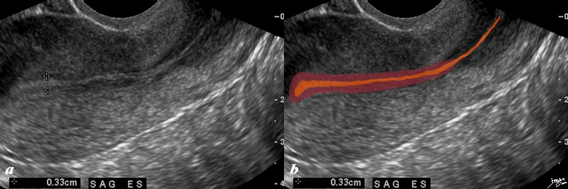
Junctional Zone In Early Proliferative Phase |
|
The normal sagittal view of the uterus is a transvaginal ultrasound, in the first week after menstruation and just prior to the next menstruation after the endometrium has been shed. It demonstrates that the endometrium becomes a single echogenic line consisting of opposing walls (orange) and is surrounded by a subendometrial halo of the junctional zone (tan). This layer is more compacted, and relatively hypovascular. This image is typical of the early proliferative phase. It is during this time that estrogen starts to rise and progesterone has fallen. The endometrium in this case measures about 3mms. Courtesy Ashley Davidoff MD Copyright 2010 All rights reserved 84698c02b.8s |
|
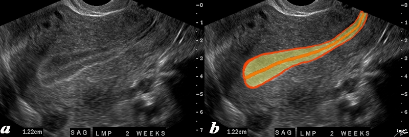
The Complex Endometrium Triple Stripe – Trilaminar Appearance – Preovulation |
|
The normal sagittal view of the uterus is a transvaginal ultrasound, 2 weeks after menstruation, and just prior to ovulation. It demonstrates that the uterus and more specifically the endometrium is more complex than just a simple columnar epithelium. This is an example of the trilaminar appearance of the endometrium and is characteristic of the appearance of the endometrium in the preovulatory phase. This pahse is also called the follicular phase, or proliferative phase. It is during this time that estrogen is the dominant hormonal influence. The appearance is also known as a “triple stripe”, and conversely, its presence is seen before progesterone is produced. If present with a 9mm+ diameter, it reflects an ideal potential lining for fertilization.. In this case it measured 1.2cms. Courtesy Ashley Davidoff MD Copyright 2010 All rights reserved 83835c02.8s |
|
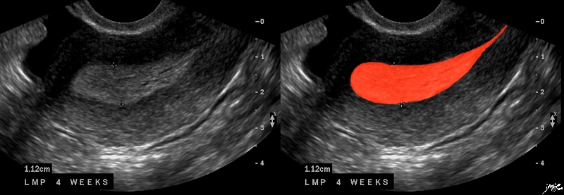
The Overipe Endometrium – Premenstrual |
|
The normal sagittal view of the uterus is a transvaginal ultrasound, about 4 weeks after menstruation, and just prior to the next menstrauation when the endometrium is overripe. It demonstrates that the uterus and more specifically the endometrium is more complex than just a simple columnar epithelium. This is an example of the hyperechoic, homogeneous, thick endometrium characteristic of the secretory phase. It is during this time that progesterone is the dominant hormonal influence and estrogen influence is minimal. The endometrium in this case measures 1.2cms. Courtesy Ashley Davidoff MD Copyright 2010 All rights reserved 46318c01.8s |
|
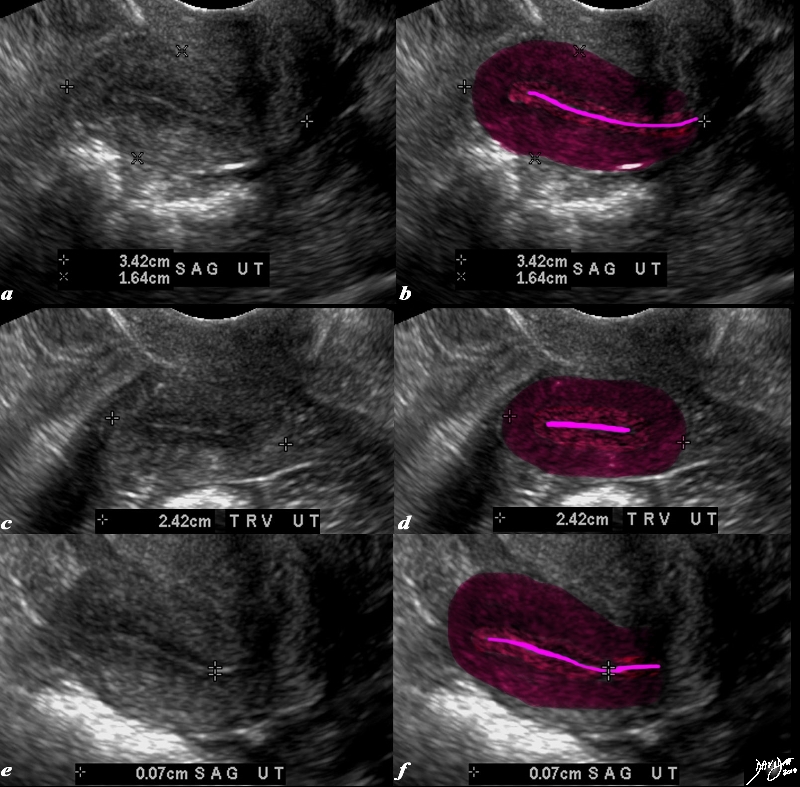
Endometrial Stripe and Junctional Zone in the Aging Uterus |
|
The transvaginal ultrasound is from a 62 year old post menopausal patient who presents with pelvic discomfort. The ultrasound shows a small atrophied uterus. The myometrium (dark red) junctional zone (light red) and endometrial cavity (pink) are outlined. In the sagittal plane the uterus measures 3.4cms in cranoicaudal dimension by 2.4cms in anteroposterior dimension (a,b) In the axial dimension (c,d) the uterus measures 2.4cms. The endometrial stripe is measured in the sagittal plane and measures .7mms. Courtesy Ashley Davidoff MD Copyright 2010 All rights reserved 84711c01.8s |
Character By CTscan
The character of the uterus is dependant on many aspects including the age, time of the month and the way the angle the uterus is scanned.
Characterization by MRI
DOMElement Object
(
    [schemaTypeInfo] =>
    [tagName] => table
    [firstElementChild] => (object value omitted)
    [lastElementChild] => (object value omitted)
    [childElementCount] => 1
    [previousElementSibling] => (object value omitted)
    [nextElementSibling] =>
    [nodeName] => table
    [nodeValue] =>


T1 Weighted with Gadolinium
Normal


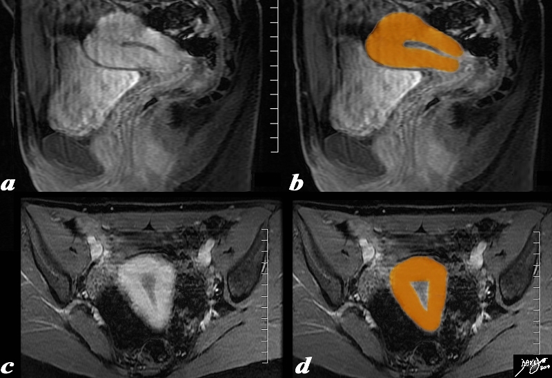
The uterus is pear shaped as depicted in this overlay sagital (a,b) and coronal (c,d) T 1weighte enhanced MRI study
Courtesy Ashley Davidoff MD Copyright 2010 96378c02.81s


    [nodeType] => 1
    [parentNode] => (object value omitted)
    [childNodes] => (object value omitted)
    [firstChild] => (object value omitted)
    [lastChild] => (object value omitted)
    [previousSibling] => (object value omitted)
    [nextSibling] => (object value omitted)
    [attributes] => (object value omitted)
    [ownerDocument] => (object value omitted)
    [namespaceURI] =>
    [prefix] =>
    [localName] => table
    [baseURI] =>
    [textContent] =>


T1 Weighted with Gadolinium
Normal


The uterus is pear shaped as depicted in this overlay sagital (a,b) and coronal (c,d) T 1weighte enhanced MRI study
Courtesy Ashley Davidoff MD Copyright 2010 96378c02.81s


)
DOMElement Object
(
    [schemaTypeInfo] =>
    [tagName] => td
    [firstElementChild] => (object value omitted)
    [lastElementChild] => (object value omitted)
    [childElementCount] => 2
    [previousElementSibling] =>
    [nextElementSibling] =>
    [nodeName] => td
    [nodeValue] =>
The uterus is pear shaped as depicted in this overlay sagital (a,b) and coronal (c,d) T 1weighte enhanced MRI study
Courtesy Ashley Davidoff MD Copyright 2010 96378c02.81s

    [nodeType] => 1
    [parentNode] => (object value omitted)
    [childNodes] => (object value omitted)
    [firstChild] => (object value omitted)
    [lastChild] => (object value omitted)
    [previousSibling] => (object value omitted)
    [nextSibling] => (object value omitted)
    [attributes] => (object value omitted)
    [ownerDocument] => (object value omitted)
    [namespaceURI] =>
    [prefix] =>
    [localName] => td
    [baseURI] =>
    [textContent] =>
The uterus is pear shaped as depicted in this overlay sagital (a,b) and coronal (c,d) T 1weighte enhanced MRI study
Courtesy Ashley Davidoff MD Copyright 2010 96378c02.81s

)
DOMElement Object
(
    [schemaTypeInfo] =>
    [tagName] => td
    [firstElementChild] => (object value omitted)
    [lastElementChild] => (object value omitted)
    [childElementCount] => 3
    [previousElementSibling] =>
    [nextElementSibling] =>
    [nodeName] => td
    [nodeValue] =>
T1 Weighted with Gadolinium
Normal
    [nodeType] => 1
    [parentNode] => (object value omitted)
    [childNodes] => (object value omitted)
    [firstChild] => (object value omitted)
    [lastChild] => (object value omitted)
    [previousSibling] => (object value omitted)
    [nextSibling] => (object value omitted)
    [attributes] => (object value omitted)
    [ownerDocument] => (object value omitted)
    [namespaceURI] =>
    [prefix] =>
    [localName] => td
    [baseURI] =>
    [textContent] =>
T1 Weighted with Gadolinium
Normal
)
DOMElement Object
(
    [schemaTypeInfo] =>
    [tagName] => table
    [firstElementChild] => (object value omitted)
    [lastElementChild] => (object value omitted)
    [childElementCount] => 1
    [previousElementSibling] => (object value omitted)
    [nextElementSibling] => (object value omitted)
    [nodeName] => table
    [nodeValue] =>


T2 Weighted MRI in the Sagittal Plane


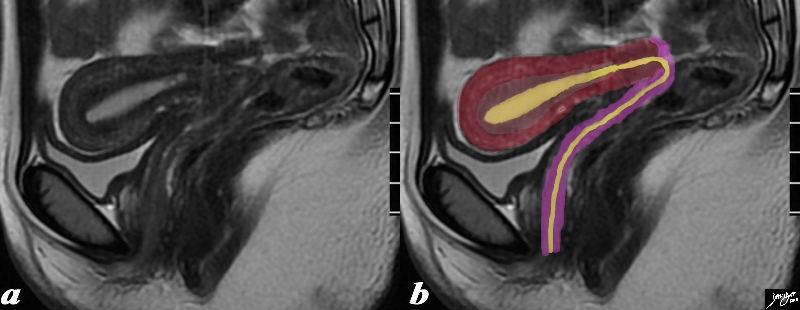
The normal sagittal view of the uterus is a T2 weighted MRI from a 16 year old female with pelvic pain. It demonstrates theta the uterus and more specifically the myometrium is more complex and consists of an outer part (dark red) and an inner more homogeneous part called the junctional zone Since a T2 weighted image is sensitive to water we understand from this image that the outer part since it has greater white signal contains more water, and likely more vascularity. The endometrial canal, cervical canal and vaginal cavity are outlined in yellow and the vaginal wall is pink.
Courtesy Ashley Davidoff MD Copyright 2010 All rights reserved 96733c01.8s


    [nodeType] => 1
    [parentNode] => (object value omitted)
    [childNodes] => (object value omitted)
    [firstChild] => (object value omitted)
    [lastChild] => (object value omitted)
    [previousSibling] => (object value omitted)
    [nextSibling] => (object value omitted)
    [attributes] => (object value omitted)
    [ownerDocument] => (object value omitted)
    [namespaceURI] =>
    [prefix] =>
    [localName] => table
    [baseURI] =>
    [textContent] =>


T2 Weighted MRI in the Sagittal Plane


The normal sagittal view of the uterus is a T2 weighted MRI from a 16 year old female with pelvic pain. It demonstrates theta the uterus and more specifically the myometrium is more complex and consists of an outer part (dark red) and an inner more homogeneous part called the junctional zone Since a T2 weighted image is sensitive to water we understand from this image that the outer part since it has greater white signal contains more water, and likely more vascularity. The endometrial canal, cervical canal and vaginal cavity are outlined in yellow and the vaginal wall is pink.
Courtesy Ashley Davidoff MD Copyright 2010 All rights reserved 96733c01.8s


)
DOMElement Object
(
    [schemaTypeInfo] =>
    [tagName] => td
    [firstElementChild] => (object value omitted)
    [lastElementChild] => (object value omitted)
    [childElementCount] => 2
    [previousElementSibling] =>
    [nextElementSibling] =>
    [nodeName] => td
    [nodeValue] =>
The normal sagittal view of the uterus is a T2 weighted MRI from a 16 year old female with pelvic pain. It demonstrates theta the uterus and more specifically the myometrium is more complex and consists of an outer part (dark red) and an inner more homogeneous part called the junctional zone Since a T2 weighted image is sensitive to water we understand from this image that the outer part since it has greater white signal contains more water, and likely more vascularity. The endometrial canal, cervical canal and vaginal cavity are outlined in yellow and the vaginal wall is pink.
Courtesy Ashley Davidoff MD Copyright 2010 All rights reserved 96733c01.8s

    [nodeType] => 1
    [parentNode] => (object value omitted)
    [childNodes] => (object value omitted)
    [firstChild] => (object value omitted)
    [lastChild] => (object value omitted)
    [previousSibling] => (object value omitted)
    [nextSibling] => (object value omitted)
    [attributes] => (object value omitted)
    [ownerDocument] => (object value omitted)
    [namespaceURI] =>
    [prefix] =>
    [localName] => td
    [baseURI] =>
    [textContent] =>
The normal sagittal view of the uterus is a T2 weighted MRI from a 16 year old female with pelvic pain. It demonstrates theta the uterus and more specifically the myometrium is more complex and consists of an outer part (dark red) and an inner more homogeneous part called the junctional zone Since a T2 weighted image is sensitive to water we understand from this image that the outer part since it has greater white signal contains more water, and likely more vascularity. The endometrial canal, cervical canal and vaginal cavity are outlined in yellow and the vaginal wall is pink.
Courtesy Ashley Davidoff MD Copyright 2010 All rights reserved 96733c01.8s

)
DOMElement Object
(
    [schemaTypeInfo] =>
    [tagName] => td
    [firstElementChild] => (object value omitted)
    [lastElementChild] => (object value omitted)
    [childElementCount] => 2
    [previousElementSibling] =>
    [nextElementSibling] =>
    [nodeName] => td
    [nodeValue] =>
T2 Weighted MRI in the Sagittal Plane
    [nodeType] => 1
    [parentNode] => (object value omitted)
    [childNodes] => (object value omitted)
    [firstChild] => (object value omitted)
    [lastChild] => (object value omitted)
    [previousSibling] => (object value omitted)
    [nextSibling] => (object value omitted)
    [attributes] => (object value omitted)
    [ownerDocument] => (object value omitted)
    [namespaceURI] =>
    [prefix] =>
    [localName] => td
    [baseURI] =>
    [textContent] =>
T2 Weighted MRI in the Sagittal Plane
)
DOMElement Object
(
    [schemaTypeInfo] =>
    [tagName] => table
    [firstElementChild] => (object value omitted)
    [lastElementChild] => (object value omitted)
    [childElementCount] => 1
    [previousElementSibling] => (object value omitted)
    [nextElementSibling] => (object value omitted)
    [nodeName] => table
    [nodeValue] =>


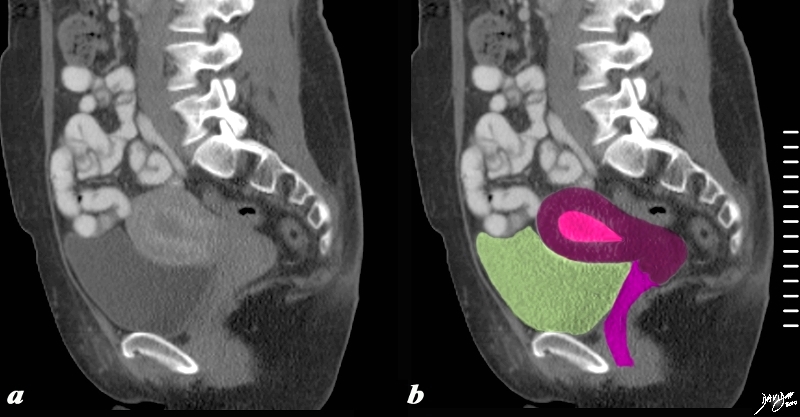
Sagittal Reconstruction


The sagittal reconstructed CT shows an anteverted uterus buoyed and cushioned by partly filled bladder (yellow) In this sagittal view the adult uterus measures about 10cms in craniocaudad span and 5cms in A-P dimension and the endometrial stripe measures about 1cms.
Courtesy Ashley Davidoff MD copyright 2010 all rights reserved 60385c03.81s


    [nodeType] => 1
    [parentNode] => (object value omitted)
    [childNodes] => (object value omitted)
    [firstChild] => (object value omitted)
    [lastChild] => (object value omitted)
    [previousSibling] => (object value omitted)
    [nextSibling] => (object value omitted)
    [attributes] => (object value omitted)
    [ownerDocument] => (object value omitted)
    [namespaceURI] =>
    [prefix] =>
    [localName] => table
    [baseURI] =>
    [textContent] =>


Sagittal Reconstruction


The sagittal reconstructed CT shows an anteverted uterus buoyed and cushioned by partly filled bladder (yellow) In this sagittal view the adult uterus measures about 10cms in craniocaudad span and 5cms in A-P dimension and the endometrial stripe measures about 1cms.
Courtesy Ashley Davidoff MD copyright 2010 all rights reserved 60385c03.81s


)
DOMElement Object
(
    [schemaTypeInfo] =>
    [tagName] => td
    [firstElementChild] => (object value omitted)
    [lastElementChild] => (object value omitted)
    [childElementCount] => 2
    [previousElementSibling] =>
    [nextElementSibling] =>
    [nodeName] => td
    [nodeValue] =>
The sagittal reconstructed CT shows an anteverted uterus buoyed and cushioned by partly filled bladder (yellow) In this sagittal view the adult uterus measures about 10cms in craniocaudad span and 5cms in A-P dimension and the endometrial stripe measures about 1cms.
Courtesy Ashley Davidoff MD copyright 2010 all rights reserved 60385c03.81s

    [nodeType] => 1
    [parentNode] => (object value omitted)
    [childNodes] => (object value omitted)
    [firstChild] => (object value omitted)
    [lastChild] => (object value omitted)
    [previousSibling] => (object value omitted)
    [nextSibling] => (object value omitted)
    [attributes] => (object value omitted)
    [ownerDocument] => (object value omitted)
    [namespaceURI] =>
    [prefix] =>
    [localName] => td
    [baseURI] =>
    [textContent] =>
The sagittal reconstructed CT shows an anteverted uterus buoyed and cushioned by partly filled bladder (yellow) In this sagittal view the adult uterus measures about 10cms in craniocaudad span and 5cms in A-P dimension and the endometrial stripe measures about 1cms.
Courtesy Ashley Davidoff MD copyright 2010 all rights reserved 60385c03.81s

)
DOMElement Object
(
    [schemaTypeInfo] =>
    [tagName] => td
    [firstElementChild] => (object value omitted)
    [lastElementChild] => (object value omitted)
    [childElementCount] => 2
    [previousElementSibling] =>
    [nextElementSibling] =>
    [nodeName] => td
    [nodeValue] =>
Sagittal Reconstruction
    [nodeType] => 1
    [parentNode] => (object value omitted)
    [childNodes] => (object value omitted)
    [firstChild] => (object value omitted)
    [lastChild] => (object value omitted)
    [previousSibling] => (object value omitted)
    [nextSibling] => (object value omitted)
    [attributes] => (object value omitted)
    [ownerDocument] => (object value omitted)
    [namespaceURI] =>
    [prefix] =>
    [localName] => td
    [baseURI] =>
    [textContent] =>
Sagittal Reconstruction
)
DOMElement Object
(
    [schemaTypeInfo] =>
    [tagName] => table
    [firstElementChild] => (object value omitted)
    [lastElementChild] => (object value omitted)
    [childElementCount] => 1
    [previousElementSibling] => (object value omitted)
    [nextElementSibling] => (object value omitted)
    [nodeName] => table
    [nodeValue] =>


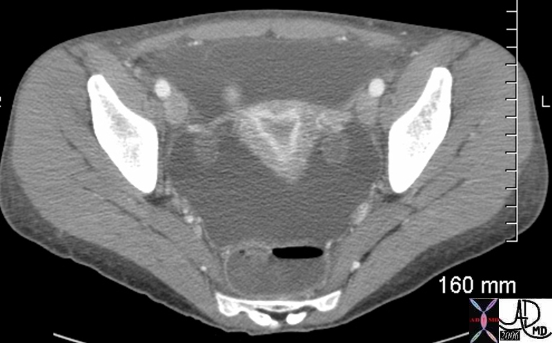
Normal Uterus in a 20 Year Old with Acute Transmural Colitis and Ascites


In this 20 yearold patient the myometrium is homogeneous and isodense with the soft tissues.  The suendometria layer is hyperemic and likely is in the latter phase of the cysle and the endometrium and endometrial cavity is hypodense.
She presented with colitis and ascites which enabled detail of the uterus to be appreciated.  Ovaries are well visualized a well
Image Courtesy Ashley Davidoff MD 45318


    [nodeType] => 1
    [parentNode] => (object value omitted)
    [childNodes] => (object value omitted)
    [firstChild] => (object value omitted)
    [lastChild] => (object value omitted)
    [previousSibling] => (object value omitted)
    [nextSibling] => (object value omitted)
    [attributes] => (object value omitted)
    [ownerDocument] => (object value omitted)
    [namespaceURI] =>
    [prefix] =>
    [localName] => table
    [baseURI] =>
    [textContent] =>


Normal Uterus in a 20 Year Old with Acute Transmural Colitis and Ascites


In this 20 yearold patient the myometrium is homogeneous and isodense with the soft tissues.  The suendometria layer is hyperemic and likely is in the latter phase of the cysle and the endometrium and endometrial cavity is hypodense.
She presented with colitis and ascites which enabled detail of the uterus to be appreciated.  Ovaries are well visualized a well
Image Courtesy Ashley Davidoff MD 45318


)
DOMElement Object
(
    [schemaTypeInfo] =>
    [tagName] => td
    [firstElementChild] => (object value omitted)
    [lastElementChild] => (object value omitted)
    [childElementCount] => 3
    [previousElementSibling] =>
    [nextElementSibling] =>
    [nodeName] => td
    [nodeValue] =>
In this 20 yearold patient the myometrium is homogeneous and isodense with the soft tissues.  The suendometria layer is hyperemic and likely is in the latter phase of the cysle and the endometrium and endometrial cavity is hypodense.
She presented with colitis and ascites which enabled detail of the uterus to be appreciated.  Ovaries are well visualized a well
Image Courtesy Ashley Davidoff MD 45318

    [nodeType] => 1
    [parentNode] => (object value omitted)
    [childNodes] => (object value omitted)
    [firstChild] => (object value omitted)
    [lastChild] => (object value omitted)
    [previousSibling] => (object value omitted)
    [nextSibling] => (object value omitted)
    [attributes] => (object value omitted)
    [ownerDocument] => (object value omitted)
    [namespaceURI] =>
    [prefix] =>
    [localName] => td
    [baseURI] =>
    [textContent] =>
In this 20 yearold patient the myometrium is homogeneous and isodense with the soft tissues.  The suendometria layer is hyperemic and likely is in the latter phase of the cysle and the endometrium and endometrial cavity is hypodense.
She presented with colitis and ascites which enabled detail of the uterus to be appreciated.  Ovaries are well visualized a well
Image Courtesy Ashley Davidoff MD 45318

)
DOMElement Object
(
    [schemaTypeInfo] =>
    [tagName] => td
    [firstElementChild] => (object value omitted)
    [lastElementChild] => (object value omitted)
    [childElementCount] => 2
    [previousElementSibling] =>
    [nextElementSibling] =>
    [nodeName] => td
    [nodeValue] =>
Normal Uterus in a 20 Year Old with Acute Transmural Colitis and Ascites
    [nodeType] => 1
    [parentNode] => (object value omitted)
    [childNodes] => (object value omitted)
    [firstChild] => (object value omitted)
    [lastChild] => (object value omitted)
    [previousSibling] => (object value omitted)
    [nextSibling] => (object value omitted)
    [attributes] => (object value omitted)
    [ownerDocument] => (object value omitted)
    [namespaceURI] =>
    [prefix] =>
    [localName] => td
    [baseURI] =>
    [textContent] =>
Normal Uterus in a 20 Year Old with Acute Transmural Colitis and Ascites
)
DOMElement Object
(
    [schemaTypeInfo] =>
    [tagName] => table
    [firstElementChild] => (object value omitted)
    [lastElementChild] => (object value omitted)
    [childElementCount] => 1
    [previousElementSibling] => (object value omitted)
    [nextElementSibling] => (object value omitted)
    [nodeName] => table
    [nodeValue] =>


Endometrial Stripe and Junctional Zone in the Aging Uterus


The transvaginal ultrasound is from a 62 year old post menopausal patient who presents with pelvic discomfort. The ultrasound shows a small atrophied uterus. The myometrium (dark red) junctional zone (light red) and endometrial cavity (pink) are outlined. In the sagittal plane the uterus measures 3.4cms in cranoicaudal dimension by 2.4cms in anteroposterior dimension (a,b) In the axial dimension (c,d) the uterus measures 2.4cms. The endometrial stripe is measured in the sagittal plane and measures .7mms.
Courtesy Ashley Davidoff MD Copyright 2010 All rights reserved 84711c01.8s


    [nodeType] => 1
    [parentNode] => (object value omitted)
    [childNodes] => (object value omitted)
    [firstChild] => (object value omitted)
    [lastChild] => (object value omitted)
    [previousSibling] => (object value omitted)
    [nextSibling] => (object value omitted)
    [attributes] => (object value omitted)
    [ownerDocument] => (object value omitted)
    [namespaceURI] =>
    [prefix] =>
    [localName] => table
    [baseURI] =>
    [textContent] =>


Endometrial Stripe and Junctional Zone in the Aging Uterus


The transvaginal ultrasound is from a 62 year old post menopausal patient who presents with pelvic discomfort. The ultrasound shows a small atrophied uterus. The myometrium (dark red) junctional zone (light red) and endometrial cavity (pink) are outlined. In the sagittal plane the uterus measures 3.4cms in cranoicaudal dimension by 2.4cms in anteroposterior dimension (a,b) In the axial dimension (c,d) the uterus measures 2.4cms. The endometrial stripe is measured in the sagittal plane and measures .7mms.
Courtesy Ashley Davidoff MD Copyright 2010 All rights reserved 84711c01.8s


)
DOMElement Object
(
    [schemaTypeInfo] =>
    [tagName] => td
    [firstElementChild] => (object value omitted)
    [lastElementChild] => (object value omitted)
    [childElementCount] => 2
    [previousElementSibling] =>
    [nextElementSibling] =>
    [nodeName] => td
    [nodeValue] =>
The transvaginal ultrasound is from a 62 year old post menopausal patient who presents with pelvic discomfort. The ultrasound shows a small atrophied uterus. The myometrium (dark red) junctional zone (light red) and endometrial cavity (pink) are outlined. In the sagittal plane the uterus measures 3.4cms in cranoicaudal dimension by 2.4cms in anteroposterior dimension (a,b) In the axial dimension (c,d) the uterus measures 2.4cms. The endometrial stripe is measured in the sagittal plane and measures .7mms.
Courtesy Ashley Davidoff MD Copyright 2010 All rights reserved 84711c01.8s

    [nodeType] => 1
    [parentNode] => (object value omitted)
    [childNodes] => (object value omitted)
    [firstChild] => (object value omitted)
    [lastChild] => (object value omitted)
    [previousSibling] => (object value omitted)
    [nextSibling] => (object value omitted)
    [attributes] => (object value omitted)
    [ownerDocument] => (object value omitted)
    [namespaceURI] =>
    [prefix] =>
    [localName] => td
    [baseURI] =>
    [textContent] =>
The transvaginal ultrasound is from a 62 year old post menopausal patient who presents with pelvic discomfort. The ultrasound shows a small atrophied uterus. The myometrium (dark red) junctional zone (light red) and endometrial cavity (pink) are outlined. In the sagittal plane the uterus measures 3.4cms in cranoicaudal dimension by 2.4cms in anteroposterior dimension (a,b) In the axial dimension (c,d) the uterus measures 2.4cms. The endometrial stripe is measured in the sagittal plane and measures .7mms.
Courtesy Ashley Davidoff MD Copyright 2010 All rights reserved 84711c01.8s

)
DOMElement Object
(
    [schemaTypeInfo] =>
    [tagName] => td
    [firstElementChild] => (object value omitted)
    [lastElementChild] => (object value omitted)
    [childElementCount] => 2
    [previousElementSibling] =>
    [nextElementSibling] =>
    [nodeName] => td
    [nodeValue] =>

Endometrial Stripe and Junctional Zone in the Aging Uterus
    [nodeType] => 1
    [parentNode] => (object value omitted)
    [childNodes] => (object value omitted)
    [firstChild] => (object value omitted)
    [lastChild] => (object value omitted)
    [previousSibling] => (object value omitted)
    [nextSibling] => (object value omitted)
    [attributes] => (object value omitted)
    [ownerDocument] => (object value omitted)
    [namespaceURI] =>
    [prefix] =>
    [localName] => td
    [baseURI] =>
    [textContent] =>

Endometrial Stripe and Junctional Zone in the Aging Uterus
)
DOMElement Object
(
    [schemaTypeInfo] =>
    [tagName] => table
    [firstElementChild] => (object value omitted)
    [lastElementChild] => (object value omitted)
    [childElementCount] => 1
    [previousElementSibling] => (object value omitted)
    [nextElementSibling] => (object value omitted)
    [nodeName] => table
    [nodeValue] =>


The Overipe Endometrium – Premenstrual


The normal sagittal view of the uterus is a transvaginal ultrasound, about 4 weeks after menstruation, and just prior to the next menstrauation when the endometrium is overripe. It demonstrates that the uterus and more specifically the endometrium is more complex than just a simple columnar epithelium. This is an example of the hyperechoic, homogeneous, thick endometrium characteristic of the secretory phase. It is during this time that progesterone is the dominant hormonal influence and estrogen influence is minimal. The endometrium in this case measures 1.2cms.
Courtesy Ashley Davidoff MD Copyright 2010 All rights reserved 46318c01.8s


    [nodeType] => 1
    [parentNode] => (object value omitted)
    [childNodes] => (object value omitted)
    [firstChild] => (object value omitted)
    [lastChild] => (object value omitted)
    [previousSibling] => (object value omitted)
    [nextSibling] => (object value omitted)
    [attributes] => (object value omitted)
    [ownerDocument] => (object value omitted)
    [namespaceURI] =>
    [prefix] =>
    [localName] => table
    [baseURI] =>
    [textContent] =>


The Overipe Endometrium – Premenstrual


The normal sagittal view of the uterus is a transvaginal ultrasound, about 4 weeks after menstruation, and just prior to the next menstrauation when the endometrium is overripe. It demonstrates that the uterus and more specifically the endometrium is more complex than just a simple columnar epithelium. This is an example of the hyperechoic, homogeneous, thick endometrium characteristic of the secretory phase. It is during this time that progesterone is the dominant hormonal influence and estrogen influence is minimal. The endometrium in this case measures 1.2cms.
Courtesy Ashley Davidoff MD Copyright 2010 All rights reserved 46318c01.8s


)
DOMElement Object
(
    [schemaTypeInfo] =>
    [tagName] => td
    [firstElementChild] => (object value omitted)
    [lastElementChild] => (object value omitted)
    [childElementCount] => 2
    [previousElementSibling] =>
    [nextElementSibling] =>
    [nodeName] => td
    [nodeValue] =>
The normal sagittal view of the uterus is a transvaginal ultrasound, about 4 weeks after menstruation, and just prior to the next menstrauation when the endometrium is overripe. It demonstrates that the uterus and more specifically the endometrium is more complex than just a simple columnar epithelium. This is an example of the hyperechoic, homogeneous, thick endometrium characteristic of the secretory phase. It is during this time that progesterone is the dominant hormonal influence and estrogen influence is minimal. The endometrium in this case measures 1.2cms.
Courtesy Ashley Davidoff MD Copyright 2010 All rights reserved 46318c01.8s

    [nodeType] => 1
    [parentNode] => (object value omitted)
    [childNodes] => (object value omitted)
    [firstChild] => (object value omitted)
    [lastChild] => (object value omitted)
    [previousSibling] => (object value omitted)
    [nextSibling] => (object value omitted)
    [attributes] => (object value omitted)
    [ownerDocument] => (object value omitted)
    [namespaceURI] =>
    [prefix] =>
    [localName] => td
    [baseURI] =>
    [textContent] =>
The normal sagittal view of the uterus is a transvaginal ultrasound, about 4 weeks after menstruation, and just prior to the next menstrauation when the endometrium is overripe. It demonstrates that the uterus and more specifically the endometrium is more complex than just a simple columnar epithelium. This is an example of the hyperechoic, homogeneous, thick endometrium characteristic of the secretory phase. It is during this time that progesterone is the dominant hormonal influence and estrogen influence is minimal. The endometrium in this case measures 1.2cms.
Courtesy Ashley Davidoff MD Copyright 2010 All rights reserved 46318c01.8s

)
DOMElement Object
(
    [schemaTypeInfo] =>
    [tagName] => td
    [firstElementChild] => (object value omitted)
    [lastElementChild] => (object value omitted)
    [childElementCount] => 2
    [previousElementSibling] =>
    [nextElementSibling] =>
    [nodeName] => td
    [nodeValue] =>

The Overipe Endometrium – Premenstrual
    [nodeType] => 1
    [parentNode] => (object value omitted)
    [childNodes] => (object value omitted)
    [firstChild] => (object value omitted)
    [lastChild] => (object value omitted)
    [previousSibling] => (object value omitted)
    [nextSibling] => (object value omitted)
    [attributes] => (object value omitted)
    [ownerDocument] => (object value omitted)
    [namespaceURI] =>
    [prefix] =>
    [localName] => td
    [baseURI] =>
    [textContent] =>

The Overipe Endometrium – Premenstrual
)
DOMElement Object
(
    [schemaTypeInfo] =>
    [tagName] => table
    [firstElementChild] => (object value omitted)
    [lastElementChild] => (object value omitted)
    [childElementCount] => 1
    [previousElementSibling] => (object value omitted)
    [nextElementSibling] => (object value omitted)
    [nodeName] => table
    [nodeValue] =>


The Complex Endometrium
Triple Stripe – Trilaminar Appearance – Preovulation


The normal sagittal view of the uterus is a transvaginal ultrasound, 2 weeks after menstruation, and just prior to ovulation. It demonstrates that the uterus and more specifically the endometrium is more complex than just a simple columnar epithelium. This is an example of the trilaminar appearance of the endometrium and is characteristic of the appearance of the endometrium in the preovulatory phase. This pahse is also called the follicular phase, or proliferative phase. It is during this time that estrogen is the dominant hormonal influence. The appearance is also known as a “triple stripe”, and conversely, its presence is seen before progesterone is produced. If present with a 9mm+ diameter, it reflects an ideal potential lining for fertilization.. In this case it measured 1.2cms.
Courtesy Ashley Davidoff MD Copyright 2010 All rights reserved  83835c02.8s


    [nodeType] => 1
    [parentNode] => (object value omitted)
    [childNodes] => (object value omitted)
    [firstChild] => (object value omitted)
    [lastChild] => (object value omitted)
    [previousSibling] => (object value omitted)
    [nextSibling] => (object value omitted)
    [attributes] => (object value omitted)
    [ownerDocument] => (object value omitted)
    [namespaceURI] =>
    [prefix] =>
    [localName] => table
    [baseURI] =>
    [textContent] =>


The Complex Endometrium
Triple Stripe – Trilaminar Appearance – Preovulation


The normal sagittal view of the uterus is a transvaginal ultrasound, 2 weeks after menstruation, and just prior to ovulation. It demonstrates that the uterus and more specifically the endometrium is more complex than just a simple columnar epithelium. This is an example of the trilaminar appearance of the endometrium and is characteristic of the appearance of the endometrium in the preovulatory phase. This pahse is also called the follicular phase, or proliferative phase. It is during this time that estrogen is the dominant hormonal influence. The appearance is also known as a “triple stripe”, and conversely, its presence is seen before progesterone is produced. If present with a 9mm+ diameter, it reflects an ideal potential lining for fertilization.. In this case it measured 1.2cms.
Courtesy Ashley Davidoff MD Copyright 2010 All rights reserved  83835c02.8s


)
DOMElement Object
(
    [schemaTypeInfo] =>
    [tagName] => td
    [firstElementChild] => (object value omitted)
    [lastElementChild] => (object value omitted)
    [childElementCount] => 2
    [previousElementSibling] =>
    [nextElementSibling] =>
    [nodeName] => td
    [nodeValue] =>
The normal sagittal view of the uterus is a transvaginal ultrasound, 2 weeks after menstruation, and just prior to ovulation. It demonstrates that the uterus and more specifically the endometrium is more complex than just a simple columnar epithelium. This is an example of the trilaminar appearance of the endometrium and is characteristic of the appearance of the endometrium in the preovulatory phase. This pahse is also called the follicular phase, or proliferative phase. It is during this time that estrogen is the dominant hormonal influence. The appearance is also known as a “triple stripe”, and conversely, its presence is seen before progesterone is produced. If present with a 9mm+ diameter, it reflects an ideal potential lining for fertilization.. In this case it measured 1.2cms.
Courtesy Ashley Davidoff MD Copyright 2010 All rights reserved  83835c02.8s

    [nodeType] => 1
    [parentNode] => (object value omitted)
    [childNodes] => (object value omitted)
    [firstChild] => (object value omitted)
    [lastChild] => (object value omitted)
    [previousSibling] => (object value omitted)
    [nextSibling] => (object value omitted)
    [attributes] => (object value omitted)
    [ownerDocument] => (object value omitted)
    [namespaceURI] =>
    [prefix] =>
    [localName] => td
    [baseURI] =>
    [textContent] =>
The normal sagittal view of the uterus is a transvaginal ultrasound, 2 weeks after menstruation, and just prior to ovulation. It demonstrates that the uterus and more specifically the endometrium is more complex than just a simple columnar epithelium. This is an example of the trilaminar appearance of the endometrium and is characteristic of the appearance of the endometrium in the preovulatory phase. This pahse is also called the follicular phase, or proliferative phase. It is during this time that estrogen is the dominant hormonal influence. The appearance is also known as a “triple stripe”, and conversely, its presence is seen before progesterone is produced. If present with a 9mm+ diameter, it reflects an ideal potential lining for fertilization.. In this case it measured 1.2cms.
Courtesy Ashley Davidoff MD Copyright 2010 All rights reserved  83835c02.8s

)
DOMElement Object
(
    [schemaTypeInfo] =>
    [tagName] => td
    [firstElementChild] => (object value omitted)
    [lastElementChild] => (object value omitted)
    [childElementCount] => 3
    [previousElementSibling] =>
    [nextElementSibling] =>
    [nodeName] => td
    [nodeValue] =>

The Complex Endometrium
Triple Stripe – Trilaminar Appearance – Preovulation
    [nodeType] => 1
    [parentNode] => (object value omitted)
    [childNodes] => (object value omitted)
    [firstChild] => (object value omitted)
    [lastChild] => (object value omitted)
    [previousSibling] => (object value omitted)
    [nextSibling] => (object value omitted)
    [attributes] => (object value omitted)
    [ownerDocument] => (object value omitted)
    [namespaceURI] =>
    [prefix] =>
    [localName] => td
    [baseURI] =>
    [textContent] =>

The Complex Endometrium
Triple Stripe – Trilaminar Appearance – Preovulation
)
DOMElement Object
(
    [schemaTypeInfo] =>
    [tagName] => table
    [firstElementChild] => (object value omitted)
    [lastElementChild] => (object value omitted)
    [childElementCount] => 1
    [previousElementSibling] => (object value omitted)
    [nextElementSibling] => (object value omitted)
    [nodeName] => table
    [nodeValue] =>


Junctional Zone In Early Proliferative Phase


The normal sagittal view of the uterus is a transvaginal ultrasound, in the first week after menstruation  and just prior to the next menstruation after the endometrium has been shed. It demonstrates that the endometrium becomes a single echogenic line consisting of opposing walls (orange) and is surrounded by a subendometrial halo of the junctional zone (tan). This layer is more compacted, and relatively hypovascular. This image is typical of the early proliferative phase. It is during this time that estrogen starts to rise and progesterone has fallen. The endometrium in this case measures about 3mms.
Courtesy Ashley Davidoff MD Copyright 2010 All rights reserved 84698c02b.8s


    [nodeType] => 1
    [parentNode] => (object value omitted)
    [childNodes] => (object value omitted)
    [firstChild] => (object value omitted)
    [lastChild] => (object value omitted)
    [previousSibling] => (object value omitted)
    [nextSibling] => (object value omitted)
    [attributes] => (object value omitted)
    [ownerDocument] => (object value omitted)
    [namespaceURI] =>
    [prefix] =>
    [localName] => table
    [baseURI] =>
    [textContent] =>


Junctional Zone In Early Proliferative Phase


The normal sagittal view of the uterus is a transvaginal ultrasound, in the first week after menstruation  and just prior to the next menstruation after the endometrium has been shed. It demonstrates that the endometrium becomes a single echogenic line consisting of opposing walls (orange) and is surrounded by a subendometrial halo of the junctional zone (tan). This layer is more compacted, and relatively hypovascular. This image is typical of the early proliferative phase. It is during this time that estrogen starts to rise and progesterone has fallen. The endometrium in this case measures about 3mms.
Courtesy Ashley Davidoff MD Copyright 2010 All rights reserved 84698c02b.8s


)
DOMElement Object
(
    [schemaTypeInfo] =>
    [tagName] => td
    [firstElementChild] => (object value omitted)
    [lastElementChild] => (object value omitted)
    [childElementCount] => 2
    [previousElementSibling] =>
    [nextElementSibling] =>
    [nodeName] => td
    [nodeValue] =>
The normal sagittal view of the uterus is a transvaginal ultrasound, in the first week after menstruation  and just prior to the next menstruation after the endometrium has been shed. It demonstrates that the endometrium becomes a single echogenic line consisting of opposing walls (orange) and is surrounded by a subendometrial halo of the junctional zone (tan). This layer is more compacted, and relatively hypovascular. This image is typical of the early proliferative phase. It is during this time that estrogen starts to rise and progesterone has fallen. The endometrium in this case measures about 3mms.
Courtesy Ashley Davidoff MD Copyright 2010 All rights reserved 84698c02b.8s

    [nodeType] => 1
    [parentNode] => (object value omitted)
    [childNodes] => (object value omitted)
    [firstChild] => (object value omitted)
    [lastChild] => (object value omitted)
    [previousSibling] => (object value omitted)
    [nextSibling] => (object value omitted)
    [attributes] => (object value omitted)
    [ownerDocument] => (object value omitted)
    [namespaceURI] =>
    [prefix] =>
    [localName] => td
    [baseURI] =>
    [textContent] =>
The normal sagittal view of the uterus is a transvaginal ultrasound, in the first week after menstruation  and just prior to the next menstruation after the endometrium has been shed. It demonstrates that the endometrium becomes a single echogenic line consisting of opposing walls (orange) and is surrounded by a subendometrial halo of the junctional zone (tan). This layer is more compacted, and relatively hypovascular. This image is typical of the early proliferative phase. It is during this time that estrogen starts to rise and progesterone has fallen. The endometrium in this case measures about 3mms.
Courtesy Ashley Davidoff MD Copyright 2010 All rights reserved 84698c02b.8s

)
DOMElement Object
(
    [schemaTypeInfo] =>
    [tagName] => td
    [firstElementChild] => (object value omitted)
    [lastElementChild] => (object value omitted)
    [childElementCount] => 2
    [previousElementSibling] =>
    [nextElementSibling] =>
    [nodeName] => td
    [nodeValue] =>

Junctional Zone In Early Proliferative Phase
    [nodeType] => 1
    [parentNode] => (object value omitted)
    [childNodes] => (object value omitted)
    [firstChild] => (object value omitted)
    [lastChild] => (object value omitted)
    [previousSibling] => (object value omitted)
    [nextSibling] => (object value omitted)
    [attributes] => (object value omitted)
    [ownerDocument] => (object value omitted)
    [namespaceURI] =>
    [prefix] =>
    [localName] => td
    [baseURI] =>
    [textContent] =>

Junctional Zone In Early Proliferative Phase
)

