The Common Vein Copyright 2011
Introduction
The thyroid is one of the largest endocrine organs in the body, but on the other hand is small compared to most other organs. It weighs about 15- 35 grams measuring approximately 4-5cms cm long, 2-2.5 cm wide and 1 -2 cm thick, in the average adult. The isthmus is 2-6mm in A-P diameter and is up to 20 mm. in width.
The size of the thyroid changes with age. At birth is weighs 2-3 grams, reaches 10-15gms at puberty and becomes full size by age 25 when it weighs 15-35gms.
The weight of the gland varies with age, sex, functional status, iodine intake, and geographic location. The thyroid tends to be larger in women and also larger during the menstrual cycle and pregnancy.
From a clinical standpoint, size is an especially important structural characteristic of the thyroid. The gland is not usually palpable on clinical examination. If palpable it is abnormal.
The enlarged gland may reflect hyperthyroidism, hypothyroidism or a nodular euthyroid gland, and further biochemical and imaging evaluation is necessary.
Fortunately, thyroid size is easily measured with ultrasound which is a safe and relatively inexpensive tool. Linear measurements are the standard for most evaluations, but measurements that portray a volume or mass are also possible using conventional imaging techniques.
|
Size of the Thyroid |
|
The thyroid gland is one of the largest glands in the body weighing 15-20grams. The right and left lobes vary slightly in size and shape and therefore each may have slightly different weights Courtesy Ashley Davidoff MD copyright 2010 all rights reserved 93852..4kd02b.8s |
Imaging the Normal Gland
|
Normal Ultrasound |
|
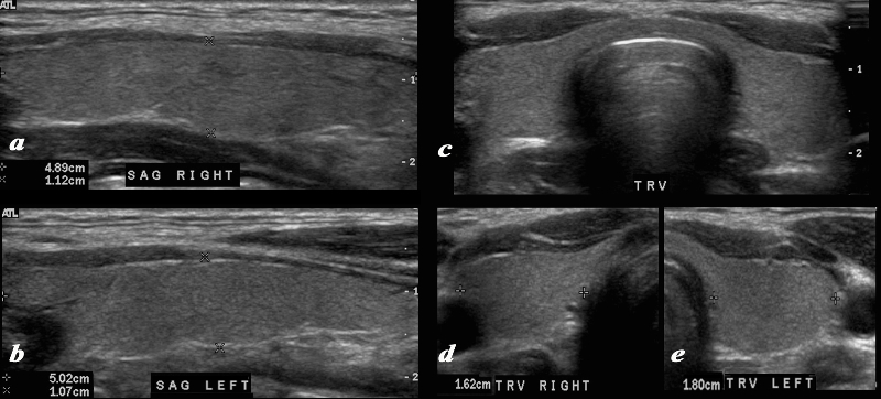
A normal ultrasound of the thyroid gland is demonstrated. Image a and b are sagittal images, image c is a transverse view of both lobes and the isthmus, and image d and e are individual measurements of the right and left lobe respectively.. The normal right lobe (a,d) measures 4.9cms (length) x 1.1cms (A-P anteroposterior) X 1.6 cms (transverse TRV) and the left lobe (b,e) 5.cms (craniocaudad), by 1.1cms (A-P) by 1.8cms (transverse). Courtesy Ashley Davidoff MD Copyright 2010 93832cL.8 |
|
Small Normal and Large Thyroid Glands |
|
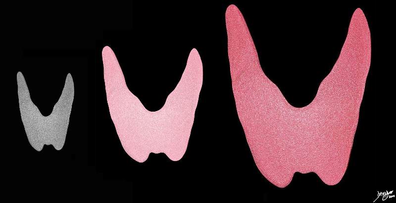
The image shows the artistic rendering of the small hypofunctioning thyroid (gray) the normal thyroid in the middle (pink), and the hyperfunctioning and enlarged thyroid of thyrotoxicosis. Courtesy Ashley Davidoff MD copyright 2010 all rights reserved 93852dc01.8s |
|
The Smooth and Enlarged Gland |
|
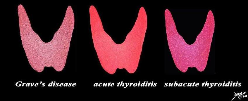
The enlarged smooth gland is usually seen in patients with thyrotoxicosis and includes Grave’s disease, acute thyroiditis and subacute thyroiditis Courtesy Ashley Davidoff MD copyright 2010 all rights reserved 93852.d03cdc06.81s |
Imaging of Goiters
A goiter implies a large gland. It may be caused by a diffusely enlarged gland, by multiple nodules or sometimes by a single large nodule
|
Large Retrosternal Goiter Compressing the Trachea |
|
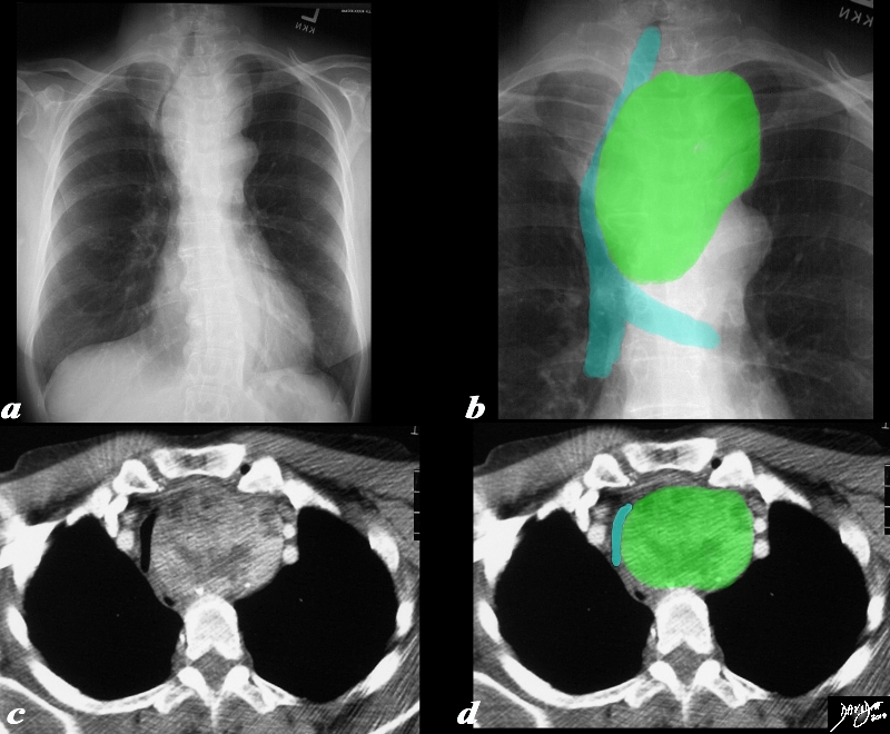
The plain film (a), with magnified view (b) shows a large mass shifting and compressing the trachea (teal) to the right. The mass is seen on cross sectional CT as an irregularly enhancing mass connected to the thyroid gland on more cranial views. Its mass effect on the trachea results in narrowing best appreciated in the transverse dimension. The findings are consistent with a retrosternal thyroid goiter Courtesy Ashley Davidoff MD Copyright 2011 31460c01.8s |
|
Retrosternal Goiter Compressing the Brachiocephalic Vein |
|
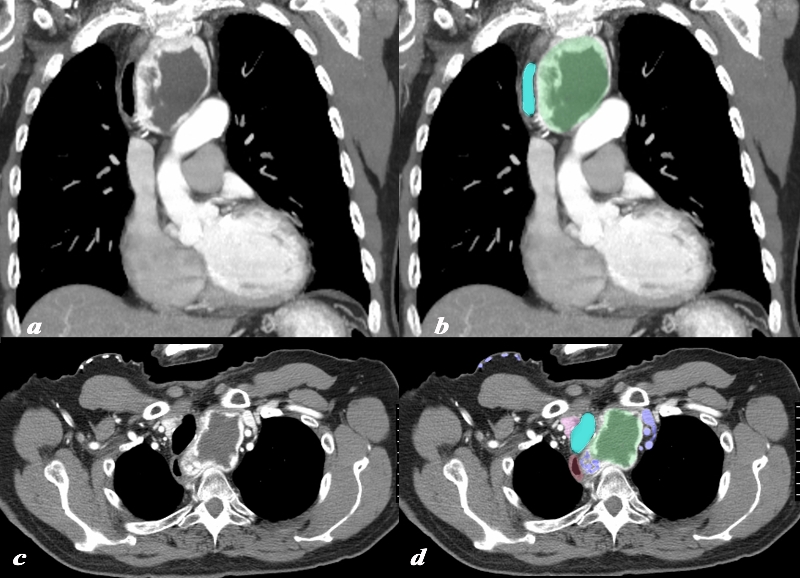
A large retrosternal goiter is seen extending down anterior to the top of the aortic arch is noted in this 74 year old female patient. In image a and b the visualized portion of the goiter measures 8.8cms, has a central avascular component and an enhancing rind. The goiter is overlaid in green and seen displacing the trachea (teal blue) to the right. On the axial cuts the goiter with central avascularity is again noted compressing on the trachea and pushing it to the right (teal blue) the esophagus is also pushed rightward. The brachiocephalic vein (blue) is compressed and there are venous varicosities both on the left and posteriorly on the right of the goiter. Anterior chest veins are also prominent. The findings are consistent with a retrosternal thyroid goiter Courtesy Ashley Davidoff MD Copyright 2010 94516c.8s |
|
Normal and Diffusely Enlarged Hypothyroid Gland – Ultrasound |
|
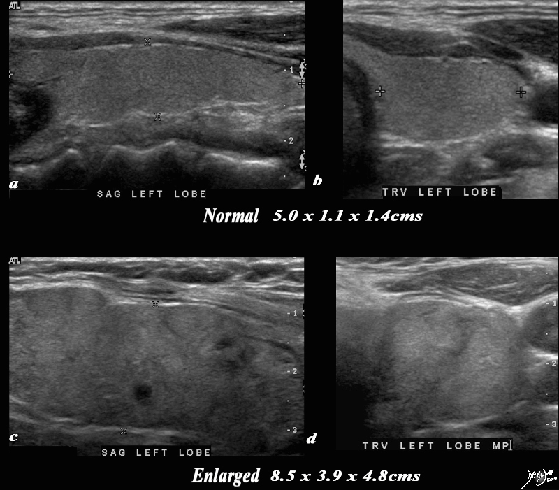
A normal ultrasound of the thyroid gland (a,b) is juxtaposed with the ultrasound of a 78 year female with hypothyroidism (c,d) A diffusely enlarged, heterogeneous thyroid gland is seen in the hypothyroid patient. The normal thyroid measures 5.0cms (length) x 1.1cms (A-P anteroposterior) X 1.4 cms (transverse TRV) and the abnormal thyroid measures 8.5cms (craniocaudad), by 3.9cms (A-P) by 4.8cms (transverse). Clinical findings were consistent with thyroiditis, with biochemical findings suggesting hypothyroidism. The enlarged gland in the transverse dimension is almost round. (d). This rounded shape in the transverse dimension is a clue to the presence of the enlarged gland, even before the measurements are taken and evaluated. The gland is heterogeneous particularly well seen on the sagittal view (c). The findings are consistent with a thyroiditis. Courtesy Ashley Davidoff MD Copyright 2010 94549c05g03.8s |
|
Amyloidosis of the Thyroid |
|
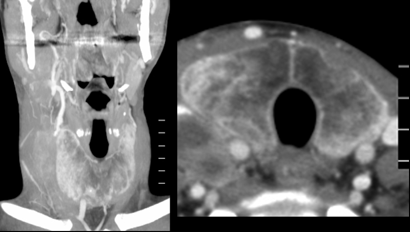
The CT is from a 35 year old man with a complex history including long standing history of Crohn?s disease and a recent history of lymphoma. He has had a goiter for many years and biopsy showed amyloidosis of the thyroid. The image on left is a coronal view of the neck and the b is a transverse view of the thyroid. The gland is enlarged measuring 6.2cms in Craniocaudal dimension (length) by 3cms in anteroposterior dimension by 3cms in transverse dimension. On the transverse view it is not quite round but the gland is full bodied reminiscent of its large size. The density appearance is puzzling. Since no non contrast CT was performed the peripheral increase in density may be due to enhancement or more likely due to iodine or calcification. The soft densities to the right of the right lobe are related to the patient?s newly diagnosed lymphoma. Courtesy Ashley Davidoff MD Copyright 2010 94553c.8 |
Mass in a Lobe Causing a Goiter
|
Goiter Caused by Large Hemorrhagic Cyst |
|
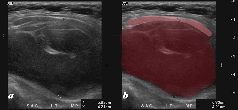
A large mostly hypoechoic mass expands the left lobe of the thyroid on the ultrasound of this 54 year female who presents with an enlarged thyroid gland. The left lobe is filled with low level echoes and contains multiple thin septal echogenic walls (maroon). The mass appears to displace and compress normal soft tissues of the neck (pink) anteriorly. Doppler showed no flow. The mass occupies the entire left lobe and measures 5.8cms in craniocaudad span and by 4.2 cms transversely The diagnosis in this patient is a hemorrhagic cyst causing a goiter. Courtesy Ashley Davidoff MD Copyright 2010 94656c01L |
The Small Gland
|
The Normal Right Lobe and Hypoplastic Left Lobe |
|
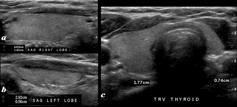
A normal ultrasound of the thyroid gland with a hypoplastic left lobe is demonstrated. Image a is a transverse image, while image b is a sagittal view of the right lobe, and image c is a sagittal view of the left lobe. The normal right lobe (a,b) measures 4.6cms (length) x 1.4cms (A-P anteroposterior) X .7 cms (transverse TRV) and the hypoplastic left lobe measures 2.9cms (craniocaudad), by .6cms (A-P) by .7cms (transverse). Ultrasound Courtesy Ashley Davidoff MD Copyright 2010 96233c01.8 |
|
Normal and Atrophied Gland |
|
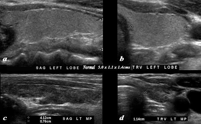
A normal thyroid gland in sagittal (a) and transverse (b) planes is juxtaposed on an abnormal atrophied gland(c,d) The images shown in c & d are from a 70year female who reveals diffuse atrophy of her thyroid by this ultrasound examination. The gland is mildly heterogeneous. The left lobe measures 4.1cms (craniocaudad), by .8 cms (A-P) by 1.1cms (transverse). The sagittal view shows mildl coarsening and heterogeneity of the echoexture. The transverse view shows a normal shape. The isthmus is normal in size. These findings suggest age related involution of the gland. Courtesy Ashley Davidoff MD Copyright 2010 94503c02L |
DOMElement Object
(
    [schemaTypeInfo] =>
    [tagName] => table
    [firstElementChild] => (object value omitted)
    [lastElementChild] => (object value omitted)
    [childElementCount] => 1
    [previousElementSibling] => (object value omitted)
    [nextElementSibling] =>
    [nodeName] => table
    [nodeValue] =>


Hypoplastic Left Lobe


A normal ultrasound of the thyroid gland with a hypoplastic left lobe is demonstrated. Image a is a transverse image, while image b is a sagittal view of the right lobe, and image c is a sagittal view of the left lobe. The normal right lobe (a,b) measures 4.6cms (length) x 1.4cms (A-P anteroposterior) X .7 cms (transverse TRV) and the hypoplastic left lobe measures 2.9cms (craniocaudad), by .6cms (A-P) by .7cms (transverse).
Courtesy Ashley Davidoff MD Copyright 2010 96233c01L.8


    [nodeType] => 1
    [parentNode] => (object value omitted)
    [childNodes] => (object value omitted)
    [firstChild] => (object value omitted)
    [lastChild] => (object value omitted)
    [previousSibling] => (object value omitted)
    [nextSibling] => (object value omitted)
    [attributes] => (object value omitted)
    [ownerDocument] => (object value omitted)
    [namespaceURI] =>
    [prefix] =>
    [localName] => table
    [baseURI] =>
    [textContent] =>


Hypoplastic Left Lobe


A normal ultrasound of the thyroid gland with a hypoplastic left lobe is demonstrated. Image a is a transverse image, while image b is a sagittal view of the right lobe, and image c is a sagittal view of the left lobe. The normal right lobe (a,b) measures 4.6cms (length) x 1.4cms (A-P anteroposterior) X .7 cms (transverse TRV) and the hypoplastic left lobe measures 2.9cms (craniocaudad), by .6cms (A-P) by .7cms (transverse).
Courtesy Ashley Davidoff MD Copyright 2010 96233c01L.8


)
DOMElement Object
(
    [schemaTypeInfo] =>
    [tagName] => td
    [firstElementChild] => (object value omitted)
    [lastElementChild] => (object value omitted)
    [childElementCount] => 2
    [previousElementSibling] =>
    [nextElementSibling] =>
    [nodeName] => td
    [nodeValue] =>
A normal ultrasound of the thyroid gland with a hypoplastic left lobe is demonstrated. Image a is a transverse image, while image b is a sagittal view of the right lobe, and image c is a sagittal view of the left lobe. The normal right lobe (a,b) measures 4.6cms (length) x 1.4cms (A-P anteroposterior) X .7 cms (transverse TRV) and the hypoplastic left lobe measures 2.9cms (craniocaudad), by .6cms (A-P) by .7cms (transverse).
Courtesy Ashley Davidoff MD Copyright 2010 96233c01L.8

    [nodeType] => 1
    [parentNode] => (object value omitted)
    [childNodes] => (object value omitted)
    [firstChild] => (object value omitted)
    [lastChild] => (object value omitted)
    [previousSibling] => (object value omitted)
    [nextSibling] => (object value omitted)
    [attributes] => (object value omitted)
    [ownerDocument] => (object value omitted)
    [namespaceURI] =>
    [prefix] =>
    [localName] => td
    [baseURI] =>
    [textContent] =>
A normal ultrasound of the thyroid gland with a hypoplastic left lobe is demonstrated. Image a is a transverse image, while image b is a sagittal view of the right lobe, and image c is a sagittal view of the left lobe. The normal right lobe (a,b) measures 4.6cms (length) x 1.4cms (A-P anteroposterior) X .7 cms (transverse TRV) and the hypoplastic left lobe measures 2.9cms (craniocaudad), by .6cms (A-P) by .7cms (transverse).
Courtesy Ashley Davidoff MD Copyright 2010 96233c01L.8

)
DOMElement Object
(
    [schemaTypeInfo] =>
    [tagName] => td
    [firstElementChild] => (object value omitted)
    [lastElementChild] => (object value omitted)
    [childElementCount] => 2
    [previousElementSibling] =>
    [nextElementSibling] =>
    [nodeName] => td
    [nodeValue] =>
Hypoplastic Left Lobe
    [nodeType] => 1
    [parentNode] => (object value omitted)
    [childNodes] => (object value omitted)
    [firstChild] => (object value omitted)
    [lastChild] => (object value omitted)
    [previousSibling] => (object value omitted)
    [nextSibling] => (object value omitted)
    [attributes] => (object value omitted)
    [ownerDocument] => (object value omitted)
    [namespaceURI] =>
    [prefix] =>
    [localName] => td
    [baseURI] =>
    [textContent] =>
Hypoplastic Left Lobe
)
DOMElement Object
(
    [schemaTypeInfo] =>
    [tagName] => table
    [firstElementChild] => (object value omitted)
    [lastElementChild] => (object value omitted)
    [childElementCount] => 1
    [previousElementSibling] => (object value omitted)
    [nextElementSibling] => (object value omitted)
    [nodeName] => table
    [nodeValue] =>


The Small Thyroid Gland


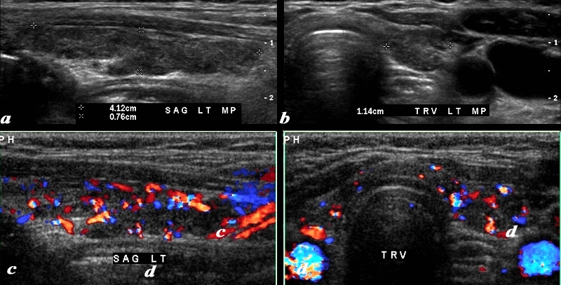
The patient is a 70 year female who shows diffuse atrophy of her thyroid by this ultrasound examination. The gland is mildly heterogeneous. The left lobe of the thyroid measures 4.1cms (craniocaudad), by .8 cms (A-P) by 1.1cms (transverse). The sagittal view shows mildly coarse heterogeneous echo texture. The transverse view shows a normal shape. The isthmus is normal in size. The color flow Doppler study seems hypervascular but the significance of this finding is not known. These findings suggest age related involution of the gland.
Courtesy Ashley Davidoff MD Copyright 2010 94503c.8L


    [nodeType] => 1
    [parentNode] => (object value omitted)
    [childNodes] => (object value omitted)
    [firstChild] => (object value omitted)
    [lastChild] => (object value omitted)
    [previousSibling] => (object value omitted)
    [nextSibling] => (object value omitted)
    [attributes] => (object value omitted)
    [ownerDocument] => (object value omitted)
    [namespaceURI] =>
    [prefix] =>
    [localName] => table
    [baseURI] =>
    [textContent] =>


The Small Thyroid Gland


The patient is a 70 year female who shows diffuse atrophy of her thyroid by this ultrasound examination. The gland is mildly heterogeneous. The left lobe of the thyroid measures 4.1cms (craniocaudad), by .8 cms (A-P) by 1.1cms (transverse). The sagittal view shows mildly coarse heterogeneous echo texture. The transverse view shows a normal shape. The isthmus is normal in size. The color flow Doppler study seems hypervascular but the significance of this finding is not known. These findings suggest age related involution of the gland.
Courtesy Ashley Davidoff MD Copyright 2010 94503c.8L


)
DOMElement Object
(
    [schemaTypeInfo] =>
    [tagName] => td
    [firstElementChild] => (object value omitted)
    [lastElementChild] => (object value omitted)
    [childElementCount] => 2
    [previousElementSibling] =>
    [nextElementSibling] =>
    [nodeName] => td
    [nodeValue] =>
The patient is a 70 year female who shows diffuse atrophy of her thyroid by this ultrasound examination. The gland is mildly heterogeneous. The left lobe of the thyroid measures 4.1cms (craniocaudad), by .8 cms (A-P) by 1.1cms (transverse). The sagittal view shows mildly coarse heterogeneous echo texture. The transverse view shows a normal shape. The isthmus is normal in size. The color flow Doppler study seems hypervascular but the significance of this finding is not known. These findings suggest age related involution of the gland.
Courtesy Ashley Davidoff MD Copyright 2010 94503c.8L

    [nodeType] => 1
    [parentNode] => (object value omitted)
    [childNodes] => (object value omitted)
    [firstChild] => (object value omitted)
    [lastChild] => (object value omitted)
    [previousSibling] => (object value omitted)
    [nextSibling] => (object value omitted)
    [attributes] => (object value omitted)
    [ownerDocument] => (object value omitted)
    [namespaceURI] =>
    [prefix] =>
    [localName] => td
    [baseURI] =>
    [textContent] =>
The patient is a 70 year female who shows diffuse atrophy of her thyroid by this ultrasound examination. The gland is mildly heterogeneous. The left lobe of the thyroid measures 4.1cms (craniocaudad), by .8 cms (A-P) by 1.1cms (transverse). The sagittal view shows mildly coarse heterogeneous echo texture. The transverse view shows a normal shape. The isthmus is normal in size. The color flow Doppler study seems hypervascular but the significance of this finding is not known. These findings suggest age related involution of the gland.
Courtesy Ashley Davidoff MD Copyright 2010 94503c.8L

)
DOMElement Object
(
    [schemaTypeInfo] =>
    [tagName] => td
    [firstElementChild] => (object value omitted)
    [lastElementChild] => (object value omitted)
    [childElementCount] => 2
    [previousElementSibling] =>
    [nextElementSibling] =>
    [nodeName] => td
    [nodeValue] =>
The Small Thyroid Gland
    [nodeType] => 1
    [parentNode] => (object value omitted)
    [childNodes] => (object value omitted)
    [firstChild] => (object value omitted)
    [lastChild] => (object value omitted)
    [previousSibling] => (object value omitted)
    [nextSibling] => (object value omitted)
    [attributes] => (object value omitted)
    [ownerDocument] => (object value omitted)
    [namespaceURI] =>
    [prefix] =>
    [localName] => td
    [baseURI] =>
    [textContent] =>
The Small Thyroid Gland
)
DOMElement Object
(
    [schemaTypeInfo] =>
    [tagName] => table
    [firstElementChild] => (object value omitted)
    [lastElementChild] => (object value omitted)
    [childElementCount] => 1
    [previousElementSibling] => (object value omitted)
    [nextElementSibling] => (object value omitted)
    [nodeName] => table
    [nodeValue] =>


Normal (a and b) and Small (c and d)


The patient is a 70year female who shows diffuse atrophy of her thyroid by this ultrasound examination. A normal thyroid gland in sagittal (a) and transverse (b) planes is juxtaposed on the atrophied gland(c,d) The gland is mildly heterogeneous. The left lobe of the thyroid measures 4.1cms (craniocaudad), by .8 cms (A-P) by 1.1cms (transverse). The sagittal view shows mildly coarse heterogeneous echo texture. The transverse view shows a normal shape. The isthmus is normal in size. These findings suggest age related involution of the gland.
Courtesy Ashley Davidoff MD Copyright 2010 94503c02L


    [nodeType] => 1
    [parentNode] => (object value omitted)
    [childNodes] => (object value omitted)
    [firstChild] => (object value omitted)
    [lastChild] => (object value omitted)
    [previousSibling] => (object value omitted)
    [nextSibling] => (object value omitted)
    [attributes] => (object value omitted)
    [ownerDocument] => (object value omitted)
    [namespaceURI] =>
    [prefix] =>
    [localName] => table
    [baseURI] =>
    [textContent] =>


Normal (a and b) and Small (c and d)


The patient is a 70year female who shows diffuse atrophy of her thyroid by this ultrasound examination. A normal thyroid gland in sagittal (a) and transverse (b) planes is juxtaposed on the atrophied gland(c,d) The gland is mildly heterogeneous. The left lobe of the thyroid measures 4.1cms (craniocaudad), by .8 cms (A-P) by 1.1cms (transverse). The sagittal view shows mildly coarse heterogeneous echo texture. The transverse view shows a normal shape. The isthmus is normal in size. These findings suggest age related involution of the gland.
Courtesy Ashley Davidoff MD Copyright 2010 94503c02L


)
DOMElement Object
(
    [schemaTypeInfo] =>
    [tagName] => td
    [firstElementChild] => (object value omitted)
    [lastElementChild] => (object value omitted)
    [childElementCount] => 2
    [previousElementSibling] =>
    [nextElementSibling] =>
    [nodeName] => td
    [nodeValue] =>
The patient is a 70year female who shows diffuse atrophy of her thyroid by this ultrasound examination. A normal thyroid gland in sagittal (a) and transverse (b) planes is juxtaposed on the atrophied gland(c,d) The gland is mildly heterogeneous. The left lobe of the thyroid measures 4.1cms (craniocaudad), by .8 cms (A-P) by 1.1cms (transverse). The sagittal view shows mildly coarse heterogeneous echo texture. The transverse view shows a normal shape. The isthmus is normal in size. These findings suggest age related involution of the gland.
Courtesy Ashley Davidoff MD Copyright 2010 94503c02L

    [nodeType] => 1
    [parentNode] => (object value omitted)
    [childNodes] => (object value omitted)
    [firstChild] => (object value omitted)
    [lastChild] => (object value omitted)
    [previousSibling] => (object value omitted)
    [nextSibling] => (object value omitted)
    [attributes] => (object value omitted)
    [ownerDocument] => (object value omitted)
    [namespaceURI] =>
    [prefix] =>
    [localName] => td
    [baseURI] =>
    [textContent] =>
The patient is a 70year female who shows diffuse atrophy of her thyroid by this ultrasound examination. A normal thyroid gland in sagittal (a) and transverse (b) planes is juxtaposed on the atrophied gland(c,d) The gland is mildly heterogeneous. The left lobe of the thyroid measures 4.1cms (craniocaudad), by .8 cms (A-P) by 1.1cms (transverse). The sagittal view shows mildly coarse heterogeneous echo texture. The transverse view shows a normal shape. The isthmus is normal in size. These findings suggest age related involution of the gland.
Courtesy Ashley Davidoff MD Copyright 2010 94503c02L

)
DOMElement Object
(
    [schemaTypeInfo] =>
    [tagName] => td
    [firstElementChild] => (object value omitted)
    [lastElementChild] => (object value omitted)
    [childElementCount] => 2
    [previousElementSibling] =>
    [nextElementSibling] =>
    [nodeName] => td
    [nodeValue] =>
Normal (a and b) and Small (c and d)
    [nodeType] => 1
    [parentNode] => (object value omitted)
    [childNodes] => (object value omitted)
    [firstChild] => (object value omitted)
    [lastChild] => (object value omitted)
    [previousSibling] => (object value omitted)
    [nextSibling] => (object value omitted)
    [attributes] => (object value omitted)
    [ownerDocument] => (object value omitted)
    [namespaceURI] =>
    [prefix] =>
    [localName] => td
    [baseURI] =>
    [textContent] =>
Normal (a and b) and Small (c and d)
)
DOMElement Object
(
    [schemaTypeInfo] =>
    [tagName] => table
    [firstElementChild] => (object value omitted)
    [lastElementChild] => (object value omitted)
    [childElementCount] => 1
    [previousElementSibling] => (object value omitted)
    [nextElementSibling] =>
    [nodeName] => table
    [nodeValue] =>


Small Thyroid Gland
The Common Vein copyright 2008
Introduction
The thyroid is one of the largest endocrine organs in the body, but on the other hand is small compared to most other organs.  It weighs about 15- 35 grams measuring approximately 4-5cms cm long, 2-2.5 cm wide and 1 -2 cm thick, in the average adult.  The isthmus is 2-6mm in A-P diameter and is up to 20 mm. in width.
The the most important dimension when considering enlargement of the gland  is the A-P diameter which should not be >2cms.  The  length is considered large when in the 4-6cms range and trannsverse should not be larger than 2.8cms.
The small gland is measures lesss than 4 cms inlength, less than 2cms wide, and less than 1cms thick.


Normal (a and b) and Small (c and d)


The patient is a 70year female who shows diffuse atrophy of her thyroid by this ultrasound examination. A normal thyroid gland in sagittal (a) and transverse (b) planes is juxtaposed on the atrophied gland(c,d) The gland is mildly heterogeneous. The left lobe of the thyroid measures 4.1cms (craniocaudad), by .8 cms (A-P) by 1.1cms (transverse). The sagittal view shows mildly coarse heterogeneous echo texture. The transverse view shows a normal shape. The isthmus is normal in size. These findings suggest age related involution of the gland.
Courtesy Ashley Davidoff MD Copyright 2010 94503c02L


The Small Thyroid Gland


The patient is a 70 year female who shows diffuse atrophy of her thyroid by this ultrasound examination. The gland is mildly heterogeneous. The left lobe of the thyroid measures 4.1cms (craniocaudad), by .8 cms (A-P) by 1.1cms (transverse). The sagittal view shows mildly coarse heterogeneous echo texture. The transverse view shows a normal shape. The isthmus is normal in size. The color flow Doppler study seems hypervascular but the significance of this finding is not known. These findings suggest age related involution of the gland.
Courtesy Ashley Davidoff MD Copyright 2010 94503c.8L


Hypoplastic Left Lobe


A normal ultrasound of the thyroid gland with a hypoplastic left lobe is demonstrated. Image a is a transverse image, while image b is a sagittal view of the right lobe, and image c is a sagittal view of the left lobe. The normal right lobe (a,b) measures 4.6cms (length) x 1.4cms (A-P anteroposterior) X .7 cms (transverse TRV) and the hypoplastic left lobe measures 2.9cms (craniocaudad), by .6cms (A-P) by .7cms (transverse).
Courtesy Ashley Davidoff MD Copyright 2010 96233c01L.8


    [nodeType] => 1
    [parentNode] => (object value omitted)
    [childNodes] => (object value omitted)
    [firstChild] => (object value omitted)
    [lastChild] => (object value omitted)
    [previousSibling] => (object value omitted)
    [nextSibling] => (object value omitted)
    [attributes] => (object value omitted)
    [ownerDocument] => (object value omitted)
    [namespaceURI] =>
    [prefix] =>
    [localName] => table
    [baseURI] =>
    [textContent] =>


Small Thyroid Gland
The Common Vein copyright 2008
Introduction
The thyroid is one of the largest endocrine organs in the body, but on the other hand is small compared to most other organs.  It weighs about 15- 35 grams measuring approximately 4-5cms cm long, 2-2.5 cm wide and 1 -2 cm thick, in the average adult.  The isthmus is 2-6mm in A-P diameter and is up to 20 mm. in width.
The the most important dimension when considering enlargement of the gland  is the A-P diameter which should not be >2cms.  The  length is considered large when in the 4-6cms range and trannsverse should not be larger than 2.8cms.
The small gland is measures lesss than 4 cms inlength, less than 2cms wide, and less than 1cms thick.


Normal (a and b) and Small (c and d)


The patient is a 70year female who shows diffuse atrophy of her thyroid by this ultrasound examination. A normal thyroid gland in sagittal (a) and transverse (b) planes is juxtaposed on the atrophied gland(c,d) The gland is mildly heterogeneous. The left lobe of the thyroid measures 4.1cms (craniocaudad), by .8 cms (A-P) by 1.1cms (transverse). The sagittal view shows mildly coarse heterogeneous echo texture. The transverse view shows a normal shape. The isthmus is normal in size. These findings suggest age related involution of the gland.
Courtesy Ashley Davidoff MD Copyright 2010 94503c02L


The Small Thyroid Gland


The patient is a 70 year female who shows diffuse atrophy of her thyroid by this ultrasound examination. The gland is mildly heterogeneous. The left lobe of the thyroid measures 4.1cms (craniocaudad), by .8 cms (A-P) by 1.1cms (transverse). The sagittal view shows mildly coarse heterogeneous echo texture. The transverse view shows a normal shape. The isthmus is normal in size. The color flow Doppler study seems hypervascular but the significance of this finding is not known. These findings suggest age related involution of the gland.
Courtesy Ashley Davidoff MD Copyright 2010 94503c.8L


Hypoplastic Left Lobe


A normal ultrasound of the thyroid gland with a hypoplastic left lobe is demonstrated. Image a is a transverse image, while image b is a sagittal view of the right lobe, and image c is a sagittal view of the left lobe. The normal right lobe (a,b) measures 4.6cms (length) x 1.4cms (A-P anteroposterior) X .7 cms (transverse TRV) and the hypoplastic left lobe measures 2.9cms (craniocaudad), by .6cms (A-P) by .7cms (transverse).
Courtesy Ashley Davidoff MD Copyright 2010 96233c01L.8


)
DOMElement Object
(
    [schemaTypeInfo] =>
    [tagName] => td
    [firstElementChild] => (object value omitted)
    [lastElementChild] => (object value omitted)
    [childElementCount] => 2
    [previousElementSibling] =>
    [nextElementSibling] =>
    [nodeName] => td
    [nodeValue] =>
A normal ultrasound of the thyroid gland with a hypoplastic left lobe is demonstrated. Image a is a transverse image, while image b is a sagittal view of the right lobe, and image c is a sagittal view of the left lobe. The normal right lobe (a,b) measures 4.6cms (length) x 1.4cms (A-P anteroposterior) X .7 cms (transverse TRV) and the hypoplastic left lobe measures 2.9cms (craniocaudad), by .6cms (A-P) by .7cms (transverse).
Courtesy Ashley Davidoff MD Copyright 2010 96233c01L.8

    [nodeType] => 1
    [parentNode] => (object value omitted)
    [childNodes] => (object value omitted)
    [firstChild] => (object value omitted)
    [lastChild] => (object value omitted)
    [previousSibling] => (object value omitted)
    [nextSibling] => (object value omitted)
    [attributes] => (object value omitted)
    [ownerDocument] => (object value omitted)
    [namespaceURI] =>
    [prefix] =>
    [localName] => td
    [baseURI] =>
    [textContent] =>
A normal ultrasound of the thyroid gland with a hypoplastic left lobe is demonstrated. Image a is a transverse image, while image b is a sagittal view of the right lobe, and image c is a sagittal view of the left lobe. The normal right lobe (a,b) measures 4.6cms (length) x 1.4cms (A-P anteroposterior) X .7 cms (transverse TRV) and the hypoplastic left lobe measures 2.9cms (craniocaudad), by .6cms (A-P) by .7cms (transverse).
Courtesy Ashley Davidoff MD Copyright 2010 96233c01L.8

)
DOMElement Object
(
    [schemaTypeInfo] =>
    [tagName] => td
    [firstElementChild] => (object value omitted)
    [lastElementChild] => (object value omitted)
    [childElementCount] => 2
    [previousElementSibling] =>
    [nextElementSibling] =>
    [nodeName] => td
    [nodeValue] =>
Hypoplastic Left Lobe
    [nodeType] => 1
    [parentNode] => (object value omitted)
    [childNodes] => (object value omitted)
    [firstChild] => (object value omitted)
    [lastChild] => (object value omitted)
    [previousSibling] => (object value omitted)
    [nextSibling] => (object value omitted)
    [attributes] => (object value omitted)
    [ownerDocument] => (object value omitted)
    [namespaceURI] =>
    [prefix] =>
    [localName] => td
    [baseURI] =>
    [textContent] =>
Hypoplastic Left Lobe
)
DOMElement Object
(
    [schemaTypeInfo] =>
    [tagName] => td
    [firstElementChild] => (object value omitted)
    [lastElementChild] => (object value omitted)
    [childElementCount] => 2
    [previousElementSibling] =>
    [nextElementSibling] =>
    [nodeName] => td
    [nodeValue] =>
The patient is a 70 year female who shows diffuse atrophy of her thyroid by this ultrasound examination. The gland is mildly heterogeneous. The left lobe of the thyroid measures 4.1cms (craniocaudad), by .8 cms (A-P) by 1.1cms (transverse). The sagittal view shows mildly coarse heterogeneous echo texture. The transverse view shows a normal shape. The isthmus is normal in size. The color flow Doppler study seems hypervascular but the significance of this finding is not known. These findings suggest age related involution of the gland.
Courtesy Ashley Davidoff MD Copyright 2010 94503c.8L

    [nodeType] => 1
    [parentNode] => (object value omitted)
    [childNodes] => (object value omitted)
    [firstChild] => (object value omitted)
    [lastChild] => (object value omitted)
    [previousSibling] => (object value omitted)
    [nextSibling] => (object value omitted)
    [attributes] => (object value omitted)
    [ownerDocument] => (object value omitted)
    [namespaceURI] =>
    [prefix] =>
    [localName] => td
    [baseURI] =>
    [textContent] =>
The patient is a 70 year female who shows diffuse atrophy of her thyroid by this ultrasound examination. The gland is mildly heterogeneous. The left lobe of the thyroid measures 4.1cms (craniocaudad), by .8 cms (A-P) by 1.1cms (transverse). The sagittal view shows mildly coarse heterogeneous echo texture. The transverse view shows a normal shape. The isthmus is normal in size. The color flow Doppler study seems hypervascular but the significance of this finding is not known. These findings suggest age related involution of the gland.
Courtesy Ashley Davidoff MD Copyright 2010 94503c.8L

)
DOMElement Object
(
    [schemaTypeInfo] =>
    [tagName] => td
    [firstElementChild] => (object value omitted)
    [lastElementChild] => (object value omitted)
    [childElementCount] => 2
    [previousElementSibling] =>
    [nextElementSibling] =>
    [nodeName] => td
    [nodeValue] =>
The Small Thyroid Gland
    [nodeType] => 1
    [parentNode] => (object value omitted)
    [childNodes] => (object value omitted)
    [firstChild] => (object value omitted)
    [lastChild] => (object value omitted)
    [previousSibling] => (object value omitted)
    [nextSibling] => (object value omitted)
    [attributes] => (object value omitted)
    [ownerDocument] => (object value omitted)
    [namespaceURI] =>
    [prefix] =>
    [localName] => td
    [baseURI] =>
    [textContent] =>
The Small Thyroid Gland
)
DOMElement Object
(
    [schemaTypeInfo] =>
    [tagName] => td
    [firstElementChild] => (object value omitted)
    [lastElementChild] => (object value omitted)
    [childElementCount] => 2
    [previousElementSibling] =>
    [nextElementSibling] =>
    [nodeName] => td
    [nodeValue] =>
The patient is a 70year female who shows diffuse atrophy of her thyroid by this ultrasound examination. A normal thyroid gland in sagittal (a) and transverse (b) planes is juxtaposed on the atrophied gland(c,d) The gland is mildly heterogeneous. The left lobe of the thyroid measures 4.1cms (craniocaudad), by .8 cms (A-P) by 1.1cms (transverse). The sagittal view shows mildly coarse heterogeneous echo texture. The transverse view shows a normal shape. The isthmus is normal in size. These findings suggest age related involution of the gland.
Courtesy Ashley Davidoff MD Copyright 2010 94503c02L

    [nodeType] => 1
    [parentNode] => (object value omitted)
    [childNodes] => (object value omitted)
    [firstChild] => (object value omitted)
    [lastChild] => (object value omitted)
    [previousSibling] => (object value omitted)
    [nextSibling] => (object value omitted)
    [attributes] => (object value omitted)
    [ownerDocument] => (object value omitted)
    [namespaceURI] =>
    [prefix] =>
    [localName] => td
    [baseURI] =>
    [textContent] =>
The patient is a 70year female who shows diffuse atrophy of her thyroid by this ultrasound examination. A normal thyroid gland in sagittal (a) and transverse (b) planes is juxtaposed on the atrophied gland(c,d) The gland is mildly heterogeneous. The left lobe of the thyroid measures 4.1cms (craniocaudad), by .8 cms (A-P) by 1.1cms (transverse). The sagittal view shows mildly coarse heterogeneous echo texture. The transverse view shows a normal shape. The isthmus is normal in size. These findings suggest age related involution of the gland.
Courtesy Ashley Davidoff MD Copyright 2010 94503c02L

)
DOMElement Object
(
    [schemaTypeInfo] =>
    [tagName] => td
    [firstElementChild] => (object value omitted)
    [lastElementChild] => (object value omitted)
    [childElementCount] => 2
    [previousElementSibling] =>
    [nextElementSibling] =>
    [nodeName] => td
    [nodeValue] =>
Normal (a and b) and Small (c and d)
    [nodeType] => 1
    [parentNode] => (object value omitted)
    [childNodes] => (object value omitted)
    [firstChild] => (object value omitted)
    [lastChild] => (object value omitted)
    [previousSibling] => (object value omitted)
    [nextSibling] => (object value omitted)
    [attributes] => (object value omitted)
    [ownerDocument] => (object value omitted)
    [namespaceURI] =>
    [prefix] =>
    [localName] => td
    [baseURI] =>
    [textContent] =>
Normal (a and b) and Small (c and d)
)
DOMElement Object
(
    [schemaTypeInfo] =>
    [tagName] => td
    [firstElementChild] => (object value omitted)
    [lastElementChild] => (object value omitted)
    [childElementCount] => 9
    [previousElementSibling] =>
    [nextElementSibling] =>
    [nodeName] => td
    [nodeValue] =>
Small Thyroid Gland
The Common Vein copyright 2008
Introduction
The thyroid is one of the largest endocrine organs in the body, but on the other hand is small compared to most other organs.  It weighs about 15- 35 grams measuring approximately 4-5cms cm long, 2-2.5 cm wide and 1 -2 cm thick, in the average adult.  The isthmus is 2-6mm in A-P diameter and is up to 20 mm. in width.
The the most important dimension when considering enlargement of the gland  is the A-P diameter which should not be >2cms.  The  length is considered large when in the 4-6cms range and trannsverse should not be larger than 2.8cms.
The small gland is measures lesss than 4 cms inlength, less than 2cms wide, and less than 1cms thick.


Normal (a and b) and Small (c and d)


The patient is a 70year female who shows diffuse atrophy of her thyroid by this ultrasound examination. A normal thyroid gland in sagittal (a) and transverse (b) planes is juxtaposed on the atrophied gland(c,d) The gland is mildly heterogeneous. The left lobe of the thyroid measures 4.1cms (craniocaudad), by .8 cms (A-P) by 1.1cms (transverse). The sagittal view shows mildly coarse heterogeneous echo texture. The transverse view shows a normal shape. The isthmus is normal in size. These findings suggest age related involution of the gland.
Courtesy Ashley Davidoff MD Copyright 2010 94503c02L


The Small Thyroid Gland


The patient is a 70 year female who shows diffuse atrophy of her thyroid by this ultrasound examination. The gland is mildly heterogeneous. The left lobe of the thyroid measures 4.1cms (craniocaudad), by .8 cms (A-P) by 1.1cms (transverse). The sagittal view shows mildly coarse heterogeneous echo texture. The transverse view shows a normal shape. The isthmus is normal in size. The color flow Doppler study seems hypervascular but the significance of this finding is not known. These findings suggest age related involution of the gland.
Courtesy Ashley Davidoff MD Copyright 2010 94503c.8L


Hypoplastic Left Lobe


A normal ultrasound of the thyroid gland with a hypoplastic left lobe is demonstrated. Image a is a transverse image, while image b is a sagittal view of the right lobe, and image c is a sagittal view of the left lobe. The normal right lobe (a,b) measures 4.6cms (length) x 1.4cms (A-P anteroposterior) X .7 cms (transverse TRV) and the hypoplastic left lobe measures 2.9cms (craniocaudad), by .6cms (A-P) by .7cms (transverse).
Courtesy Ashley Davidoff MD Copyright 2010 96233c01L.8


    [nodeType] => 1
    [parentNode] => (object value omitted)
    [childNodes] => (object value omitted)
    [firstChild] => (object value omitted)
    [lastChild] => (object value omitted)
    [previousSibling] => (object value omitted)
    [nextSibling] => (object value omitted)
    [attributes] => (object value omitted)
    [ownerDocument] => (object value omitted)
    [namespaceURI] =>
    [prefix] =>
    [localName] => td
    [baseURI] =>
    [textContent] =>
Small Thyroid Gland
The Common Vein copyright 2008
Introduction
The thyroid is one of the largest endocrine organs in the body, but on the other hand is small compared to most other organs.  It weighs about 15- 35 grams measuring approximately 4-5cms cm long, 2-2.5 cm wide and 1 -2 cm thick, in the average adult.  The isthmus is 2-6mm in A-P diameter and is up to 20 mm. in width.
The the most important dimension when considering enlargement of the gland  is the A-P diameter which should not be >2cms.  The  length is considered large when in the 4-6cms range and trannsverse should not be larger than 2.8cms.
The small gland is measures lesss than 4 cms inlength, less than 2cms wide, and less than 1cms thick.


Normal (a and b) and Small (c and d)


The patient is a 70year female who shows diffuse atrophy of her thyroid by this ultrasound examination. A normal thyroid gland in sagittal (a) and transverse (b) planes is juxtaposed on the atrophied gland(c,d) The gland is mildly heterogeneous. The left lobe of the thyroid measures 4.1cms (craniocaudad), by .8 cms (A-P) by 1.1cms (transverse). The sagittal view shows mildly coarse heterogeneous echo texture. The transverse view shows a normal shape. The isthmus is normal in size. These findings suggest age related involution of the gland.
Courtesy Ashley Davidoff MD Copyright 2010 94503c02L


The Small Thyroid Gland


The patient is a 70 year female who shows diffuse atrophy of her thyroid by this ultrasound examination. The gland is mildly heterogeneous. The left lobe of the thyroid measures 4.1cms (craniocaudad), by .8 cms (A-P) by 1.1cms (transverse). The sagittal view shows mildly coarse heterogeneous echo texture. The transverse view shows a normal shape. The isthmus is normal in size. The color flow Doppler study seems hypervascular but the significance of this finding is not known. These findings suggest age related involution of the gland.
Courtesy Ashley Davidoff MD Copyright 2010 94503c.8L


Hypoplastic Left Lobe


A normal ultrasound of the thyroid gland with a hypoplastic left lobe is demonstrated. Image a is a transverse image, while image b is a sagittal view of the right lobe, and image c is a sagittal view of the left lobe. The normal right lobe (a,b) measures 4.6cms (length) x 1.4cms (A-P anteroposterior) X .7 cms (transverse TRV) and the hypoplastic left lobe measures 2.9cms (craniocaudad), by .6cms (A-P) by .7cms (transverse).
Courtesy Ashley Davidoff MD Copyright 2010 96233c01L.8


)
DOMElement Object
(
    [schemaTypeInfo] =>
    [tagName] => td
    [firstElementChild] => (object value omitted)
    [lastElementChild] => (object value omitted)
    [childElementCount] => 2
    [previousElementSibling] =>
    [nextElementSibling] =>
    [nodeName] => td
    [nodeValue] =>

Normal and Atrophied Gland

    [nodeType] => 1
    [parentNode] => (object value omitted)
    [childNodes] => (object value omitted)
    [firstChild] => (object value omitted)
    [lastChild] => (object value omitted)
    [previousSibling] => (object value omitted)
    [nextSibling] => (object value omitted)
    [attributes] => (object value omitted)
    [ownerDocument] => (object value omitted)
    [namespaceURI] =>
    [prefix] =>
    [localName] => td
    [baseURI] =>
    [textContent] =>

Normal and Atrophied Gland

)
DOMElement Object
(
    [schemaTypeInfo] =>
    [tagName] => table
    [firstElementChild] => (object value omitted)
    [lastElementChild] => (object value omitted)
    [childElementCount] => 1
    [previousElementSibling] => (object value omitted)
    [nextElementSibling] => (object value omitted)
    [nodeName] => table
    [nodeValue] =>


The Normal Right Lobe and Hypoplastic Left Lobe


A normal ultrasound of the thyroid gland with a hypoplastic left lobe is demonstrated. Image a is a transverse image, while image b is a sagittal view of the right lobe, and image c is a sagittal view of the left lobe. The normal right lobe (a,b) measures 4.6cms (length) x 1.4cms (A-P anteroposterior) X .7 cms (transverse TRV) and the hypoplastic left lobe measures 2.9cms (craniocaudad), by .6cms (A-P) by .7cms (transverse).
Ultrasound Courtesy Ashley Davidoff MD Copyright 2010 96233c01.8


    [nodeType] => 1
    [parentNode] => (object value omitted)
    [childNodes] => (object value omitted)
    [firstChild] => (object value omitted)
    [lastChild] => (object value omitted)
    [previousSibling] => (object value omitted)
    [nextSibling] => (object value omitted)
    [attributes] => (object value omitted)
    [ownerDocument] => (object value omitted)
    [namespaceURI] =>
    [prefix] =>
    [localName] => table
    [baseURI] =>
    [textContent] =>


The Normal Right Lobe and Hypoplastic Left Lobe


A normal ultrasound of the thyroid gland with a hypoplastic left lobe is demonstrated. Image a is a transverse image, while image b is a sagittal view of the right lobe, and image c is a sagittal view of the left lobe. The normal right lobe (a,b) measures 4.6cms (length) x 1.4cms (A-P anteroposterior) X .7 cms (transverse TRV) and the hypoplastic left lobe measures 2.9cms (craniocaudad), by .6cms (A-P) by .7cms (transverse).
Ultrasound Courtesy Ashley Davidoff MD Copyright 2010 96233c01.8


)
DOMElement Object
(
    [schemaTypeInfo] =>
    [tagName] => td
    [firstElementChild] => (object value omitted)
    [lastElementChild] => (object value omitted)
    [childElementCount] => 2
    [previousElementSibling] =>
    [nextElementSibling] =>
    [nodeName] => td
    [nodeValue] =>
A normal ultrasound of the thyroid gland with a hypoplastic left lobe is demonstrated. Image a is a transverse image, while image b is a sagittal view of the right lobe, and image c is a sagittal view of the left lobe. The normal right lobe (a,b) measures 4.6cms (length) x 1.4cms (A-P anteroposterior) X .7 cms (transverse TRV) and the hypoplastic left lobe measures 2.9cms (craniocaudad), by .6cms (A-P) by .7cms (transverse).
Ultrasound Courtesy Ashley Davidoff MD Copyright 2010 96233c01.8

    [nodeType] => 1
    [parentNode] => (object value omitted)
    [childNodes] => (object value omitted)
    [firstChild] => (object value omitted)
    [lastChild] => (object value omitted)
    [previousSibling] => (object value omitted)
    [nextSibling] => (object value omitted)
    [attributes] => (object value omitted)
    [ownerDocument] => (object value omitted)
    [namespaceURI] =>
    [prefix] =>
    [localName] => td
    [baseURI] =>
    [textContent] =>
A normal ultrasound of the thyroid gland with a hypoplastic left lobe is demonstrated. Image a is a transverse image, while image b is a sagittal view of the right lobe, and image c is a sagittal view of the left lobe. The normal right lobe (a,b) measures 4.6cms (length) x 1.4cms (A-P anteroposterior) X .7 cms (transverse TRV) and the hypoplastic left lobe measures 2.9cms (craniocaudad), by .6cms (A-P) by .7cms (transverse).
Ultrasound Courtesy Ashley Davidoff MD Copyright 2010 96233c01.8

)
DOMElement Object
(
    [schemaTypeInfo] =>
    [tagName] => td
    [firstElementChild] => (object value omitted)
    [lastElementChild] => (object value omitted)
    [childElementCount] => 2
    [previousElementSibling] =>
    [nextElementSibling] =>
    [nodeName] => td
    [nodeValue] =>

The Normal Right Lobe and Hypoplastic Left Lobe

    [nodeType] => 1
    [parentNode] => (object value omitted)
    [childNodes] => (object value omitted)
    [firstChild] => (object value omitted)
    [lastChild] => (object value omitted)
    [previousSibling] => (object value omitted)
    [nextSibling] => (object value omitted)
    [attributes] => (object value omitted)
    [ownerDocument] => (object value omitted)
    [namespaceURI] =>
    [prefix] =>
    [localName] => td
    [baseURI] =>
    [textContent] =>

The Normal Right Lobe and Hypoplastic Left Lobe

)
DOMElement Object
(
    [schemaTypeInfo] =>
    [tagName] => table
    [firstElementChild] => (object value omitted)
    [lastElementChild] => (object value omitted)
    [childElementCount] => 1
    [previousElementSibling] => (object value omitted)
    [nextElementSibling] => (object value omitted)
    [nodeName] => table
    [nodeValue] =>


Goiter Caused by Large Hemorrhagic Cyst


A large mostly hypoechoic mass expands the left lobe of the thyroid on the ultrasound of this 54 year female who presents with an enlarged thyroid gland. The left lobe is filled with low level echoes and contains multiple thin septal echogenic walls (maroon). The mass appears to displace and compress normal soft tissues of the neck (pink) anteriorly. Doppler showed no flow. The mass occupies the entire left lobe and measures 5.8cms in craniocaudad span and by 4.2 cms transversely The diagnosis in this patient is a hemorrhagic cyst causing a goiter.
Courtesy Ashley Davidoff MD Copyright 2010 94656c01L


    [nodeType] => 1
    [parentNode] => (object value omitted)
    [childNodes] => (object value omitted)
    [firstChild] => (object value omitted)
    [lastChild] => (object value omitted)
    [previousSibling] => (object value omitted)
    [nextSibling] => (object value omitted)
    [attributes] => (object value omitted)
    [ownerDocument] => (object value omitted)
    [namespaceURI] =>
    [prefix] =>
    [localName] => table
    [baseURI] =>
    [textContent] =>


Goiter Caused by Large Hemorrhagic Cyst


A large mostly hypoechoic mass expands the left lobe of the thyroid on the ultrasound of this 54 year female who presents with an enlarged thyroid gland. The left lobe is filled with low level echoes and contains multiple thin septal echogenic walls (maroon). The mass appears to displace and compress normal soft tissues of the neck (pink) anteriorly. Doppler showed no flow. The mass occupies the entire left lobe and measures 5.8cms in craniocaudad span and by 4.2 cms transversely The diagnosis in this patient is a hemorrhagic cyst causing a goiter.
Courtesy Ashley Davidoff MD Copyright 2010 94656c01L


)
DOMElement Object
(
    [schemaTypeInfo] =>
    [tagName] => td
    [firstElementChild] => (object value omitted)
    [lastElementChild] => (object value omitted)
    [childElementCount] => 2
    [previousElementSibling] =>
    [nextElementSibling] =>
    [nodeName] => td
    [nodeValue] =>
A large mostly hypoechoic mass expands the left lobe of the thyroid on the ultrasound of this 54 year female who presents with an enlarged thyroid gland. The left lobe is filled with low level echoes and contains multiple thin septal echogenic walls (maroon). The mass appears to displace and compress normal soft tissues of the neck (pink) anteriorly. Doppler showed no flow. The mass occupies the entire left lobe and measures 5.8cms in craniocaudad span and by 4.2 cms transversely The diagnosis in this patient is a hemorrhagic cyst causing a goiter.
Courtesy Ashley Davidoff MD Copyright 2010 94656c01L

    [nodeType] => 1
    [parentNode] => (object value omitted)
    [childNodes] => (object value omitted)
    [firstChild] => (object value omitted)
    [lastChild] => (object value omitted)
    [previousSibling] => (object value omitted)
    [nextSibling] => (object value omitted)
    [attributes] => (object value omitted)
    [ownerDocument] => (object value omitted)
    [namespaceURI] =>
    [prefix] =>
    [localName] => td
    [baseURI] =>
    [textContent] =>
A large mostly hypoechoic mass expands the left lobe of the thyroid on the ultrasound of this 54 year female who presents with an enlarged thyroid gland. The left lobe is filled with low level echoes and contains multiple thin septal echogenic walls (maroon). The mass appears to displace and compress normal soft tissues of the neck (pink) anteriorly. Doppler showed no flow. The mass occupies the entire left lobe and measures 5.8cms in craniocaudad span and by 4.2 cms transversely The diagnosis in this patient is a hemorrhagic cyst causing a goiter.
Courtesy Ashley Davidoff MD Copyright 2010 94656c01L

)
DOMElement Object
(
    [schemaTypeInfo] =>
    [tagName] => td
    [firstElementChild] => (object value omitted)
    [lastElementChild] => (object value omitted)
    [childElementCount] => 2
    [previousElementSibling] =>
    [nextElementSibling] =>
    [nodeName] => td
    [nodeValue] =>

Goiter Caused by Large Hemorrhagic Cyst

    [nodeType] => 1
    [parentNode] => (object value omitted)
    [childNodes] => (object value omitted)
    [firstChild] => (object value omitted)
    [lastChild] => (object value omitted)
    [previousSibling] => (object value omitted)
    [nextSibling] => (object value omitted)
    [attributes] => (object value omitted)
    [ownerDocument] => (object value omitted)
    [namespaceURI] =>
    [prefix] =>
    [localName] => td
    [baseURI] =>
    [textContent] =>

Goiter Caused by Large Hemorrhagic Cyst

)
DOMElement Object
(
    [schemaTypeInfo] =>
    [tagName] => table
    [firstElementChild] => (object value omitted)
    [lastElementChild] => (object value omitted)
    [childElementCount] => 1
    [previousElementSibling] => (object value omitted)
    [nextElementSibling] => (object value omitted)
    [nodeName] => table
    [nodeValue] =>


Amyloidosis of the Thyroid


The CT is from a 35 year old man with a complex history including long standing history of Crohn?s disease and a recent history of lymphoma. He has had a goiter for many years and biopsy showed amyloidosis of the thyroid. The image on left is a coronal view of the neck and the b is a transverse view of the thyroid. The gland is enlarged measuring 6.2cms in Craniocaudal dimension (length) by 3cms in anteroposterior dimension by 3cms in transverse dimension. On the transverse view it is not quite round but the gland is full bodied reminiscent of its large size. The density appearance is puzzling. Since no non contrast CT was performed the peripheral increase in density may be due to enhancement or more likely due to iodine or calcification. The soft densities to the right of the right lobe are related to the patient?s newly diagnosed lymphoma.
Courtesy Ashley Davidoff MD Copyright 2010 94553c.8


    [nodeType] => 1
    [parentNode] => (object value omitted)
    [childNodes] => (object value omitted)
    [firstChild] => (object value omitted)
    [lastChild] => (object value omitted)
    [previousSibling] => (object value omitted)
    [nextSibling] => (object value omitted)
    [attributes] => (object value omitted)
    [ownerDocument] => (object value omitted)
    [namespaceURI] =>
    [prefix] =>
    [localName] => table
    [baseURI] =>
    [textContent] =>


Amyloidosis of the Thyroid


The CT is from a 35 year old man with a complex history including long standing history of Crohn?s disease and a recent history of lymphoma. He has had a goiter for many years and biopsy showed amyloidosis of the thyroid. The image on left is a coronal view of the neck and the b is a transverse view of the thyroid. The gland is enlarged measuring 6.2cms in Craniocaudal dimension (length) by 3cms in anteroposterior dimension by 3cms in transverse dimension. On the transverse view it is not quite round but the gland is full bodied reminiscent of its large size. The density appearance is puzzling. Since no non contrast CT was performed the peripheral increase in density may be due to enhancement or more likely due to iodine or calcification. The soft densities to the right of the right lobe are related to the patient?s newly diagnosed lymphoma.
Courtesy Ashley Davidoff MD Copyright 2010 94553c.8


)
DOMElement Object
(
    [schemaTypeInfo] =>
    [tagName] => td
    [firstElementChild] => (object value omitted)
    [lastElementChild] => (object value omitted)
    [childElementCount] => 2
    [previousElementSibling] =>
    [nextElementSibling] =>
    [nodeName] => td
    [nodeValue] =>
The CT is from a 35 year old man with a complex history including long standing history of Crohn?s disease and a recent history of lymphoma. He has had a goiter for many years and biopsy showed amyloidosis of the thyroid. The image on left is a coronal view of the neck and the b is a transverse view of the thyroid. The gland is enlarged measuring 6.2cms in Craniocaudal dimension (length) by 3cms in anteroposterior dimension by 3cms in transverse dimension. On the transverse view it is not quite round but the gland is full bodied reminiscent of its large size. The density appearance is puzzling. Since no non contrast CT was performed the peripheral increase in density may be due to enhancement or more likely due to iodine or calcification. The soft densities to the right of the right lobe are related to the patient?s newly diagnosed lymphoma.
Courtesy Ashley Davidoff MD Copyright 2010 94553c.8

    [nodeType] => 1
    [parentNode] => (object value omitted)
    [childNodes] => (object value omitted)
    [firstChild] => (object value omitted)
    [lastChild] => (object value omitted)
    [previousSibling] => (object value omitted)
    [nextSibling] => (object value omitted)
    [attributes] => (object value omitted)
    [ownerDocument] => (object value omitted)
    [namespaceURI] =>
    [prefix] =>
    [localName] => td
    [baseURI] =>
    [textContent] =>
The CT is from a 35 year old man with a complex history including long standing history of Crohn?s disease and a recent history of lymphoma. He has had a goiter for many years and biopsy showed amyloidosis of the thyroid. The image on left is a coronal view of the neck and the b is a transverse view of the thyroid. The gland is enlarged measuring 6.2cms in Craniocaudal dimension (length) by 3cms in anteroposterior dimension by 3cms in transverse dimension. On the transverse view it is not quite round but the gland is full bodied reminiscent of its large size. The density appearance is puzzling. Since no non contrast CT was performed the peripheral increase in density may be due to enhancement or more likely due to iodine or calcification. The soft densities to the right of the right lobe are related to the patient?s newly diagnosed lymphoma.
Courtesy Ashley Davidoff MD Copyright 2010 94553c.8

)
DOMElement Object
(
    [schemaTypeInfo] =>
    [tagName] => td
    [firstElementChild] => (object value omitted)
    [lastElementChild] => (object value omitted)
    [childElementCount] => 2
    [previousElementSibling] =>
    [nextElementSibling] =>
    [nodeName] => td
    [nodeValue] =>

Amyloidosis of the Thyroid

    [nodeType] => 1
    [parentNode] => (object value omitted)
    [childNodes] => (object value omitted)
    [firstChild] => (object value omitted)
    [lastChild] => (object value omitted)
    [previousSibling] => (object value omitted)
    [nextSibling] => (object value omitted)
    [attributes] => (object value omitted)
    [ownerDocument] => (object value omitted)
    [namespaceURI] =>
    [prefix] =>
    [localName] => td
    [baseURI] =>
    [textContent] =>

Amyloidosis of the Thyroid

)
DOMElement Object
(
    [schemaTypeInfo] =>
    [tagName] => table
    [firstElementChild] => (object value omitted)
    [lastElementChild] => (object value omitted)
    [childElementCount] => 1
    [previousElementSibling] => (object value omitted)
    [nextElementSibling] => (object value omitted)
    [nodeName] => table
    [nodeValue] =>


Normal and Diffusely Enlarged Hypothyroid Gland – Ultrasound


A normal ultrasound of the thyroid gland (a,b) is juxtaposed with the ultrasound of a 78 year female with hypothyroidism (c,d) A diffusely enlarged, heterogeneous thyroid gland is seen in the hypothyroid patient. The normal thyroid measures 5.0cms (length) x 1.1cms (A-P anteroposterior) X 1.4 cms (transverse TRV) and the abnormal thyroid measures 8.5cms (craniocaudad), by 3.9cms (A-P) by 4.8cms (transverse). Clinical findings were consistent with thyroiditis, with biochemical findings suggesting hypothyroidism. The enlarged gland in the transverse dimension is almost round. (d). This rounded shape in the transverse dimension is a clue to the presence of the enlarged gland, even before the measurements are taken and evaluated. The gland is heterogeneous particularly well seen on the sagittal view (c). The findings are consistent with a thyroiditis.
Courtesy Ashley Davidoff MD Copyright 2010 94549c05g03.8s


    [nodeType] => 1
    [parentNode] => (object value omitted)
    [childNodes] => (object value omitted)
    [firstChild] => (object value omitted)
    [lastChild] => (object value omitted)
    [previousSibling] => (object value omitted)
    [nextSibling] => (object value omitted)
    [attributes] => (object value omitted)
    [ownerDocument] => (object value omitted)
    [namespaceURI] =>
    [prefix] =>
    [localName] => table
    [baseURI] =>
    [textContent] =>


Normal and Diffusely Enlarged Hypothyroid Gland – Ultrasound


A normal ultrasound of the thyroid gland (a,b) is juxtaposed with the ultrasound of a 78 year female with hypothyroidism (c,d) A diffusely enlarged, heterogeneous thyroid gland is seen in the hypothyroid patient. The normal thyroid measures 5.0cms (length) x 1.1cms (A-P anteroposterior) X 1.4 cms (transverse TRV) and the abnormal thyroid measures 8.5cms (craniocaudad), by 3.9cms (A-P) by 4.8cms (transverse). Clinical findings were consistent with thyroiditis, with biochemical findings suggesting hypothyroidism. The enlarged gland in the transverse dimension is almost round. (d). This rounded shape in the transverse dimension is a clue to the presence of the enlarged gland, even before the measurements are taken and evaluated. The gland is heterogeneous particularly well seen on the sagittal view (c). The findings are consistent with a thyroiditis.
Courtesy Ashley Davidoff MD Copyright 2010 94549c05g03.8s


)
DOMElement Object
(
    [schemaTypeInfo] =>
    [tagName] => td
    [firstElementChild] => (object value omitted)
    [lastElementChild] => (object value omitted)
    [childElementCount] => 2
    [previousElementSibling] =>
    [nextElementSibling] =>
    [nodeName] => td
    [nodeValue] =>
A normal ultrasound of the thyroid gland (a,b) is juxtaposed with the ultrasound of a 78 year female with hypothyroidism (c,d) A diffusely enlarged, heterogeneous thyroid gland is seen in the hypothyroid patient. The normal thyroid measures 5.0cms (length) x 1.1cms (A-P anteroposterior) X 1.4 cms (transverse TRV) and the abnormal thyroid measures 8.5cms (craniocaudad), by 3.9cms (A-P) by 4.8cms (transverse). Clinical findings were consistent with thyroiditis, with biochemical findings suggesting hypothyroidism. The enlarged gland in the transverse dimension is almost round. (d). This rounded shape in the transverse dimension is a clue to the presence of the enlarged gland, even before the measurements are taken and evaluated. The gland is heterogeneous particularly well seen on the sagittal view (c). The findings are consistent with a thyroiditis.
Courtesy Ashley Davidoff MD Copyright 2010 94549c05g03.8s

    [nodeType] => 1
    [parentNode] => (object value omitted)
    [childNodes] => (object value omitted)
    [firstChild] => (object value omitted)
    [lastChild] => (object value omitted)
    [previousSibling] => (object value omitted)
    [nextSibling] => (object value omitted)
    [attributes] => (object value omitted)
    [ownerDocument] => (object value omitted)
    [namespaceURI] =>
    [prefix] =>
    [localName] => td
    [baseURI] =>
    [textContent] =>
A normal ultrasound of the thyroid gland (a,b) is juxtaposed with the ultrasound of a 78 year female with hypothyroidism (c,d) A diffusely enlarged, heterogeneous thyroid gland is seen in the hypothyroid patient. The normal thyroid measures 5.0cms (length) x 1.1cms (A-P anteroposterior) X 1.4 cms (transverse TRV) and the abnormal thyroid measures 8.5cms (craniocaudad), by 3.9cms (A-P) by 4.8cms (transverse). Clinical findings were consistent with thyroiditis, with biochemical findings suggesting hypothyroidism. The enlarged gland in the transverse dimension is almost round. (d). This rounded shape in the transverse dimension is a clue to the presence of the enlarged gland, even before the measurements are taken and evaluated. The gland is heterogeneous particularly well seen on the sagittal view (c). The findings are consistent with a thyroiditis.
Courtesy Ashley Davidoff MD Copyright 2010 94549c05g03.8s

)
DOMElement Object
(
    [schemaTypeInfo] =>
    [tagName] => td
    [firstElementChild] => (object value omitted)
    [lastElementChild] => (object value omitted)
    [childElementCount] => 2
    [previousElementSibling] =>
    [nextElementSibling] =>
    [nodeName] => td
    [nodeValue] =>

Normal and Diffusely Enlarged Hypothyroid Gland – Ultrasound

    [nodeType] => 1
    [parentNode] => (object value omitted)
    [childNodes] => (object value omitted)
    [firstChild] => (object value omitted)
    [lastChild] => (object value omitted)
    [previousSibling] => (object value omitted)
    [nextSibling] => (object value omitted)
    [attributes] => (object value omitted)
    [ownerDocument] => (object value omitted)
    [namespaceURI] =>
    [prefix] =>
    [localName] => td
    [baseURI] =>
    [textContent] =>

Normal and Diffusely Enlarged Hypothyroid Gland – Ultrasound

)
DOMElement Object
(
    [schemaTypeInfo] =>
    [tagName] => table
    [firstElementChild] => (object value omitted)
    [lastElementChild] => (object value omitted)
    [childElementCount] => 1
    [previousElementSibling] => (object value omitted)
    [nextElementSibling] => (object value omitted)
    [nodeName] => table
    [nodeValue] =>


Retrosternal Goiter Compressing the Brachiocephalic Vein


A large retrosternal goiter is seen extending down anterior to the top of the aortic arch is noted in this 74 year old female patient. In image a and b the visualized portion of the goiter measures 8.8cms, has a central avascular component and an enhancing rind. The goiter is overlaid in green and seen displacing the trachea (teal blue) to the right. On the axial cuts the goiter with central avascularity is again noted compressing on the trachea and pushing it to the right (teal blue) the esophagus is also pushed rightward. The brachiocephalic vein (blue) is compressed and there are venous varicosities both on the left and posteriorly on the right of the goiter. Anterior chest veins are also prominent. The findings are consistent with a retrosternal thyroid goiter
Courtesy Ashley Davidoff MD Copyright 2010 94516c.8s


    [nodeType] => 1
    [parentNode] => (object value omitted)
    [childNodes] => (object value omitted)
    [firstChild] => (object value omitted)
    [lastChild] => (object value omitted)
    [previousSibling] => (object value omitted)
    [nextSibling] => (object value omitted)
    [attributes] => (object value omitted)
    [ownerDocument] => (object value omitted)
    [namespaceURI] =>
    [prefix] =>
    [localName] => table
    [baseURI] =>
    [textContent] =>


Retrosternal Goiter Compressing the Brachiocephalic Vein


A large retrosternal goiter is seen extending down anterior to the top of the aortic arch is noted in this 74 year old female patient. In image a and b the visualized portion of the goiter measures 8.8cms, has a central avascular component and an enhancing rind. The goiter is overlaid in green and seen displacing the trachea (teal blue) to the right. On the axial cuts the goiter with central avascularity is again noted compressing on the trachea and pushing it to the right (teal blue) the esophagus is also pushed rightward. The brachiocephalic vein (blue) is compressed and there are venous varicosities both on the left and posteriorly on the right of the goiter. Anterior chest veins are also prominent. The findings are consistent with a retrosternal thyroid goiter
Courtesy Ashley Davidoff MD Copyright 2010 94516c.8s


)
DOMElement Object
(
    [schemaTypeInfo] =>
    [tagName] => td
    [firstElementChild] => (object value omitted)
    [lastElementChild] => (object value omitted)
    [childElementCount] => 2
    [previousElementSibling] =>
    [nextElementSibling] =>
    [nodeName] => td
    [nodeValue] =>
A large retrosternal goiter is seen extending down anterior to the top of the aortic arch is noted in this 74 year old female patient. In image a and b the visualized portion of the goiter measures 8.8cms, has a central avascular component and an enhancing rind. The goiter is overlaid in green and seen displacing the trachea (teal blue) to the right. On the axial cuts the goiter with central avascularity is again noted compressing on the trachea and pushing it to the right (teal blue) the esophagus is also pushed rightward. The brachiocephalic vein (blue) is compressed and there are venous varicosities both on the left and posteriorly on the right of the goiter. Anterior chest veins are also prominent. The findings are consistent with a retrosternal thyroid goiter
Courtesy Ashley Davidoff MD Copyright 2010 94516c.8s

    [nodeType] => 1
    [parentNode] => (object value omitted)
    [childNodes] => (object value omitted)
    [firstChild] => (object value omitted)
    [lastChild] => (object value omitted)
    [previousSibling] => (object value omitted)
    [nextSibling] => (object value omitted)
    [attributes] => (object value omitted)
    [ownerDocument] => (object value omitted)
    [namespaceURI] =>
    [prefix] =>
    [localName] => td
    [baseURI] =>
    [textContent] =>
A large retrosternal goiter is seen extending down anterior to the top of the aortic arch is noted in this 74 year old female patient. In image a and b the visualized portion of the goiter measures 8.8cms, has a central avascular component and an enhancing rind. The goiter is overlaid in green and seen displacing the trachea (teal blue) to the right. On the axial cuts the goiter with central avascularity is again noted compressing on the trachea and pushing it to the right (teal blue) the esophagus is also pushed rightward. The brachiocephalic vein (blue) is compressed and there are venous varicosities both on the left and posteriorly on the right of the goiter. Anterior chest veins are also prominent. The findings are consistent with a retrosternal thyroid goiter
Courtesy Ashley Davidoff MD Copyright 2010 94516c.8s

)
DOMElement Object
(
    [schemaTypeInfo] =>
    [tagName] => td
    [firstElementChild] => (object value omitted)
    [lastElementChild] => (object value omitted)
    [childElementCount] => 2
    [previousElementSibling] =>
    [nextElementSibling] =>
    [nodeName] => td
    [nodeValue] =>

Retrosternal Goiter Compressing the Brachiocephalic Vein

    [nodeType] => 1
    [parentNode] => (object value omitted)
    [childNodes] => (object value omitted)
    [firstChild] => (object value omitted)
    [lastChild] => (object value omitted)
    [previousSibling] => (object value omitted)
    [nextSibling] => (object value omitted)
    [attributes] => (object value omitted)
    [ownerDocument] => (object value omitted)
    [namespaceURI] =>
    [prefix] =>
    [localName] => td
    [baseURI] =>
    [textContent] =>

Retrosternal Goiter Compressing the Brachiocephalic Vein

)
DOMElement Object
(
    [schemaTypeInfo] =>
    [tagName] => table
    [firstElementChild] => (object value omitted)
    [lastElementChild] => (object value omitted)
    [childElementCount] => 1
    [previousElementSibling] => (object value omitted)
    [nextElementSibling] => (object value omitted)
    [nodeName] => table
    [nodeValue] =>


Large Retrosternal Goiter Compressing the Trachea


The plain film (a), with magnified view (b) shows a large mass shifting and compressing the trachea (teal) to the right. The mass is seen on cross sectional CT as an irregularly enhancing mass connected to the thyroid gland on more cranial views. Its mass effect on the trachea results in narrowing best appreciated in the transverse dimension. The findings are consistent with a retrosternal thyroid goiter
Courtesy Ashley Davidoff MD Copyright 2011 31460c01.8s


    [nodeType] => 1
    [parentNode] => (object value omitted)
    [childNodes] => (object value omitted)
    [firstChild] => (object value omitted)
    [lastChild] => (object value omitted)
    [previousSibling] => (object value omitted)
    [nextSibling] => (object value omitted)
    [attributes] => (object value omitted)
    [ownerDocument] => (object value omitted)
    [namespaceURI] =>
    [prefix] =>
    [localName] => table
    [baseURI] =>
    [textContent] =>


Large Retrosternal Goiter Compressing the Trachea


The plain film (a), with magnified view (b) shows a large mass shifting and compressing the trachea (teal) to the right. The mass is seen on cross sectional CT as an irregularly enhancing mass connected to the thyroid gland on more cranial views. Its mass effect on the trachea results in narrowing best appreciated in the transverse dimension. The findings are consistent with a retrosternal thyroid goiter
Courtesy Ashley Davidoff MD Copyright 2011 31460c01.8s


)
DOMElement Object
(
    [schemaTypeInfo] =>
    [tagName] => td
    [firstElementChild] => (object value omitted)
    [lastElementChild] => (object value omitted)
    [childElementCount] => 2
    [previousElementSibling] =>
    [nextElementSibling] =>
    [nodeName] => td
    [nodeValue] =>
The plain film (a), with magnified view (b) shows a large mass shifting and compressing the trachea (teal) to the right. The mass is seen on cross sectional CT as an irregularly enhancing mass connected to the thyroid gland on more cranial views. Its mass effect on the trachea results in narrowing best appreciated in the transverse dimension. The findings are consistent with a retrosternal thyroid goiter
Courtesy Ashley Davidoff MD Copyright 2011 31460c01.8s

    [nodeType] => 1
    [parentNode] => (object value omitted)
    [childNodes] => (object value omitted)
    [firstChild] => (object value omitted)
    [lastChild] => (object value omitted)
    [previousSibling] => (object value omitted)
    [nextSibling] => (object value omitted)
    [attributes] => (object value omitted)
    [ownerDocument] => (object value omitted)
    [namespaceURI] =>
    [prefix] =>
    [localName] => td
    [baseURI] =>
    [textContent] =>
The plain film (a), with magnified view (b) shows a large mass shifting and compressing the trachea (teal) to the right. The mass is seen on cross sectional CT as an irregularly enhancing mass connected to the thyroid gland on more cranial views. Its mass effect on the trachea results in narrowing best appreciated in the transverse dimension. The findings are consistent with a retrosternal thyroid goiter
Courtesy Ashley Davidoff MD Copyright 2011 31460c01.8s

)
DOMElement Object
(
    [schemaTypeInfo] =>
    [tagName] => td
    [firstElementChild] => (object value omitted)
    [lastElementChild] => (object value omitted)
    [childElementCount] => 2
    [previousElementSibling] =>
    [nextElementSibling] =>
    [nodeName] => td
    [nodeValue] =>

Large Retrosternal Goiter Compressing the Trachea

    [nodeType] => 1
    [parentNode] => (object value omitted)
    [childNodes] => (object value omitted)
    [firstChild] => (object value omitted)
    [lastChild] => (object value omitted)
    [previousSibling] => (object value omitted)
    [nextSibling] => (object value omitted)
    [attributes] => (object value omitted)
    [ownerDocument] => (object value omitted)
    [namespaceURI] =>
    [prefix] =>
    [localName] => td
    [baseURI] =>
    [textContent] =>

Large Retrosternal Goiter Compressing the Trachea

)
DOMElement Object
(
    [schemaTypeInfo] =>
    [tagName] => table
    [firstElementChild] => (object value omitted)
    [lastElementChild] => (object value omitted)
    [childElementCount] => 1
    [previousElementSibling] => (object value omitted)
    [nextElementSibling] => (object value omitted)
    [nodeName] => table
    [nodeValue] =>


The Smooth and Enlarged Gland


The enlarged smooth gland is usually seen in patients with thyrotoxicosis and includes Grave’s disease, acute thyroiditis and subacute thyroiditis
Courtesy Ashley Davidoff MD copyright 2010 all rights reserved 93852.d03cdc06.81s


    [nodeType] => 1
    [parentNode] => (object value omitted)
    [childNodes] => (object value omitted)
    [firstChild] => (object value omitted)
    [lastChild] => (object value omitted)
    [previousSibling] => (object value omitted)
    [nextSibling] => (object value omitted)
    [attributes] => (object value omitted)
    [ownerDocument] => (object value omitted)
    [namespaceURI] =>
    [prefix] =>
    [localName] => table
    [baseURI] =>
    [textContent] =>


The Smooth and Enlarged Gland


The enlarged smooth gland is usually seen in patients with thyrotoxicosis and includes Grave’s disease, acute thyroiditis and subacute thyroiditis
Courtesy Ashley Davidoff MD copyright 2010 all rights reserved 93852.d03cdc06.81s


)
DOMElement Object
(
    [schemaTypeInfo] =>
    [tagName] => td
    [firstElementChild] => (object value omitted)
    [lastElementChild] => (object value omitted)
    [childElementCount] => 2
    [previousElementSibling] =>
    [nextElementSibling] =>
    [nodeName] => td
    [nodeValue] =>
The enlarged smooth gland is usually seen in patients with thyrotoxicosis and includes Grave’s disease, acute thyroiditis and subacute thyroiditis
Courtesy Ashley Davidoff MD copyright 2010 all rights reserved 93852.d03cdc06.81s

    [nodeType] => 1
    [parentNode] => (object value omitted)
    [childNodes] => (object value omitted)
    [firstChild] => (object value omitted)
    [lastChild] => (object value omitted)
    [previousSibling] => (object value omitted)
    [nextSibling] => (object value omitted)
    [attributes] => (object value omitted)
    [ownerDocument] => (object value omitted)
    [namespaceURI] =>
    [prefix] =>
    [localName] => td
    [baseURI] =>
    [textContent] =>
The enlarged smooth gland is usually seen in patients with thyrotoxicosis and includes Grave’s disease, acute thyroiditis and subacute thyroiditis
Courtesy Ashley Davidoff MD copyright 2010 all rights reserved 93852.d03cdc06.81s

)
DOMElement Object
(
    [schemaTypeInfo] =>
    [tagName] => td
    [firstElementChild] => (object value omitted)
    [lastElementChild] => (object value omitted)
    [childElementCount] => 2
    [previousElementSibling] =>
    [nextElementSibling] =>
    [nodeName] => td
    [nodeValue] =>

The Smooth and Enlarged Gland

    [nodeType] => 1
    [parentNode] => (object value omitted)
    [childNodes] => (object value omitted)
    [firstChild] => (object value omitted)
    [lastChild] => (object value omitted)
    [previousSibling] => (object value omitted)
    [nextSibling] => (object value omitted)
    [attributes] => (object value omitted)
    [ownerDocument] => (object value omitted)
    [namespaceURI] =>
    [prefix] =>
    [localName] => td
    [baseURI] =>
    [textContent] =>

The Smooth and Enlarged Gland

)
DOMElement Object
(
    [schemaTypeInfo] =>
    [tagName] => table
    [firstElementChild] => (object value omitted)
    [lastElementChild] => (object value omitted)
    [childElementCount] => 1
    [previousElementSibling] => (object value omitted)
    [nextElementSibling] => (object value omitted)
    [nodeName] => table
    [nodeValue] =>


Small Normal and Large Thyroid Glands


The image shows the artistic rendering of the small hypofunctioning thyroid (gray) the normal thyroid in the middle (pink), and the hyperfunctioning and enlarged thyroid of thyrotoxicosis.
Courtesy Ashley Davidoff MD copyright 2010 all rights reserved 93852dc01.8s


    [nodeType] => 1
    [parentNode] => (object value omitted)
    [childNodes] => (object value omitted)
    [firstChild] => (object value omitted)
    [lastChild] => (object value omitted)
    [previousSibling] => (object value omitted)
    [nextSibling] => (object value omitted)
    [attributes] => (object value omitted)
    [ownerDocument] => (object value omitted)
    [namespaceURI] =>
    [prefix] =>
    [localName] => table
    [baseURI] =>
    [textContent] =>


Small Normal and Large Thyroid Glands


The image shows the artistic rendering of the small hypofunctioning thyroid (gray) the normal thyroid in the middle (pink), and the hyperfunctioning and enlarged thyroid of thyrotoxicosis.
Courtesy Ashley Davidoff MD copyright 2010 all rights reserved 93852dc01.8s


)
DOMElement Object
(
    [schemaTypeInfo] =>
    [tagName] => td
    [firstElementChild] => (object value omitted)
    [lastElementChild] => (object value omitted)
    [childElementCount] => 2
    [previousElementSibling] =>
    [nextElementSibling] =>
    [nodeName] => td
    [nodeValue] =>
The image shows the artistic rendering of the small hypofunctioning thyroid (gray) the normal thyroid in the middle (pink), and the hyperfunctioning and enlarged thyroid of thyrotoxicosis.
Courtesy Ashley Davidoff MD copyright 2010 all rights reserved 93852dc01.8s

    [nodeType] => 1
    [parentNode] => (object value omitted)
    [childNodes] => (object value omitted)
    [firstChild] => (object value omitted)
    [lastChild] => (object value omitted)
    [previousSibling] => (object value omitted)
    [nextSibling] => (object value omitted)
    [attributes] => (object value omitted)
    [ownerDocument] => (object value omitted)
    [namespaceURI] =>
    [prefix] =>
    [localName] => td
    [baseURI] =>
    [textContent] =>
The image shows the artistic rendering of the small hypofunctioning thyroid (gray) the normal thyroid in the middle (pink), and the hyperfunctioning and enlarged thyroid of thyrotoxicosis.
Courtesy Ashley Davidoff MD copyright 2010 all rights reserved 93852dc01.8s

)
DOMElement Object
(
    [schemaTypeInfo] =>
    [tagName] => td
    [firstElementChild] => (object value omitted)
    [lastElementChild] => (object value omitted)
    [childElementCount] => 2
    [previousElementSibling] =>
    [nextElementSibling] =>
    [nodeName] => td
    [nodeValue] =>

Small Normal and Large Thyroid Glands

    [nodeType] => 1
    [parentNode] => (object value omitted)
    [childNodes] => (object value omitted)
    [firstChild] => (object value omitted)
    [lastChild] => (object value omitted)
    [previousSibling] => (object value omitted)
    [nextSibling] => (object value omitted)
    [attributes] => (object value omitted)
    [ownerDocument] => (object value omitted)
    [namespaceURI] =>
    [prefix] =>
    [localName] => td
    [baseURI] =>
    [textContent] =>

Small Normal and Large Thyroid Glands

)
DOMElement Object
(
    [schemaTypeInfo] =>
    [tagName] => table
    [firstElementChild] => (object value omitted)
    [lastElementChild] => (object value omitted)
    [childElementCount] => 1
    [previousElementSibling] => (object value omitted)
    [nextElementSibling] => (object value omitted)
    [nodeName] => table
    [nodeValue] =>


Normal Ultrasound


A normal ultrasound of the thyroid gland is demonstrated. Image a and b are sagittal images, image c is a transverse view of both lobes and the isthmus, and image d and e are individual measurements of the right and left lobe respectively.. The normal right lobe (a,d) measures 4.9cms (length) x 1.1cms (A-P anteroposterior) X 1.6 cms (transverse TRV) and the left lobe (b,e) 5.cms (craniocaudad), by 1.1cms (A-P) by 1.8cms (transverse).
Courtesy Ashley Davidoff MD Copyright 2010 93832cL.8


    [nodeType] => 1
    [parentNode] => (object value omitted)
    [childNodes] => (object value omitted)
    [firstChild] => (object value omitted)
    [lastChild] => (object value omitted)
    [previousSibling] => (object value omitted)
    [nextSibling] => (object value omitted)
    [attributes] => (object value omitted)
    [ownerDocument] => (object value omitted)
    [namespaceURI] =>
    [prefix] =>
    [localName] => table
    [baseURI] =>
    [textContent] =>


Normal Ultrasound


A normal ultrasound of the thyroid gland is demonstrated. Image a and b are sagittal images, image c is a transverse view of both lobes and the isthmus, and image d and e are individual measurements of the right and left lobe respectively.. The normal right lobe (a,d) measures 4.9cms (length) x 1.1cms (A-P anteroposterior) X 1.6 cms (transverse TRV) and the left lobe (b,e) 5.cms (craniocaudad), by 1.1cms (A-P) by 1.8cms (transverse).
Courtesy Ashley Davidoff MD Copyright 2010 93832cL.8


)
DOMElement Object
(
    [schemaTypeInfo] =>
    [tagName] => td
    [firstElementChild] => (object value omitted)
    [lastElementChild] => (object value omitted)
    [childElementCount] => 2
    [previousElementSibling] =>
    [nextElementSibling] =>
    [nodeName] => td
    [nodeValue] =>
A normal ultrasound of the thyroid gland is demonstrated. Image a and b are sagittal images, image c is a transverse view of both lobes and the isthmus, and image d and e are individual measurements of the right and left lobe respectively.. The normal right lobe (a,d) measures 4.9cms (length) x 1.1cms (A-P anteroposterior) X 1.6 cms (transverse TRV) and the left lobe (b,e) 5.cms (craniocaudad), by 1.1cms (A-P) by 1.8cms (transverse).
Courtesy Ashley Davidoff MD Copyright 2010 93832cL.8

    [nodeType] => 1
    [parentNode] => (object value omitted)
    [childNodes] => (object value omitted)
    [firstChild] => (object value omitted)
    [lastChild] => (object value omitted)
    [previousSibling] => (object value omitted)
    [nextSibling] => (object value omitted)
    [attributes] => (object value omitted)
    [ownerDocument] => (object value omitted)
    [namespaceURI] =>
    [prefix] =>
    [localName] => td
    [baseURI] =>
    [textContent] =>
A normal ultrasound of the thyroid gland is demonstrated. Image a and b are sagittal images, image c is a transverse view of both lobes and the isthmus, and image d and e are individual measurements of the right and left lobe respectively.. The normal right lobe (a,d) measures 4.9cms (length) x 1.1cms (A-P anteroposterior) X 1.6 cms (transverse TRV) and the left lobe (b,e) 5.cms (craniocaudad), by 1.1cms (A-P) by 1.8cms (transverse).
Courtesy Ashley Davidoff MD Copyright 2010 93832cL.8

)
DOMElement Object
(
    [schemaTypeInfo] =>
    [tagName] => td
    [firstElementChild] => (object value omitted)
    [lastElementChild] => (object value omitted)
    [childElementCount] => 2
    [previousElementSibling] =>
    [nextElementSibling] =>
    [nodeName] => td
    [nodeValue] =>

Normal Ultrasound

    [nodeType] => 1
    [parentNode] => (object value omitted)
    [childNodes] => (object value omitted)
    [firstChild] => (object value omitted)
    [lastChild] => (object value omitted)
    [previousSibling] => (object value omitted)
    [nextSibling] => (object value omitted)
    [attributes] => (object value omitted)
    [ownerDocument] => (object value omitted)
    [namespaceURI] =>
    [prefix] =>
    [localName] => td
    [baseURI] =>
    [textContent] =>

Normal Ultrasound

)
DOMElement Object
(
    [schemaTypeInfo] =>
    [tagName] => table
    [firstElementChild] => (object value omitted)
    [lastElementChild] => (object value omitted)
    [childElementCount] => 1
    [previousElementSibling] => (object value omitted)
    [nextElementSibling] => (object value omitted)
    [nodeName] => table
    [nodeValue] =>


Size of the Thyroid


The thyroid gland is one of the largest glands in the body weighing 15-20grams.  The right and left lobes vary slightly in size and shape and therefore each may have slightly different weights
Courtesy Ashley Davidoff MD copyright 2010 all rights reserved 93852..4kd02b.8s


    [nodeType] => 1
    [parentNode] => (object value omitted)
    [childNodes] => (object value omitted)
    [firstChild] => (object value omitted)
    [lastChild] => (object value omitted)
    [previousSibling] => (object value omitted)
    [nextSibling] => (object value omitted)
    [attributes] => (object value omitted)
    [ownerDocument] => (object value omitted)
    [namespaceURI] =>
    [prefix] =>
    [localName] => table
    [baseURI] =>
    [textContent] =>


Size of the Thyroid


The thyroid gland is one of the largest glands in the body weighing 15-20grams.  The right and left lobes vary slightly in size and shape and therefore each may have slightly different weights
Courtesy Ashley Davidoff MD copyright 2010 all rights reserved 93852..4kd02b.8s


)
DOMElement Object
(
    [schemaTypeInfo] =>
    [tagName] => td
    [firstElementChild] => (object value omitted)
    [lastElementChild] => (object value omitted)
    [childElementCount] => 2
    [previousElementSibling] =>
    [nextElementSibling] =>
    [nodeName] => td
    [nodeValue] =>
The thyroid gland is one of the largest glands in the body weighing 15-20grams.  The right and left lobes vary slightly in size and shape and therefore each may have slightly different weights
Courtesy Ashley Davidoff MD copyright 2010 all rights reserved 93852..4kd02b.8s

    [nodeType] => 1
    [parentNode] => (object value omitted)
    [childNodes] => (object value omitted)
    [firstChild] => (object value omitted)
    [lastChild] => (object value omitted)
    [previousSibling] => (object value omitted)
    [nextSibling] => (object value omitted)
    [attributes] => (object value omitted)
    [ownerDocument] => (object value omitted)
    [namespaceURI] =>
    [prefix] =>
    [localName] => td
    [baseURI] =>
    [textContent] =>
The thyroid gland is one of the largest glands in the body weighing 15-20grams.  The right and left lobes vary slightly in size and shape and therefore each may have slightly different weights
Courtesy Ashley Davidoff MD copyright 2010 all rights reserved 93852..4kd02b.8s

)
DOMElement Object
(
    [schemaTypeInfo] =>
    [tagName] => td
    [firstElementChild] => (object value omitted)
    [lastElementChild] => (object value omitted)
    [childElementCount] => 2
    [previousElementSibling] =>
    [nextElementSibling] =>
    [nodeName] => td
    [nodeValue] =>

Size of the Thyroid

    [nodeType] => 1
    [parentNode] => (object value omitted)
    [childNodes] => (object value omitted)
    [firstChild] => (object value omitted)
    [lastChild] => (object value omitted)
    [previousSibling] => (object value omitted)
    [nextSibling] => (object value omitted)
    [attributes] => (object value omitted)
    [ownerDocument] => (object value omitted)
    [namespaceURI] =>
    [prefix] =>
    [localName] => td
    [baseURI] =>
    [textContent] =>

Size of the Thyroid

)

