Ashley Davidoff MD
The Common Vein Copyright 2010
Introduction
The Corpus
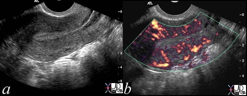
Normal Gray Scale and Power Doppler |
| 46314c01 uterus endometrium blood flow myometrium perfusion normal anatomy USscan Davidoff MD |
Cervix
|
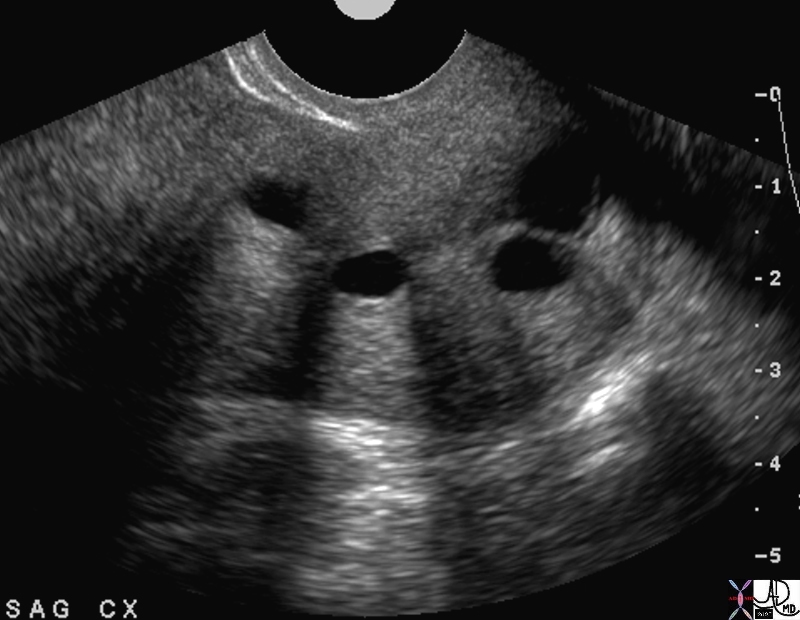
Nabothian Cysts Sagittal View of the Cervix |
|
The ultrasound is from a 40 year old patient who presents with pelvic discomfort . Nabothian cysts in the cervix are of incidental note. They are cystic in nature demonstaring anechogenicity, through transmission, and backwall enhancement. The examination was otherwise normal. Courtesy Ashley Davidoff MD Copyright 2010 49463 |
The Endometrial Cavity
Thin Stripe
|
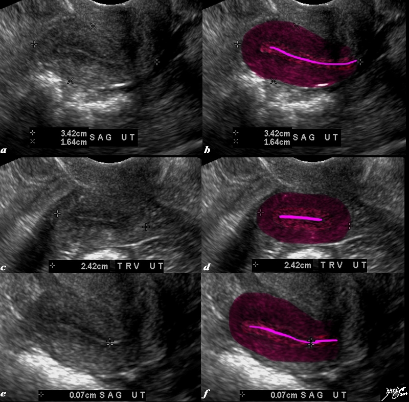
Post Menopausal Atrophy |
|
The transvaginal ultrasound is from a 62 year old post menopausal patient who presents with pelvic discomfort. The ultrasound shows a small atrophied uterus. The myometrium (dark red) junctional zone (light red) and endometrial cavity (pink) are outlined. In the sagittal plane the uterus measures 3.4cms in cranoicaudal dimension by 2.4cms in anteroposterior dimension (a,b) In the axial dimension (c,d) the uterus measures 2.4cms. The endometrial stripe is measured in the sagittal plane and measures .7mms. Courtesy Ashley Davidoff MD Copyright 2010 All rights reserved 84711c01.8s |
|
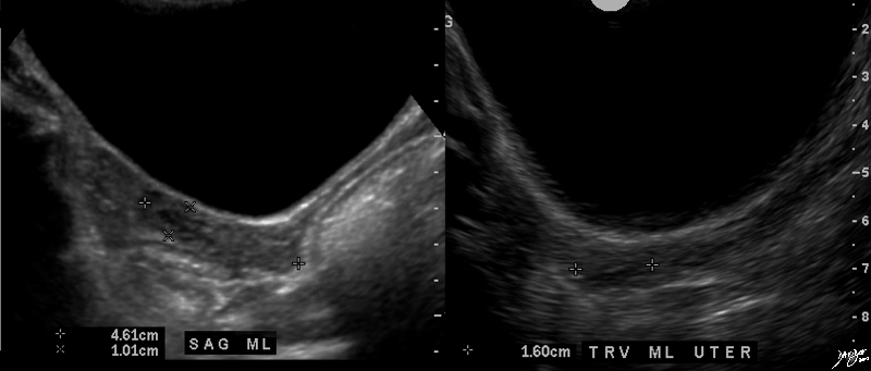
Turners Syndrome Small uterus Small Stripe |
|
The ultrasound is from a patient with Turners syndromerevealing an exteremely small uterus measuring 4.6X 1×1.6cms. The endometrial stripe measures about 1mms uterus Turner’s syndrome size congenital small USscan ultrasound Courtesy Ashley Davidoff MD copyright 2010 all rights reserved 85678c.8s |
Thick Stripe
|
Endometrial Carcinoma and Uterine Obstruction Use of Doppler |
|
The ultrasound is from a 70 year old post menopausal female who presents with an enlarged uterus. The endometrial stripe is enlarged and is filled with fluid and an enhancing soft tissue mass consistent with an endometrial carcinoma. Note blood flow as depicted by Doppler exam (c) characterizing the soft tissue as tumor rather than a clot. Courtesy Ashley Davidoff MD copyright 2009 all rights reserved 86206c.8s |
Obstruction
Cervical Stenosis
|
Cervical Stenosis and Metrorrhagia |
|
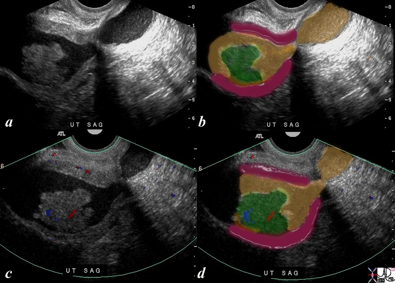
The transvaginal ultrasound is from a 50 year old perimenopausal female with metrorhagia. The uterine cavity and cervical cavity are filled with fluid, and soft tissue elements are identified in the expanded cervical canal. The findings are consistent with cervical stenosis, burt the cause of the metrirhagia is not obvious. The stenosis was relieved and follow up ultrasound showed resolution. No cervical mass was identified. Courtesy Ashley Davidoff MD copyright 2010 all rights reserved 85921c04.8s |
Polyp
|
Polyp as a Cause of Obstruction Presumed Benign Disease |
|
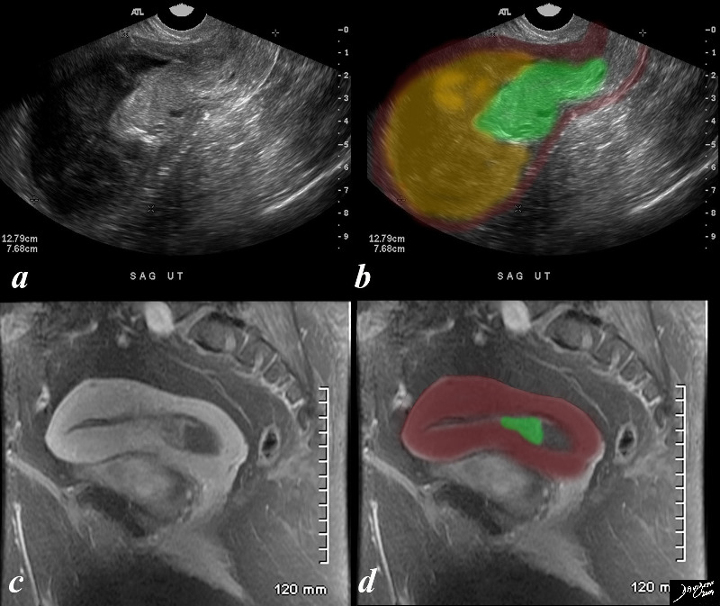
The ultrasound and MRIscan are from a 67 year old female, with an LMP of 10 years ago and with gynecologic history G9 and current history of bleeding for one month, a fever and low hematocrit. She has not had follow up gynecological examination for many years. The studies reveal a mass in the lower uterine segment and an obstructed endometrial cavity tha contains complex material which is presumed combination of blood and pus. ie hematometria and pyometria. The ultrasound is far more impressive than the MRI in that the mass (green) looks more impressive in size and there is prominent upstream accumulation of of complex material and fluids. The MRI shows a enhancing lesion arising from the endometrial mucosa, and at this time the fundal portion is relatively decompressed. The mass is presumably a benign polyp since her subsequent history to the hospital revealed only follow up for chronic renal failure and no pathological report of a malignancy has been n noted. Courtesy Ashley Davidoff MD copyright 2009 all rights reserved 84127c01.8s |
DOMElement Object
(
    [schemaTypeInfo] =>
    [tagName] => table
    [firstElementChild] => (object value omitted)
    [lastElementChild] => (object value omitted)
    [childElementCount] => 1
    [previousElementSibling] => (object value omitted)
    [nextElementSibling] =>
    [nodeName] => table
    [nodeValue] =>


Polyp as a Cause of Obstruction
Presumed Benign Disease


The ultrasound and MRIscan are from a 67 year old female, with an LMP of 10 years ago and with gynecologic history G9 and current history of bleeding for one month, a fever and low hematocrit. She has not had follow up gynecological examination for many years. The studies reveal a mass in the lower uterine segment and an obstructed endometrial cavity tha contains complex material which is presumed combination of blood and pus. ie hematometria and pyometria. The ultrasound is far more impressive than the MRI in that the mass (green) looks more impressive in size and there is prominent upstream accumulation of of complex material and fluids. The MRI shows a enhancing lesion arising from the endometrial mucosa, and at this time the fundal portion is relatively decompressed. The mass is presumably a benign polyp since her subsequent history to the hospital revealed only follow up for chronic renal failure and no pathological report of a malignancy has been n noted.
Courtesy Ashley Davidoff MD copyright 2009 all rights reserved 84127c01.8s


    [nodeType] => 1
    [parentNode] => (object value omitted)
    [childNodes] => (object value omitted)
    [firstChild] => (object value omitted)
    [lastChild] => (object value omitted)
    [previousSibling] => (object value omitted)
    [nextSibling] => (object value omitted)
    [attributes] => (object value omitted)
    [ownerDocument] => (object value omitted)
    [namespaceURI] =>
    [prefix] =>
    [localName] => table
    [baseURI] =>
    [textContent] =>


Polyp as a Cause of Obstruction
Presumed Benign Disease


The ultrasound and MRIscan are from a 67 year old female, with an LMP of 10 years ago and with gynecologic history G9 and current history of bleeding for one month, a fever and low hematocrit. She has not had follow up gynecological examination for many years. The studies reveal a mass in the lower uterine segment and an obstructed endometrial cavity tha contains complex material which is presumed combination of blood and pus. ie hematometria and pyometria. The ultrasound is far more impressive than the MRI in that the mass (green) looks more impressive in size and there is prominent upstream accumulation of of complex material and fluids. The MRI shows a enhancing lesion arising from the endometrial mucosa, and at this time the fundal portion is relatively decompressed. The mass is presumably a benign polyp since her subsequent history to the hospital revealed only follow up for chronic renal failure and no pathological report of a malignancy has been n noted.
Courtesy Ashley Davidoff MD copyright 2009 all rights reserved 84127c01.8s


)
DOMElement Object
(
    [schemaTypeInfo] =>
    [tagName] => td
    [firstElementChild] => (object value omitted)
    [lastElementChild] => (object value omitted)
    [childElementCount] => 2
    [previousElementSibling] =>
    [nextElementSibling] =>
    [nodeName] => td
    [nodeValue] =>
The ultrasound and MRIscan are from a 67 year old female, with an LMP of 10 years ago and with gynecologic history G9 and current history of bleeding for one month, a fever and low hematocrit. She has not had follow up gynecological examination for many years. The studies reveal a mass in the lower uterine segment and an obstructed endometrial cavity tha contains complex material which is presumed combination of blood and pus. ie hematometria and pyometria. The ultrasound is far more impressive than the MRI in that the mass (green) looks more impressive in size and there is prominent upstream accumulation of of complex material and fluids. The MRI shows a enhancing lesion arising from the endometrial mucosa, and at this time the fundal portion is relatively decompressed. The mass is presumably a benign polyp since her subsequent history to the hospital revealed only follow up for chronic renal failure and no pathological report of a malignancy has been n noted.
Courtesy Ashley Davidoff MD copyright 2009 all rights reserved 84127c01.8s

    [nodeType] => 1
    [parentNode] => (object value omitted)
    [childNodes] => (object value omitted)
    [firstChild] => (object value omitted)
    [lastChild] => (object value omitted)
    [previousSibling] => (object value omitted)
    [nextSibling] => (object value omitted)
    [attributes] => (object value omitted)
    [ownerDocument] => (object value omitted)
    [namespaceURI] =>
    [prefix] =>
    [localName] => td
    [baseURI] =>
    [textContent] =>
The ultrasound and MRIscan are from a 67 year old female, with an LMP of 10 years ago and with gynecologic history G9 and current history of bleeding for one month, a fever and low hematocrit. She has not had follow up gynecological examination for many years. The studies reveal a mass in the lower uterine segment and an obstructed endometrial cavity tha contains complex material which is presumed combination of blood and pus. ie hematometria and pyometria. The ultrasound is far more impressive than the MRI in that the mass (green) looks more impressive in size and there is prominent upstream accumulation of of complex material and fluids. The MRI shows a enhancing lesion arising from the endometrial mucosa, and at this time the fundal portion is relatively decompressed. The mass is presumably a benign polyp since her subsequent history to the hospital revealed only follow up for chronic renal failure and no pathological report of a malignancy has been n noted.
Courtesy Ashley Davidoff MD copyright 2009 all rights reserved 84127c01.8s

)
DOMElement Object
(
    [schemaTypeInfo] =>
    [tagName] => td
    [firstElementChild] => (object value omitted)
    [lastElementChild] => (object value omitted)
    [childElementCount] => 3
    [previousElementSibling] =>
    [nextElementSibling] =>
    [nodeName] => td
    [nodeValue] =>

Polyp as a Cause of Obstruction
Presumed Benign Disease
    [nodeType] => 1
    [parentNode] => (object value omitted)
    [childNodes] => (object value omitted)
    [firstChild] => (object value omitted)
    [lastChild] => (object value omitted)
    [previousSibling] => (object value omitted)
    [nextSibling] => (object value omitted)
    [attributes] => (object value omitted)
    [ownerDocument] => (object value omitted)
    [namespaceURI] =>
    [prefix] =>
    [localName] => td
    [baseURI] =>
    [textContent] =>

Polyp as a Cause of Obstruction
Presumed Benign Disease
)
DOMElement Object
(
    [schemaTypeInfo] =>
    [tagName] => table
    [firstElementChild] => (object value omitted)
    [lastElementChild] => (object value omitted)
    [childElementCount] => 1
    [previousElementSibling] => (object value omitted)
    [nextElementSibling] => (object value omitted)
    [nodeName] => table
    [nodeValue] =>


Cervical Stenosis and Metrorrhagia


The transvaginal ultrasound is from a 50 year old perimenopausal female with metrorhagia. The uterine cavity and cervical cavity are filled with fluid, and soft tissue elements are identified in the expanded cervical canal. The findings are consistent with cervical stenosis, burt the cause of the metrirhagia is not obvious. The stenosis was relieved and follow up ultrasound showed resolution. No cervical mass was identified.
Courtesy Ashley Davidoff MD copyright 2010 all rights reserved 85921c04.8s


    [nodeType] => 1
    [parentNode] => (object value omitted)
    [childNodes] => (object value omitted)
    [firstChild] => (object value omitted)
    [lastChild] => (object value omitted)
    [previousSibling] => (object value omitted)
    [nextSibling] => (object value omitted)
    [attributes] => (object value omitted)
    [ownerDocument] => (object value omitted)
    [namespaceURI] =>
    [prefix] =>
    [localName] => table
    [baseURI] =>
    [textContent] =>


Cervical Stenosis and Metrorrhagia


The transvaginal ultrasound is from a 50 year old perimenopausal female with metrorhagia. The uterine cavity and cervical cavity are filled with fluid, and soft tissue elements are identified in the expanded cervical canal. The findings are consistent with cervical stenosis, burt the cause of the metrirhagia is not obvious. The stenosis was relieved and follow up ultrasound showed resolution. No cervical mass was identified.
Courtesy Ashley Davidoff MD copyright 2010 all rights reserved 85921c04.8s


)
DOMElement Object
(
    [schemaTypeInfo] =>
    [tagName] => td
    [firstElementChild] => (object value omitted)
    [lastElementChild] => (object value omitted)
    [childElementCount] => 2
    [previousElementSibling] =>
    [nextElementSibling] =>
    [nodeName] => td
    [nodeValue] =>
The transvaginal ultrasound is from a 50 year old perimenopausal female with metrorhagia. The uterine cavity and cervical cavity are filled with fluid, and soft tissue elements are identified in the expanded cervical canal. The findings are consistent with cervical stenosis, burt the cause of the metrirhagia is not obvious. The stenosis was relieved and follow up ultrasound showed resolution. No cervical mass was identified.
Courtesy Ashley Davidoff MD copyright 2010 all rights reserved 85921c04.8s

    [nodeType] => 1
    [parentNode] => (object value omitted)
    [childNodes] => (object value omitted)
    [firstChild] => (object value omitted)
    [lastChild] => (object value omitted)
    [previousSibling] => (object value omitted)
    [nextSibling] => (object value omitted)
    [attributes] => (object value omitted)
    [ownerDocument] => (object value omitted)
    [namespaceURI] =>
    [prefix] =>
    [localName] => td
    [baseURI] =>
    [textContent] =>
The transvaginal ultrasound is from a 50 year old perimenopausal female with metrorhagia. The uterine cavity and cervical cavity are filled with fluid, and soft tissue elements are identified in the expanded cervical canal. The findings are consistent with cervical stenosis, burt the cause of the metrirhagia is not obvious. The stenosis was relieved and follow up ultrasound showed resolution. No cervical mass was identified.
Courtesy Ashley Davidoff MD copyright 2010 all rights reserved 85921c04.8s

)
DOMElement Object
(
    [schemaTypeInfo] =>
    [tagName] => td
    [firstElementChild] => (object value omitted)
    [lastElementChild] => (object value omitted)
    [childElementCount] => 2
    [previousElementSibling] =>
    [nextElementSibling] =>
    [nodeName] => td
    [nodeValue] =>

Cervical Stenosis and Metrorrhagia
    [nodeType] => 1
    [parentNode] => (object value omitted)
    [childNodes] => (object value omitted)
    [firstChild] => (object value omitted)
    [lastChild] => (object value omitted)
    [previousSibling] => (object value omitted)
    [nextSibling] => (object value omitted)
    [attributes] => (object value omitted)
    [ownerDocument] => (object value omitted)
    [namespaceURI] =>
    [prefix] =>
    [localName] => td
    [baseURI] =>
    [textContent] =>

Cervical Stenosis and Metrorrhagia
)
DOMElement Object
(
    [schemaTypeInfo] =>
    [tagName] => table
    [firstElementChild] => (object value omitted)
    [lastElementChild] => (object value omitted)
    [childElementCount] => 1
    [previousElementSibling] => (object value omitted)
    [nextElementSibling] => (object value omitted)
    [nodeName] => table
    [nodeValue] =>


Endometrial Carcinoma and Uterine Obstruction
Use of Doppler


The ultrasound is from a 70 year old post menopausal female who presents with an enlarged uterus. The endometrial stripe is enlarged and is filled with fluid and an enhancing soft tissue mass consistent with an endometrial carcinoma. Note blood flow as depicted by Doppler exam (c) characterizing the soft tissue as tumor rather than a clot.
Courtesy Ashley Davidoff MD copyright 2009 all rights reserved 86206c.8s


    [nodeType] => 1
    [parentNode] => (object value omitted)
    [childNodes] => (object value omitted)
    [firstChild] => (object value omitted)
    [lastChild] => (object value omitted)
    [previousSibling] => (object value omitted)
    [nextSibling] => (object value omitted)
    [attributes] => (object value omitted)
    [ownerDocument] => (object value omitted)
    [namespaceURI] =>
    [prefix] =>
    [localName] => table
    [baseURI] =>
    [textContent] =>


Endometrial Carcinoma and Uterine Obstruction
Use of Doppler


The ultrasound is from a 70 year old post menopausal female who presents with an enlarged uterus. The endometrial stripe is enlarged and is filled with fluid and an enhancing soft tissue mass consistent with an endometrial carcinoma. Note blood flow as depicted by Doppler exam (c) characterizing the soft tissue as tumor rather than a clot.
Courtesy Ashley Davidoff MD copyright 2009 all rights reserved 86206c.8s


)
DOMElement Object
(
    [schemaTypeInfo] =>
    [tagName] => td
    [firstElementChild] => (object value omitted)
    [lastElementChild] => (object value omitted)
    [childElementCount] => 2
    [previousElementSibling] =>
    [nextElementSibling] =>
    [nodeName] => td
    [nodeValue] =>
The ultrasound is from a 70 year old post menopausal female who presents with an enlarged uterus. The endometrial stripe is enlarged and is filled with fluid and an enhancing soft tissue mass consistent with an endometrial carcinoma. Note blood flow as depicted by Doppler exam (c) characterizing the soft tissue as tumor rather than a clot.
Courtesy Ashley Davidoff MD copyright 2009 all rights reserved 86206c.8s

    [nodeType] => 1
    [parentNode] => (object value omitted)
    [childNodes] => (object value omitted)
    [firstChild] => (object value omitted)
    [lastChild] => (object value omitted)
    [previousSibling] => (object value omitted)
    [nextSibling] => (object value omitted)
    [attributes] => (object value omitted)
    [ownerDocument] => (object value omitted)
    [namespaceURI] =>
    [prefix] =>
    [localName] => td
    [baseURI] =>
    [textContent] =>
The ultrasound is from a 70 year old post menopausal female who presents with an enlarged uterus. The endometrial stripe is enlarged and is filled with fluid and an enhancing soft tissue mass consistent with an endometrial carcinoma. Note blood flow as depicted by Doppler exam (c) characterizing the soft tissue as tumor rather than a clot.
Courtesy Ashley Davidoff MD copyright 2009 all rights reserved 86206c.8s

)
DOMElement Object
(
    [schemaTypeInfo] =>
    [tagName] => td
    [firstElementChild] => (object value omitted)
    [lastElementChild] => (object value omitted)
    [childElementCount] => 3
    [previousElementSibling] =>
    [nextElementSibling] =>
    [nodeName] => td
    [nodeValue] =>

Endometrial Carcinoma and Uterine Obstruction
Use of Doppler
    [nodeType] => 1
    [parentNode] => (object value omitted)
    [childNodes] => (object value omitted)
    [firstChild] => (object value omitted)
    [lastChild] => (object value omitted)
    [previousSibling] => (object value omitted)
    [nextSibling] => (object value omitted)
    [attributes] => (object value omitted)
    [ownerDocument] => (object value omitted)
    [namespaceURI] =>
    [prefix] =>
    [localName] => td
    [baseURI] =>
    [textContent] =>

Endometrial Carcinoma and Uterine Obstruction
Use of Doppler
)
DOMElement Object
(
    [schemaTypeInfo] =>
    [tagName] => table
    [firstElementChild] => (object value omitted)
    [lastElementChild] => (object value omitted)
    [childElementCount] => 1
    [previousElementSibling] => (object value omitted)
    [nextElementSibling] => (object value omitted)
    [nodeName] => table
    [nodeValue] =>


Hyperplasia


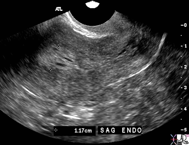
The transvaginal ultrasound is from a 60 year old female who presents with spotting Ultrasound reveals a heterogeneous endometrial stripe consistent with endometrial hyperplasia though endometrial carcinoma is a possibility. Malignant neoplasia is a a les likely possibility
Courtesy Ashley DAvidoff MD copyright  83301.81s


    [nodeType] => 1
    [parentNode] => (object value omitted)
    [childNodes] => (object value omitted)
    [firstChild] => (object value omitted)
    [lastChild] => (object value omitted)
    [previousSibling] => (object value omitted)
    [nextSibling] => (object value omitted)
    [attributes] => (object value omitted)
    [ownerDocument] => (object value omitted)
    [namespaceURI] =>
    [prefix] =>
    [localName] => table
    [baseURI] =>
    [textContent] =>


Hyperplasia


The transvaginal ultrasound is from a 60 year old female who presents with spotting Ultrasound reveals a heterogeneous endometrial stripe consistent with endometrial hyperplasia though endometrial carcinoma is a possibility. Malignant neoplasia is a a les likely possibility
Courtesy Ashley DAvidoff MD copyright  83301.81s


)
DOMElement Object
(
    [schemaTypeInfo] =>
    [tagName] => td
    [firstElementChild] => (object value omitted)
    [lastElementChild] => (object value omitted)
    [childElementCount] => 2
    [previousElementSibling] =>
    [nextElementSibling] =>
    [nodeName] => td
    [nodeValue] =>
The transvaginal ultrasound is from a 60 year old female who presents with spotting Ultrasound reveals a heterogeneous endometrial stripe consistent with endometrial hyperplasia though endometrial carcinoma is a possibility. Malignant neoplasia is a a les likely possibility
Courtesy Ashley DAvidoff MD copyright  83301.81s

    [nodeType] => 1
    [parentNode] => (object value omitted)
    [childNodes] => (object value omitted)
    [firstChild] => (object value omitted)
    [lastChild] => (object value omitted)
    [previousSibling] => (object value omitted)
    [nextSibling] => (object value omitted)
    [attributes] => (object value omitted)
    [ownerDocument] => (object value omitted)
    [namespaceURI] =>
    [prefix] =>
    [localName] => td
    [baseURI] =>
    [textContent] =>
The transvaginal ultrasound is from a 60 year old female who presents with spotting Ultrasound reveals a heterogeneous endometrial stripe consistent with endometrial hyperplasia though endometrial carcinoma is a possibility. Malignant neoplasia is a a les likely possibility
Courtesy Ashley DAvidoff MD copyright  83301.81s

)
DOMElement Object
(
    [schemaTypeInfo] =>
    [tagName] => td
    [firstElementChild] => (object value omitted)
    [lastElementChild] => (object value omitted)
    [childElementCount] => 2
    [previousElementSibling] =>
    [nextElementSibling] =>
    [nodeName] => td
    [nodeValue] =>
Hyperplasia
    [nodeType] => 1
    [parentNode] => (object value omitted)
    [childNodes] => (object value omitted)
    [firstChild] => (object value omitted)
    [lastChild] => (object value omitted)
    [previousSibling] => (object value omitted)
    [nextSibling] => (object value omitted)
    [attributes] => (object value omitted)
    [ownerDocument] => (object value omitted)
    [namespaceURI] =>
    [prefix] =>
    [localName] => td
    [baseURI] =>
    [textContent] =>
Hyperplasia
)
DOMElement Object
(
    [schemaTypeInfo] =>
    [tagName] => table
    [firstElementChild] => (object value omitted)
    [lastElementChild] => (object value omitted)
    [childElementCount] => 1
    [previousElementSibling] => (object value omitted)
    [nextElementSibling] => (object value omitted)
    [nodeName] => table
    [nodeValue] =>


Turners Syndrome
Small uterus Small Stripe


The ultrasound is from a patient with Turners syndromerevealing an exteremely small uterus measuring 4.6X 1×1.6cms. The endometrial stripe measures about 1mms uterus Turner’s syndrome size congenital small USscan ultrasound
Courtesy Ashley Davidoff MD copyright 2010 all rights reserved 85678c.8s


    [nodeType] => 1
    [parentNode] => (object value omitted)
    [childNodes] => (object value omitted)
    [firstChild] => (object value omitted)
    [lastChild] => (object value omitted)
    [previousSibling] => (object value omitted)
    [nextSibling] => (object value omitted)
    [attributes] => (object value omitted)
    [ownerDocument] => (object value omitted)
    [namespaceURI] =>
    [prefix] =>
    [localName] => table
    [baseURI] =>
    [textContent] =>


Turners Syndrome
Small uterus Small Stripe


The ultrasound is from a patient with Turners syndromerevealing an exteremely small uterus measuring 4.6X 1×1.6cms. The endometrial stripe measures about 1mms uterus Turner’s syndrome size congenital small USscan ultrasound
Courtesy Ashley Davidoff MD copyright 2010 all rights reserved 85678c.8s


)
DOMElement Object
(
    [schemaTypeInfo] =>
    [tagName] => td
    [firstElementChild] => (object value omitted)
    [lastElementChild] => (object value omitted)
    [childElementCount] => 2
    [previousElementSibling] =>
    [nextElementSibling] =>
    [nodeName] => td
    [nodeValue] =>
The ultrasound is from a patient with Turners syndromerevealing an exteremely small uterus measuring 4.6X 1×1.6cms. The endometrial stripe measures about 1mms uterus Turner’s syndrome size congenital small USscan ultrasound
Courtesy Ashley Davidoff MD copyright 2010 all rights reserved 85678c.8s

    [nodeType] => 1
    [parentNode] => (object value omitted)
    [childNodes] => (object value omitted)
    [firstChild] => (object value omitted)
    [lastChild] => (object value omitted)
    [previousSibling] => (object value omitted)
    [nextSibling] => (object value omitted)
    [attributes] => (object value omitted)
    [ownerDocument] => (object value omitted)
    [namespaceURI] =>
    [prefix] =>
    [localName] => td
    [baseURI] =>
    [textContent] =>
The ultrasound is from a patient with Turners syndromerevealing an exteremely small uterus measuring 4.6X 1×1.6cms. The endometrial stripe measures about 1mms uterus Turner’s syndrome size congenital small USscan ultrasound
Courtesy Ashley Davidoff MD copyright 2010 all rights reserved 85678c.8s

)
DOMElement Object
(
    [schemaTypeInfo] =>
    [tagName] => td
    [firstElementChild] => (object value omitted)
    [lastElementChild] => (object value omitted)
    [childElementCount] => 3
    [previousElementSibling] =>
    [nextElementSibling] =>
    [nodeName] => td
    [nodeValue] =>

Turners Syndrome
Small uterus Small Stripe
    [nodeType] => 1
    [parentNode] => (object value omitted)
    [childNodes] => (object value omitted)
    [firstChild] => (object value omitted)
    [lastChild] => (object value omitted)
    [previousSibling] => (object value omitted)
    [nextSibling] => (object value omitted)
    [attributes] => (object value omitted)
    [ownerDocument] => (object value omitted)
    [namespaceURI] =>
    [prefix] =>
    [localName] => td
    [baseURI] =>
    [textContent] =>

Turners Syndrome
Small uterus Small Stripe
)
DOMElement Object
(
    [schemaTypeInfo] =>
    [tagName] => table
    [firstElementChild] => (object value omitted)
    [lastElementChild] => (object value omitted)
    [childElementCount] => 1
    [previousElementSibling] => (object value omitted)
    [nextElementSibling] => (object value omitted)
    [nodeName] => table
    [nodeValue] =>


Post Menopausal Atrophy


The transvaginal ultrasound is from a 62 year old post menopausal patient who presents with pelvic discomfort. The ultrasound shows a small atrophied uterus. The myometrium (dark red) junctional zone (light red) and endometrial cavity (pink) are outlined. In the sagittal plane the uterus measures 3.4cms in cranoicaudal dimension by 2.4cms in anteroposterior dimension (a,b) In the axial dimension (c,d) the uterus measures 2.4cms. The endometrial stripe is measured in the sagittal plane and measures .7mms.
Courtesy Ashley Davidoff MD Copyright 2010 All rights reserved 84711c01.8s


    [nodeType] => 1
    [parentNode] => (object value omitted)
    [childNodes] => (object value omitted)
    [firstChild] => (object value omitted)
    [lastChild] => (object value omitted)
    [previousSibling] => (object value omitted)
    [nextSibling] => (object value omitted)
    [attributes] => (object value omitted)
    [ownerDocument] => (object value omitted)
    [namespaceURI] =>
    [prefix] =>
    [localName] => table
    [baseURI] =>
    [textContent] =>


Post Menopausal Atrophy


The transvaginal ultrasound is from a 62 year old post menopausal patient who presents with pelvic discomfort. The ultrasound shows a small atrophied uterus. The myometrium (dark red) junctional zone (light red) and endometrial cavity (pink) are outlined. In the sagittal plane the uterus measures 3.4cms in cranoicaudal dimension by 2.4cms in anteroposterior dimension (a,b) In the axial dimension (c,d) the uterus measures 2.4cms. The endometrial stripe is measured in the sagittal plane and measures .7mms.
Courtesy Ashley Davidoff MD Copyright 2010 All rights reserved 84711c01.8s


)
DOMElement Object
(
    [schemaTypeInfo] =>
    [tagName] => td
    [firstElementChild] => (object value omitted)
    [lastElementChild] => (object value omitted)
    [childElementCount] => 2
    [previousElementSibling] =>
    [nextElementSibling] =>
    [nodeName] => td
    [nodeValue] =>
The transvaginal ultrasound is from a 62 year old post menopausal patient who presents with pelvic discomfort. The ultrasound shows a small atrophied uterus. The myometrium (dark red) junctional zone (light red) and endometrial cavity (pink) are outlined. In the sagittal plane the uterus measures 3.4cms in cranoicaudal dimension by 2.4cms in anteroposterior dimension (a,b) In the axial dimension (c,d) the uterus measures 2.4cms. The endometrial stripe is measured in the sagittal plane and measures .7mms.
Courtesy Ashley Davidoff MD Copyright 2010 All rights reserved 84711c01.8s

    [nodeType] => 1
    [parentNode] => (object value omitted)
    [childNodes] => (object value omitted)
    [firstChild] => (object value omitted)
    [lastChild] => (object value omitted)
    [previousSibling] => (object value omitted)
    [nextSibling] => (object value omitted)
    [attributes] => (object value omitted)
    [ownerDocument] => (object value omitted)
    [namespaceURI] =>
    [prefix] =>
    [localName] => td
    [baseURI] =>
    [textContent] =>
The transvaginal ultrasound is from a 62 year old post menopausal patient who presents with pelvic discomfort. The ultrasound shows a small atrophied uterus. The myometrium (dark red) junctional zone (light red) and endometrial cavity (pink) are outlined. In the sagittal plane the uterus measures 3.4cms in cranoicaudal dimension by 2.4cms in anteroposterior dimension (a,b) In the axial dimension (c,d) the uterus measures 2.4cms. The endometrial stripe is measured in the sagittal plane and measures .7mms.
Courtesy Ashley Davidoff MD Copyright 2010 All rights reserved 84711c01.8s

)
DOMElement Object
(
    [schemaTypeInfo] =>
    [tagName] => td
    [firstElementChild] => (object value omitted)
    [lastElementChild] => (object value omitted)
    [childElementCount] => 2
    [previousElementSibling] =>
    [nextElementSibling] =>
    [nodeName] => td
    [nodeValue] =>

Post Menopausal Atrophy
    [nodeType] => 1
    [parentNode] => (object value omitted)
    [childNodes] => (object value omitted)
    [firstChild] => (object value omitted)
    [lastChild] => (object value omitted)
    [previousSibling] => (object value omitted)
    [nextSibling] => (object value omitted)
    [attributes] => (object value omitted)
    [ownerDocument] => (object value omitted)
    [namespaceURI] =>
    [prefix] =>
    [localName] => td
    [baseURI] =>
    [textContent] =>

Post Menopausal Atrophy
)
DOMElement Object
(
    [schemaTypeInfo] =>
    [tagName] => table
    [firstElementChild] => (object value omitted)
    [lastElementChild] => (object value omitted)
    [childElementCount] => 1
    [previousElementSibling] => (object value omitted)
    [nextElementSibling] => (object value omitted)
    [nodeName] => table
    [nodeValue] =>


Nabothian Cysts
Sagittal View of the Cervix


The ultrasound is from a 40 year old patient who presents with pelvic discomfort . Nabothian cysts in the cervix are of incidental note. They are cystic in nature demonstaring anechogenicity, through transmission, and backwall enhancement. The examination was otherwise normal.
Courtesy Ashley Davidoff MD Copyright 2010 49463


    [nodeType] => 1
    [parentNode] => (object value omitted)
    [childNodes] => (object value omitted)
    [firstChild] => (object value omitted)
    [lastChild] => (object value omitted)
    [previousSibling] => (object value omitted)
    [nextSibling] => (object value omitted)
    [attributes] => (object value omitted)
    [ownerDocument] => (object value omitted)
    [namespaceURI] =>
    [prefix] =>
    [localName] => table
    [baseURI] =>
    [textContent] =>


Nabothian Cysts
Sagittal View of the Cervix


The ultrasound is from a 40 year old patient who presents with pelvic discomfort . Nabothian cysts in the cervix are of incidental note. They are cystic in nature demonstaring anechogenicity, through transmission, and backwall enhancement. The examination was otherwise normal.
Courtesy Ashley Davidoff MD Copyright 2010 49463


)
DOMElement Object
(
    [schemaTypeInfo] =>
    [tagName] => td
    [firstElementChild] => (object value omitted)
    [lastElementChild] => (object value omitted)
    [childElementCount] => 2
    [previousElementSibling] =>
    [nextElementSibling] =>
    [nodeName] => td
    [nodeValue] =>
The ultrasound is from a 40 year old patient who presents with pelvic discomfort . Nabothian cysts in the cervix are of incidental note. They are cystic in nature demonstaring anechogenicity, through transmission, and backwall enhancement. The examination was otherwise normal.
Courtesy Ashley Davidoff MD Copyright 2010 49463

    [nodeType] => 1
    [parentNode] => (object value omitted)
    [childNodes] => (object value omitted)
    [firstChild] => (object value omitted)
    [lastChild] => (object value omitted)
    [previousSibling] => (object value omitted)
    [nextSibling] => (object value omitted)
    [attributes] => (object value omitted)
    [ownerDocument] => (object value omitted)
    [namespaceURI] =>
    [prefix] =>
    [localName] => td
    [baseURI] =>
    [textContent] =>
The ultrasound is from a 40 year old patient who presents with pelvic discomfort . Nabothian cysts in the cervix are of incidental note. They are cystic in nature demonstaring anechogenicity, through transmission, and backwall enhancement. The examination was otherwise normal.
Courtesy Ashley Davidoff MD Copyright 2010 49463

)
DOMElement Object
(
    [schemaTypeInfo] =>
    [tagName] => td
    [firstElementChild] => (object value omitted)
    [lastElementChild] => (object value omitted)
    [childElementCount] => 3
    [previousElementSibling] =>
    [nextElementSibling] =>
    [nodeName] => td
    [nodeValue] =>

Nabothian Cysts
Sagittal View of the Cervix
    [nodeType] => 1
    [parentNode] => (object value omitted)
    [childNodes] => (object value omitted)
    [firstChild] => (object value omitted)
    [lastChild] => (object value omitted)
    [previousSibling] => (object value omitted)
    [nextSibling] => (object value omitted)
    [attributes] => (object value omitted)
    [ownerDocument] => (object value omitted)
    [namespaceURI] =>
    [prefix] =>
    [localName] => td
    [baseURI] =>
    [textContent] =>

Nabothian Cysts
Sagittal View of the Cervix
)
DOMElement Object
(
    [schemaTypeInfo] =>
    [tagName] => table
    [firstElementChild] => (object value omitted)
    [lastElementChild] => (object value omitted)
    [childElementCount] => 1
    [previousElementSibling] => (object value omitted)
    [nextElementSibling] => (object value omitted)
    [nodeName] => table
    [nodeValue] =>


Normal Gray Scale and Power Doppler


46314c01 uterus endometrium blood flow myometrium perfusion normal anatomy USscan Davidoff MD


    [nodeType] => 1
    [parentNode] => (object value omitted)
    [childNodes] => (object value omitted)
    [firstChild] => (object value omitted)
    [lastChild] => (object value omitted)
    [previousSibling] => (object value omitted)
    [nextSibling] => (object value omitted)
    [attributes] => (object value omitted)
    [ownerDocument] => (object value omitted)
    [namespaceURI] =>
    [prefix] =>
    [localName] => table
    [baseURI] =>
    [textContent] =>


Normal Gray Scale and Power Doppler


46314c01 uterus endometrium blood flow myometrium perfusion normal anatomy USscan Davidoff MD


)
DOMElement Object
(
    [schemaTypeInfo] =>
    [tagName] => td
    [firstElementChild] =>
    [lastElementChild] =>
    [childElementCount] => 0
    [previousElementSibling] =>
    [nextElementSibling] =>
    [nodeName] => td
    [nodeValue] => 46314c01 uterus endometrium blood flow myometrium perfusion normal anatomy USscan Davidoff MD
    [nodeType] => 1
    [parentNode] => (object value omitted)
    [childNodes] => (object value omitted)
    [firstChild] => (object value omitted)
    [lastChild] => (object value omitted)
    [previousSibling] => (object value omitted)
    [nextSibling] => (object value omitted)
    [attributes] => (object value omitted)
    [ownerDocument] => (object value omitted)
    [namespaceURI] =>
    [prefix] =>
    [localName] => td
    [baseURI] =>
    [textContent] => 46314c01 uterus endometrium blood flow myometrium perfusion normal anatomy USscan Davidoff MD
)
DOMElement Object
(
    [schemaTypeInfo] =>
    [tagName] => td
    [firstElementChild] => (object value omitted)
    [lastElementChild] => (object value omitted)
    [childElementCount] => 2
    [previousElementSibling] =>
    [nextElementSibling] =>
    [nodeName] => td
    [nodeValue] =>
Normal Gray Scale and Power Doppler
    [nodeType] => 1
    [parentNode] => (object value omitted)
    [childNodes] => (object value omitted)
    [firstChild] => (object value omitted)
    [lastChild] => (object value omitted)
    [previousSibling] => (object value omitted)
    [nextSibling] => (object value omitted)
    [attributes] => (object value omitted)
    [ownerDocument] => (object value omitted)
    [namespaceURI] =>
    [prefix] =>
    [localName] => td
    [baseURI] =>
    [textContent] =>
Normal Gray Scale and Power Doppler
)

