Structure
The Common Vein Copyright 2008
Introduction
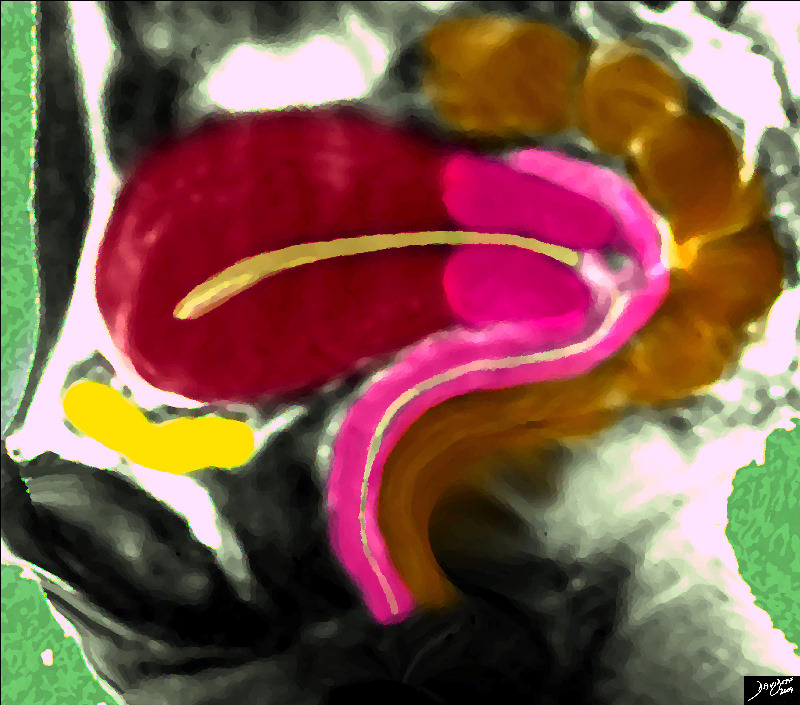
In virgin state the uterus is flattened antero-posteriorly and is pyriform in shape, with the apex directed downward and backward. The bladder is to anterior and rectum is related to it posteriorly. It is a pelvic organ suspended by broad ligament and round ligament superiorly to pelvic fibrous tissue inferiorly. The uterus measures about 7.5 cm. in length, 5 cm. in breadth, at its upper part, and nearly 2.5 cm. in thickness; it weighs from 30 to 40 gms. It is divisible into two portions. On the surface, about midway between the apex and base, is a slight constriction, known as the isthmus, and corresponding to this in the interior is a narrowing of the uterine cavity, the internal orifice of the uterus. The portion above the isthmus is termed thebody, and that below, the cervix. The part of the body which lies above a plane passing through the points of entrance of the uterine tubes is known as the fundus.
The uterine cavity is lined by a special mucosal surface called endometrium. The muscular layer of uterus is called myometrium has different layers in various orientation of fibers. Myometrium usually proliferates during pregnancy. The peritoneal area of uterus is covered by a serosal layer.
Applied anatomy: The developmental uterine anomalies may hinder with conception and normal child birth. The changes in position could give rise to chronic pelvic pain. During childbirth there is a risk of injury to urinary bladder as well as anal sphincter as uterus is anatomically closely related to these vital structures. Uterus may lose its supports with age, repeated pregnancies and post menopause and may give rise to uterovaginal prolapse. Uterine fibroids are most common benign tumors arising from uterine myometrium. Uterine endometrium is ectopic place like in myometrium may give rise to adenomyosis and endometriosis when it is in ovary and pelvis.

The Pathway to Conception
|
| Image courtesy Ashley Davidoff MD copyright 2009 14707.2kb04i06.s.4k.8s |
DOMElement Object
(
    [schemaTypeInfo] =>
    [tagName] => table
    [firstElementChild] => (object value omitted)
    [lastElementChild] => (object value omitted)
    [childElementCount] => 1
    [previousElementSibling] => (object value omitted)
    [nextElementSibling] =>
    [nodeName] => table
    [nodeValue] =>


The Pathway to Conception


Image  courtesy Ashley Davidoff MD copyright 2009 14707.2kb04i06.s.4k.8s


    [nodeType] => 1
    [parentNode] => (object value omitted)
    [childNodes] => (object value omitted)
    [firstChild] => (object value omitted)
    [lastChild] => (object value omitted)
    [previousSibling] => (object value omitted)
    [nextSibling] => (object value omitted)
    [attributes] => (object value omitted)
    [ownerDocument] => (object value omitted)
    [namespaceURI] =>
    [prefix] =>
    [localName] => table
    [baseURI] =>
    [textContent] =>


The Pathway to Conception


Image  courtesy Ashley Davidoff MD copyright 2009 14707.2kb04i06.s.4k.8s


)
DOMElement Object
(
    [schemaTypeInfo] =>
    [tagName] => td
    [firstElementChild] => (object value omitted)
    [lastElementChild] => (object value omitted)
    [childElementCount] => 2
    [previousElementSibling] =>
    [nextElementSibling] =>
    [nodeName] => td
    [nodeValue] => Image  courtesy Ashley Davidoff MD copyright 2009 14707.2kb04i06.s.4k.8s
    [nodeType] => 1
    [parentNode] => (object value omitted)
    [childNodes] => (object value omitted)
    [firstChild] => (object value omitted)
    [lastChild] => (object value omitted)
    [previousSibling] => (object value omitted)
    [nextSibling] => (object value omitted)
    [attributes] => (object value omitted)
    [ownerDocument] => (object value omitted)
    [namespaceURI] =>
    [prefix] =>
    [localName] => td
    [baseURI] =>
    [textContent] => Image  courtesy Ashley Davidoff MD copyright 2009 14707.2kb04i06.s.4k.8s
)
DOMElement Object
(
    [schemaTypeInfo] =>
    [tagName] => td
    [firstElementChild] => (object value omitted)
    [lastElementChild] => (object value omitted)
    [childElementCount] => 2
    [previousElementSibling] =>
    [nextElementSibling] =>
    [nodeName] => td
    [nodeValue] =>
The Pathway to Conception

    [nodeType] => 1
    [parentNode] => (object value omitted)
    [childNodes] => (object value omitted)
    [firstChild] => (object value omitted)
    [lastChild] => (object value omitted)
    [previousSibling] => (object value omitted)
    [nextSibling] => (object value omitted)
    [attributes] => (object value omitted)
    [ownerDocument] => (object value omitted)
    [namespaceURI] =>
    [prefix] =>
    [localName] => td
    [baseURI] =>
    [textContent] =>
The Pathway to Conception

)

