The Common Vein Copyright 2009
Myometrium
Endometrium
Cervix
Tubes
DOMElement Object
(
    [schemaTypeInfo] =>
    [tagName] => table
    [firstElementChild] => (object value omitted)
    [lastElementChild] => (object value omitted)
    [childElementCount] => 1
    [previousElementSibling] => (object value omitted)
    [nextElementSibling] => (object value omitted)
    [nodeName] => table
    [nodeValue] =>


83301.8s


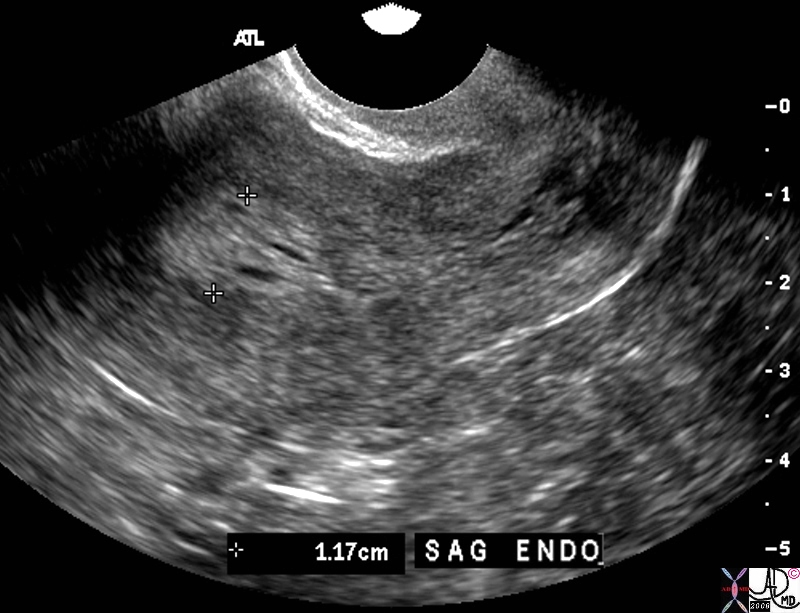
83301.81s The transvaginal ultrasound is from a 60 year old female who presents with spotting Ultrasound reveals a heterogeneous endometrial stripe consistent with endometrial hyperplasia thug endometrial carcinoma is a possibility. Malignant neoplasia is a a les likely possibility uterus endometrium lack of homogeneity hyperplasia vs metaplasia dysplasia carcinoma of the uterus US Courtesy Ashley DAvidoff MD copyright 2009


    [nodeType] => 1
    [parentNode] => (object value omitted)
    [childNodes] => (object value omitted)
    [firstChild] => (object value omitted)
    [lastChild] => (object value omitted)
    [previousSibling] => (object value omitted)
    [nextSibling] => (object value omitted)
    [attributes] => (object value omitted)
    [ownerDocument] => (object value omitted)
    [namespaceURI] =>
    [prefix] =>
    [localName] => table
    [baseURI] =>
    [textContent] =>


83301.8s


83301.81s The transvaginal ultrasound is from a 60 year old female who presents with spotting Ultrasound reveals a heterogeneous endometrial stripe consistent with endometrial hyperplasia thug endometrial carcinoma is a possibility. Malignant neoplasia is a a les likely possibility uterus endometrium lack of homogeneity hyperplasia vs metaplasia dysplasia carcinoma of the uterus US Courtesy Ashley DAvidoff MD copyright 2009


)
DOMElement Object
(
    [schemaTypeInfo] =>
    [tagName] => td
    [firstElementChild] => (object value omitted)
    [lastElementChild] => (object value omitted)
    [childElementCount] => 1
    [previousElementSibling] =>
    [nextElementSibling] =>
    [nodeName] => td
    [nodeValue] => 83301.81s The transvaginal ultrasound is from a 60 year old female who presents with spotting Ultrasound reveals a heterogeneous endometrial stripe consistent with endometrial hyperplasia thug endometrial carcinoma is a possibility. Malignant neoplasia is a a les likely possibility uterus endometrium lack of homogeneity hyperplasia vs metaplasia dysplasia carcinoma of the uterus US Courtesy Ashley DAvidoff MD copyright 2009
    [nodeType] => 1
    [parentNode] => (object value omitted)
    [childNodes] => (object value omitted)
    [firstChild] => (object value omitted)
    [lastChild] => (object value omitted)
    [previousSibling] => (object value omitted)
    [nextSibling] => (object value omitted)
    [attributes] => (object value omitted)
    [ownerDocument] => (object value omitted)
    [namespaceURI] =>
    [prefix] =>
    [localName] => td
    [baseURI] =>
    [textContent] => 83301.81s The transvaginal ultrasound is from a 60 year old female who presents with spotting Ultrasound reveals a heterogeneous endometrial stripe consistent with endometrial hyperplasia thug endometrial carcinoma is a possibility. Malignant neoplasia is a a les likely possibility uterus endometrium lack of homogeneity hyperplasia vs metaplasia dysplasia carcinoma of the uterus US Courtesy Ashley DAvidoff MD copyright 2009
)
DOMElement Object
(
    [schemaTypeInfo] =>
    [tagName] => td
    [firstElementChild] => (object value omitted)
    [lastElementChild] => (object value omitted)
    [childElementCount] => 2
    [previousElementSibling] =>
    [nextElementSibling] =>
    [nodeName] => td
    [nodeValue] =>
83301.8s
    [nodeType] => 1
    [parentNode] => (object value omitted)
    [childNodes] => (object value omitted)
    [firstChild] => (object value omitted)
    [lastChild] => (object value omitted)
    [previousSibling] => (object value omitted)
    [nextSibling] => (object value omitted)
    [attributes] => (object value omitted)
    [ownerDocument] => (object value omitted)
    [namespaceURI] =>
    [prefix] =>
    [localName] => td
    [baseURI] =>
    [textContent] =>
83301.8s
)
DOMElement Object
(
    [schemaTypeInfo] =>
    [tagName] => table
    [firstElementChild] => (object value omitted)
    [lastElementChild] => (object value omitted)
    [childElementCount] => 1
    [previousElementSibling] => (object value omitted)
    [nextElementSibling] => (object value omitted)
    [nodeName] => table
    [nodeValue] =>


83298c.81s


83298c.81s This T2 weighted MRI of a 41 year old female shows thickened junctional zone of the uterus measuring up to 12 mms characteristic of adenomyosis uterus junctional zone thickened enlarged MRI T2 weighted Adenomyosis the uterus Courrtesy Ashey Davidoff MD copyright 2009 ectopic tissue


    [nodeType] => 1
    [parentNode] => (object value omitted)
    [childNodes] => (object value omitted)
    [firstChild] => (object value omitted)
    [lastChild] => (object value omitted)
    [previousSibling] => (object value omitted)
    [nextSibling] => (object value omitted)
    [attributes] => (object value omitted)
    [ownerDocument] => (object value omitted)
    [namespaceURI] =>
    [prefix] =>
    [localName] => table
    [baseURI] =>
    [textContent] =>


83298c.81s


83298c.81s This T2 weighted MRI of a 41 year old female shows thickened junctional zone of the uterus measuring up to 12 mms characteristic of adenomyosis uterus junctional zone thickened enlarged MRI T2 weighted Adenomyosis the uterus Courrtesy Ashey Davidoff MD copyright 2009 ectopic tissue


)
DOMElement Object
(
    [schemaTypeInfo] =>
    [tagName] => td
    [firstElementChild] => (object value omitted)
    [lastElementChild] => (object value omitted)
    [childElementCount] => 1
    [previousElementSibling] =>
    [nextElementSibling] =>
    [nodeName] => td
    [nodeValue] => 83298c.81s This T2 weighted MRI of a 41 year old female shows thickened junctional zone of the uterus measuring up to 12 mms characteristic of adenomyosis uterus junctional zone thickened enlarged MRI T2 weighted Adenomyosis the uterus Courrtesy Ashey Davidoff MD copyright 2009 ectopic tissue
    [nodeType] => 1
    [parentNode] => (object value omitted)
    [childNodes] => (object value omitted)
    [firstChild] => (object value omitted)
    [lastChild] => (object value omitted)
    [previousSibling] => (object value omitted)
    [nextSibling] => (object value omitted)
    [attributes] => (object value omitted)
    [ownerDocument] => (object value omitted)
    [namespaceURI] =>
    [prefix] =>
    [localName] => td
    [baseURI] =>
    [textContent] => 83298c.81s This T2 weighted MRI of a 41 year old female shows thickened junctional zone of the uterus measuring up to 12 mms characteristic of adenomyosis uterus junctional zone thickened enlarged MRI T2 weighted Adenomyosis the uterus Courrtesy Ashey Davidoff MD copyright 2009 ectopic tissue
)
DOMElement Object
(
    [schemaTypeInfo] =>
    [tagName] => td
    [firstElementChild] => (object value omitted)
    [lastElementChild] => (object value omitted)
    [childElementCount] => 2
    [previousElementSibling] =>
    [nextElementSibling] =>
    [nodeName] => td
    [nodeValue] =>
83298c.81s
    [nodeType] => 1
    [parentNode] => (object value omitted)
    [childNodes] => (object value omitted)
    [firstChild] => (object value omitted)
    [lastChild] => (object value omitted)
    [previousSibling] => (object value omitted)
    [nextSibling] => (object value omitted)
    [attributes] => (object value omitted)
    [ownerDocument] => (object value omitted)
    [namespaceURI] =>
    [prefix] =>
    [localName] => td
    [baseURI] =>
    [textContent] =>
83298c.81s
)


