Cystic Lesions of the Uterus
Ashley Davidoff MD
The Common Vein Copyright 2010
Introduction
|
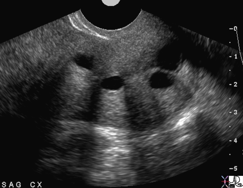
Nabothian Cysts Sagittal View of the Cervix |
|
The ultrasound is from a 40 year old patient who presents with pelvic discomfort . Nabothian cysts in the cervix are of incidental note. They are cystic in nature demonstaring anechogenicity, through transmission, and backwall enhancement. The examination was otherwise normal. Courtesy Ashley Davidoff MD Copyright 2010 49463 |
|
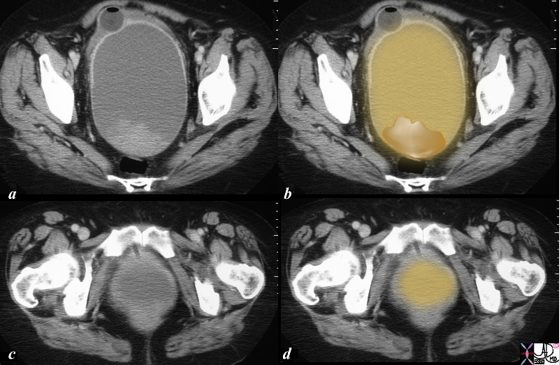
Mucocele of the Cervix |
|
The CTscan is from a 64 year old female who had a cervical mass by clinical examination. The study shows a large cystic mass in the expected location of the cervix. Within the mass there are soft tissue elements attached to the roof of the mass as well as lying dependantly on the floor of the cyst. A diagnosis of mucocele of the cervix was made following surgery. Courtesy Ashley Davidoff MD copyright 2010 all rights reserved 85931c01.8s |
|
Cervical Stenosis and Metrorrhagia |
|
The transvaginal ultrasound is from a 50 year old perimenopausal female with metrorhagia. The uterine cavity and cervical cavity are filled with fluid, and soft tissue elements are identified in the expanded cervical canal. The findings are consistent with cervical stenosis, burt the cause of the metrirhagia is not obvious. The stenosis was relieved and follow up ultrasound showed resolution. No cervical mass was identified. Courtesy Ashley Davidoff MD copyright 2010 all rights reserved 85921c04.8s |
DOMElement Object
(
    [schemaTypeInfo] =>
    [tagName] => table
    [firstElementChild] => (object value omitted)
    [lastElementChild] => (object value omitted)
    [childElementCount] => 1
    [previousElementSibling] => (object value omitted)
    [nextElementSibling] =>
    [nodeName] => table
    [nodeValue] =>


Cervical Stenosis and Metrorrhagia


The transvaginal ultrasound is from a 50 year old perimenopausal female with metrorhagia. The uterine cavity and cervical cavity are filled with fluid, and soft tissue elements are identified in the expanded cervical canal. The findings are consistent with cervical stenosis, burt the cause of the metrirhagia is not obvious. The stenosis was relieved and follow up ultrasound showed resolution. No cervical mass was identified.
Courtesy Ashley Davidoff MD copyright 2010 all rights reserved 85921c04.8s


    [nodeType] => 1
    [parentNode] => (object value omitted)
    [childNodes] => (object value omitted)
    [firstChild] => (object value omitted)
    [lastChild] => (object value omitted)
    [previousSibling] => (object value omitted)
    [nextSibling] => (object value omitted)
    [attributes] => (object value omitted)
    [ownerDocument] => (object value omitted)
    [namespaceURI] =>
    [prefix] =>
    [localName] => table
    [baseURI] =>
    [textContent] =>


Cervical Stenosis and Metrorrhagia


The transvaginal ultrasound is from a 50 year old perimenopausal female with metrorhagia. The uterine cavity and cervical cavity are filled with fluid, and soft tissue elements are identified in the expanded cervical canal. The findings are consistent with cervical stenosis, burt the cause of the metrirhagia is not obvious. The stenosis was relieved and follow up ultrasound showed resolution. No cervical mass was identified.
Courtesy Ashley Davidoff MD copyright 2010 all rights reserved 85921c04.8s


)
DOMElement Object
(
    [schemaTypeInfo] =>
    [tagName] => td
    [firstElementChild] => (object value omitted)
    [lastElementChild] => (object value omitted)
    [childElementCount] => 2
    [previousElementSibling] =>
    [nextElementSibling] =>
    [nodeName] => td
    [nodeValue] =>
The transvaginal ultrasound is from a 50 year old perimenopausal female with metrorhagia. The uterine cavity and cervical cavity are filled with fluid, and soft tissue elements are identified in the expanded cervical canal. The findings are consistent with cervical stenosis, burt the cause of the metrirhagia is not obvious. The stenosis was relieved and follow up ultrasound showed resolution. No cervical mass was identified.
Courtesy Ashley Davidoff MD copyright 2010 all rights reserved 85921c04.8s

    [nodeType] => 1
    [parentNode] => (object value omitted)
    [childNodes] => (object value omitted)
    [firstChild] => (object value omitted)
    [lastChild] => (object value omitted)
    [previousSibling] => (object value omitted)
    [nextSibling] => (object value omitted)
    [attributes] => (object value omitted)
    [ownerDocument] => (object value omitted)
    [namespaceURI] =>
    [prefix] =>
    [localName] => td
    [baseURI] =>
    [textContent] =>
The transvaginal ultrasound is from a 50 year old perimenopausal female with metrorhagia. The uterine cavity and cervical cavity are filled with fluid, and soft tissue elements are identified in the expanded cervical canal. The findings are consistent with cervical stenosis, burt the cause of the metrirhagia is not obvious. The stenosis was relieved and follow up ultrasound showed resolution. No cervical mass was identified.
Courtesy Ashley Davidoff MD copyright 2010 all rights reserved 85921c04.8s

)
DOMElement Object
(
    [schemaTypeInfo] =>
    [tagName] => td
    [firstElementChild] => (object value omitted)
    [lastElementChild] => (object value omitted)
    [childElementCount] => 2
    [previousElementSibling] =>
    [nextElementSibling] =>
    [nodeName] => td
    [nodeValue] =>

Cervical Stenosis and Metrorrhagia
    [nodeType] => 1
    [parentNode] => (object value omitted)
    [childNodes] => (object value omitted)
    [firstChild] => (object value omitted)
    [lastChild] => (object value omitted)
    [previousSibling] => (object value omitted)
    [nextSibling] => (object value omitted)
    [attributes] => (object value omitted)
    [ownerDocument] => (object value omitted)
    [namespaceURI] =>
    [prefix] =>
    [localName] => td
    [baseURI] =>
    [textContent] =>

Cervical Stenosis and Metrorrhagia
)
DOMElement Object
(
    [schemaTypeInfo] =>
    [tagName] => table
    [firstElementChild] => (object value omitted)
    [lastElementChild] => (object value omitted)
    [childElementCount] => 1
    [previousElementSibling] => (object value omitted)
    [nextElementSibling] => (object value omitted)
    [nodeName] => table
    [nodeValue] =>


Mucocele of the Cervix


The CTscan is from a 64 year old female who had a cervical mass by clinical examination. The study shows a large cystic mass in the expected location of the cervix. Within the mass there are soft tissue elements attached to the roof of the mass as well as lying dependantly on the floor of the cyst. A diagnosis of mucocele of the cervix was made following surgery.
Courtesy Ashley Davidoff MD copyright 2010 all rights reserved 85931c01.8s


    [nodeType] => 1
    [parentNode] => (object value omitted)
    [childNodes] => (object value omitted)
    [firstChild] => (object value omitted)
    [lastChild] => (object value omitted)
    [previousSibling] => (object value omitted)
    [nextSibling] => (object value omitted)
    [attributes] => (object value omitted)
    [ownerDocument] => (object value omitted)
    [namespaceURI] =>
    [prefix] =>
    [localName] => table
    [baseURI] =>
    [textContent] =>


Mucocele of the Cervix


The CTscan is from a 64 year old female who had a cervical mass by clinical examination. The study shows a large cystic mass in the expected location of the cervix. Within the mass there are soft tissue elements attached to the roof of the mass as well as lying dependantly on the floor of the cyst. A diagnosis of mucocele of the cervix was made following surgery.
Courtesy Ashley Davidoff MD copyright 2010 all rights reserved 85931c01.8s


)
DOMElement Object
(
    [schemaTypeInfo] =>
    [tagName] => td
    [firstElementChild] => (object value omitted)
    [lastElementChild] => (object value omitted)
    [childElementCount] => 2
    [previousElementSibling] =>
    [nextElementSibling] =>
    [nodeName] => td
    [nodeValue] =>
The CTscan is from a 64 year old female who had a cervical mass by clinical examination. The study shows a large cystic mass in the expected location of the cervix. Within the mass there are soft tissue elements attached to the roof of the mass as well as lying dependantly on the floor of the cyst. A diagnosis of mucocele of the cervix was made following surgery.
Courtesy Ashley Davidoff MD copyright 2010 all rights reserved 85931c01.8s

    [nodeType] => 1
    [parentNode] => (object value omitted)
    [childNodes] => (object value omitted)
    [firstChild] => (object value omitted)
    [lastChild] => (object value omitted)
    [previousSibling] => (object value omitted)
    [nextSibling] => (object value omitted)
    [attributes] => (object value omitted)
    [ownerDocument] => (object value omitted)
    [namespaceURI] =>
    [prefix] =>
    [localName] => td
    [baseURI] =>
    [textContent] =>
The CTscan is from a 64 year old female who had a cervical mass by clinical examination. The study shows a large cystic mass in the expected location of the cervix. Within the mass there are soft tissue elements attached to the roof of the mass as well as lying dependantly on the floor of the cyst. A diagnosis of mucocele of the cervix was made following surgery.
Courtesy Ashley Davidoff MD copyright 2010 all rights reserved 85931c01.8s

)
DOMElement Object
(
    [schemaTypeInfo] =>
    [tagName] => td
    [firstElementChild] => (object value omitted)
    [lastElementChild] => (object value omitted)
    [childElementCount] => 2
    [previousElementSibling] =>
    [nextElementSibling] =>
    [nodeName] => td
    [nodeValue] =>

Mucocele of the Cervix
    [nodeType] => 1
    [parentNode] => (object value omitted)
    [childNodes] => (object value omitted)
    [firstChild] => (object value omitted)
    [lastChild] => (object value omitted)
    [previousSibling] => (object value omitted)
    [nextSibling] => (object value omitted)
    [attributes] => (object value omitted)
    [ownerDocument] => (object value omitted)
    [namespaceURI] =>
    [prefix] =>
    [localName] => td
    [baseURI] =>
    [textContent] =>

Mucocele of the Cervix
)
DOMElement Object
(
    [schemaTypeInfo] =>
    [tagName] => table
    [firstElementChild] => (object value omitted)
    [lastElementChild] => (object value omitted)
    [childElementCount] => 1
    [previousElementSibling] => (object value omitted)
    [nextElementSibling] => (object value omitted)
    [nodeName] => table
    [nodeValue] =>


Nabothian Cysts
Sagittal View of the Cervix


The ultrasound is from a 40 year old patient who presents with pelvic discomfort . Nabothian cysts in the cervix are of incidental note. They are cystic in nature demonstaring anechogenicity, through transmission, and backwall enhancement. The examination was otherwise normal.
Courtesy Ashley Davidoff MD Copyright 2010 49463


    [nodeType] => 1
    [parentNode] => (object value omitted)
    [childNodes] => (object value omitted)
    [firstChild] => (object value omitted)
    [lastChild] => (object value omitted)
    [previousSibling] => (object value omitted)
    [nextSibling] => (object value omitted)
    [attributes] => (object value omitted)
    [ownerDocument] => (object value omitted)
    [namespaceURI] =>
    [prefix] =>
    [localName] => table
    [baseURI] =>
    [textContent] =>


Nabothian Cysts
Sagittal View of the Cervix


The ultrasound is from a 40 year old patient who presents with pelvic discomfort . Nabothian cysts in the cervix are of incidental note. They are cystic in nature demonstaring anechogenicity, through transmission, and backwall enhancement. The examination was otherwise normal.
Courtesy Ashley Davidoff MD Copyright 2010 49463


)
DOMElement Object
(
    [schemaTypeInfo] =>
    [tagName] => td
    [firstElementChild] => (object value omitted)
    [lastElementChild] => (object value omitted)
    [childElementCount] => 2
    [previousElementSibling] =>
    [nextElementSibling] =>
    [nodeName] => td
    [nodeValue] =>
The ultrasound is from a 40 year old patient who presents with pelvic discomfort . Nabothian cysts in the cervix are of incidental note. They are cystic in nature demonstaring anechogenicity, through transmission, and backwall enhancement. The examination was otherwise normal.
Courtesy Ashley Davidoff MD Copyright 2010 49463

    [nodeType] => 1
    [parentNode] => (object value omitted)
    [childNodes] => (object value omitted)
    [firstChild] => (object value omitted)
    [lastChild] => (object value omitted)
    [previousSibling] => (object value omitted)
    [nextSibling] => (object value omitted)
    [attributes] => (object value omitted)
    [ownerDocument] => (object value omitted)
    [namespaceURI] =>
    [prefix] =>
    [localName] => td
    [baseURI] =>
    [textContent] =>
The ultrasound is from a 40 year old patient who presents with pelvic discomfort . Nabothian cysts in the cervix are of incidental note. They are cystic in nature demonstaring anechogenicity, through transmission, and backwall enhancement. The examination was otherwise normal.
Courtesy Ashley Davidoff MD Copyright 2010 49463

)
DOMElement Object
(
    [schemaTypeInfo] =>
    [tagName] => td
    [firstElementChild] => (object value omitted)
    [lastElementChild] => (object value omitted)
    [childElementCount] => 3
    [previousElementSibling] =>
    [nextElementSibling] =>
    [nodeName] => td
    [nodeValue] =>

Nabothian Cysts
Sagittal View of the Cervix
    [nodeType] => 1
    [parentNode] => (object value omitted)
    [childNodes] => (object value omitted)
    [firstChild] => (object value omitted)
    [lastChild] => (object value omitted)
    [previousSibling] => (object value omitted)
    [nextSibling] => (object value omitted)
    [attributes] => (object value omitted)
    [ownerDocument] => (object value omitted)
    [namespaceURI] =>
    [prefix] =>
    [localName] => td
    [baseURI] =>
    [textContent] =>

Nabothian Cysts
Sagittal View of the Cervix
)


