Blood Supply
Ashley Davidoff MD
The Common Vein Copyright 2010
Introduction
The blood supply of the uterus is by the uterine artery, a branch of the internal Iliac artery and ovarian artery a branch of aorta. The uterine artery passes inferiorly from its origin into the pelvic fascia and reaches junction of cervix and uterus by passing superiorly. It is superiorly related to ureter which passes the pelvic brim to the urinary bladder and crosses bifurcation of common iliac artery. uterine artey anastomoses with ovarian artery superiorly and vaginal artery inferiorly.
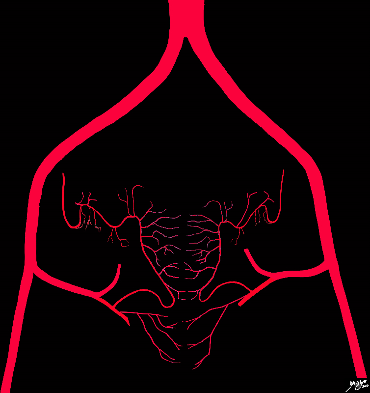
Blood Supply – Skeleton |
|
The blood supply to the uterus is from the uterine arteries which take their origin from the iliac system. The uterine artery collateralizes with the ovarian artery and there are also rich anastomoses between the left and right sides both anterior to and posterior to the uterus
Courtesy Ashley Davidoff MD Copyright 2010 All rights reserved 96269b35b01d02a.8s
|
|
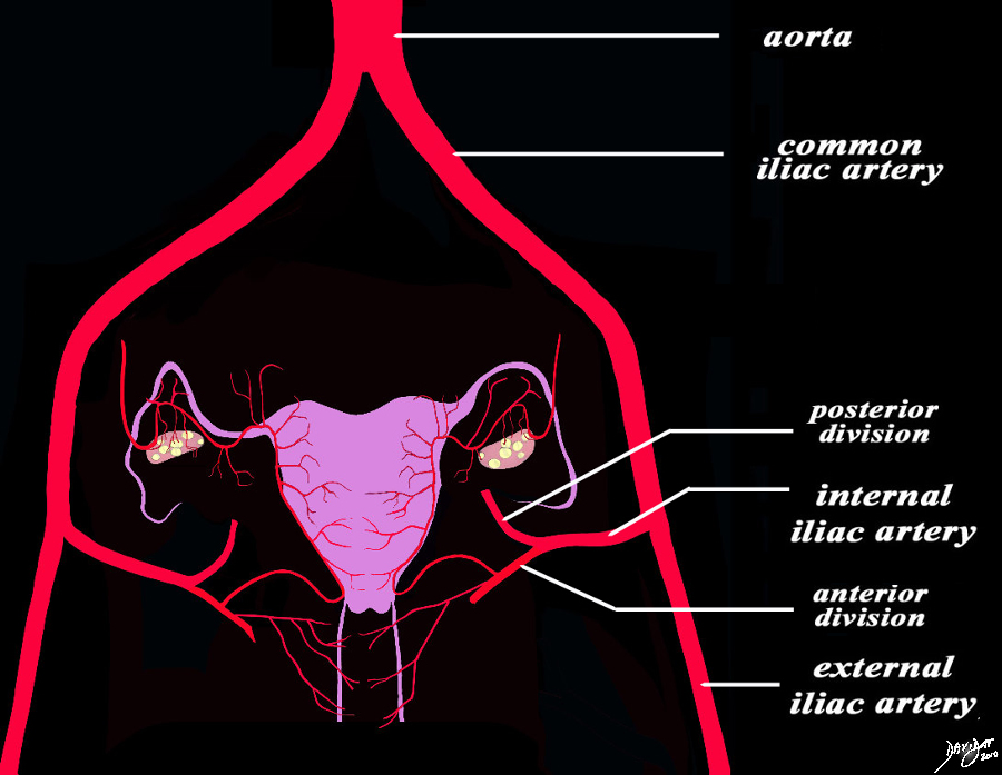
Inflow intto the Pelvis
The Iliac System |
|
The diagram of the blood supply to the uterus in its most basic form and shows the large inflow arteries into the pelvis. The main artery is the aorta which branches into the common iliac arteries which in turn branch into the external and internal iliac arteries. The internal iliac branches into an anterior division and a posterior division. The uterine and vaginal arteries arise from the posterior division.
Courtesy Ashley Davidoff MD Copyright 2010 All rights reserved 96269b35b01dL01.9s
|
|
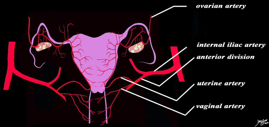
Blood Supply |
|
The diagram of the blood supply to the uterus in its most basic form shows that the uterine artery is a branch of the anterior branch of the internal iliac artery, and the uterine artery collateralizes and in fact is continuous with the ovarian artery which takes its origin directly from the abdominal aorta. The main branches include the uterine branches, vaginal branches and the ovarian branches
Courtesy Ashley Davidoff MD Copyright 2010 All rights reserved 96269b35b01dLb01L02.9s
|
|
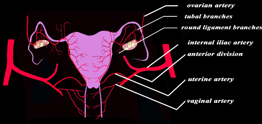
Smaller Branches and the Collateralization of the Vessels |
|
The diagram of the blood supply to the uterus displays the uterine artery with a few more of the smaller vessels including the branch to the Fallopian tube, round ligament, and the smaller branches to the vagina.
The uterine artery is a branch of the anterior branch of the internal iliac artery, and the uterine artery collateralizes and in fact is continuous with the ovarian artery which takes its origin directly from the abdominal aorta. The main branches include the uterine branches, vaginal branches and the ovarian branches The branches of the uterine and vaginal arteries spread anteriorly and posteriorly to the respective organs and result in extensive collateralization with the vessels from the contralateral side.
Courtesy Ashley Davidoff MD Copyright 2010 All rights reserved 96269b35b01dLb01L03.9s
|
|
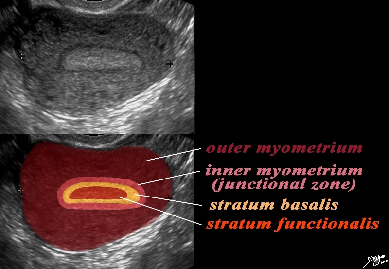
The Layers in the Premenstrual Uterus
Stratum Basalis and Stratum Functionalis are Not Distinguished by this US and appear as One LAyer |
|
In this 26 year premenstrual female a transvaginal ultrasound reveals a normal transverse view of the uterus with characteristic premenstrual appearance. The stripe is homogeneously echogenic and thick but also shows a hypoechoic halo of the junctional zone or inner myometrium. (salmon) The homogeneous stripe is made up from two histological layers (not distinguished by this ultrasound)? the inner stratum functionalis (deep orange) that will shed once the spiral arteries vasoconstrict, and the outer stratum basalis (deep yellow) that will not shed, and will be the basis for regenerating the endometrium in the next cycle. The next layer as stated above is the compact myometrium – the junctional zone (aka inner myometrium) , and is followed by the thicker outer myometrium (maroon).
Courtesy Ashley Davidoff MD Copyright 2010 All rights reserved 84540cc06.8s
|
|
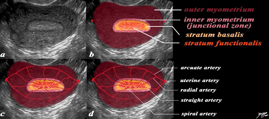
Spiral Arteries Staratum Functionalis and Menstruation |
|
In this 26 year premenstrual female a transvaginal ultrasound reveals a normal transverse view of the uterus with characteristic premenstrual appearance. (a) The stripe is homogeneously echogenic and thick but also shows a hypoechoic halo of the junctional zone or inner myometrium. (salmon) The homogeneous stripe is made up from two histological layers (not distinguished by this ultrasound)? the inner stratum functionalis (deep orange) that will shed once the spiral arteries vasoconstrict, and the outer stratum basalis (deep yellow) that will not shed, and will be the basis for regenerating the endometrium in the next cycle. The next layer as stated above is the compact myometrium – the junctional zone (aka inner myometrium) , and is followed by the thicker outer myometrium (maroon). (b) In the second series of images (c,d) the blood supply of the uterus is exemplified to demonstrate the branches that lead to the spiral arteries which undergo vasoconstriction at the time of the menses resulting in ischemia of the functionalis and subsequent shedding . The uterine arteries give rise to the arcuate arteries which in turn give rise to the radial arteries, straight arteries and finally the spiral arteries.
Courtesy Ashley Davidoff MD Copyright 2010 All rights reserved 84540.2kc07L.9s
|
|
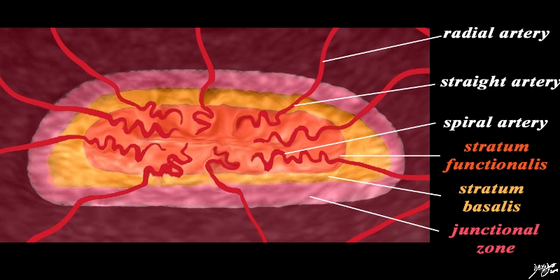
The Spiral Arteries and the Stratum Functionalis |
|
The magnifies view of the spiral arteries demonstrates its relationship to the stratum functionalis (deep orange). The uterine arteries give rise to the arcuate arteries which in turn give rise to the radial arteries, straight arteries and finally the spiral arteries. The spiral arteries undergo vasoconstriction at the time of the menses, resulting in ischemia of the stratum functionalis and subsequent shedding of this layer leaving stratum basalis intact.
Courtesy Ashley Davidoff MD Copyright 2010 All rights reserved 84540.33b02.8s
|
Histological Correlation
|
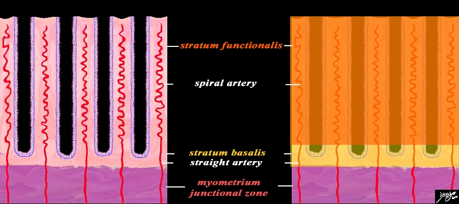
Functional Layer (Stratum Functionalis) Basal Layer (Stratum Basalis) and the Arteries
Premenstrual Phase |
|
A closer view of the endometrium as seen in the premenstrual phase exemplified by the characteristic spiral (helical) arteries running in the stroma (pink) between the simple tubular test tube shaped glands (purple) The spiral arteries supply the functional layer (stratum functionalis deep orange) and the straight arteries supply the basal layer (stratum basalis- light orange). Beneath the basal layer is the myometrium- Subendometrial smooth muscle or junctional zone (dark pink).
Courtesy Ashley Davidoff MD Copyright 2010 All rights reserved 32347f08cL.9s
|
|
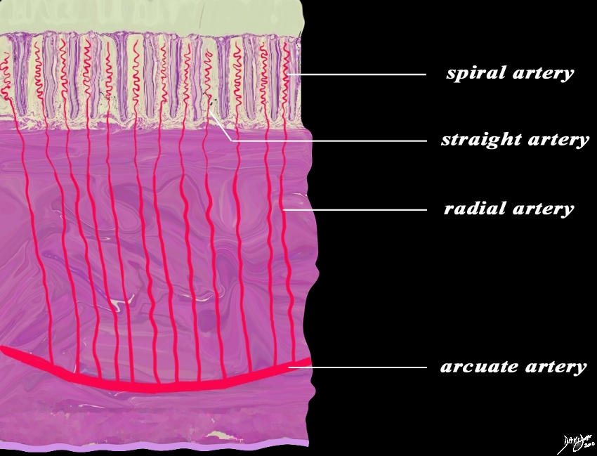
Distribution of the Arterial Supply Histologic Level |
|
This diagram exemplifies the position the arteries in the epithelial and muscular layers. The spiral arteries run in the stratum functionalis, the straight arteries run in the stratum basalis, while the radial arteries and the arcuate arteries run in the muscularis.
Courtesy Ashley Davidoff MD Copyright 2010 All rights reserved 12047e04b04L02.8s
|
Cyclical Changes of the Endometrium
The three major phases of the cycle that manifest in the endometrium are the proliferative (follicular), secretory (luteal), and menstrual phases.
|
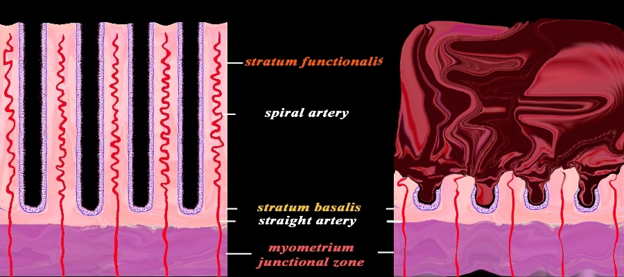
Premenstrual (left) and Active Menstruation (right) |
|
The diagram reflects the premenstrual endometrium (left) and the post menstrual endometrium right, revealing the necrosis of the functional layer with sloughing and hemorrhage. The basal layer with the staright arteries and a small portion of the spiral artery remains intact. The hemorrhage is controlled by spasm of the arteries and contraction of the myometrium.
Courtesy Ashley Davidoff MD Copyright 2010 All rights reserved 32347f08cL.92s
|
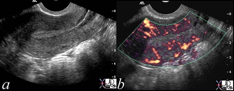
Uterine Perfusion |
| 46314c01 uterus endometrium blood flow myometrium perfusion normal anatomy USscan Davidoff MD |
The uterine artery runs along the lateral wall of the uterus, and then penetrates the myometrium and divides in general into an anterior and posterior branches. As they course toward th center they are called the arcuate arteries whose direction in general is circumferential around the the uterus. They give rise to the radial arteries which course almost at right angles toward the endometrium.
Branches from these arteries pass to the endometrium. As they enter the endometrium they run a straight course and supply the stratum basalis and as they enter the superficval layer of the stratume functionalis they become coiled and are called the spiral arteries. The straight arteries do not undergo cyclic changes, and hey supply stable unchanging blood supply to the stratum basalis which does not shed at the menses. The spiral arteries in the startum functionalis on the other hand are subject to significant change with hormonal fluxews. Just before menstruation the spiral arteries coil, and initially vasodialte and then there is an intense vasoconstriction causing ischemia and subsequent ischemia and destruction of the stratum functionalis which sheds as a result.
As they enter the endometrium they become the spiral arteries and their capillaries. The superficial layer of the endometrium (functionalis) is shed during menstruation. A basal layer, the basalis is permanent and it regenerates after each menstruation. At the onset of menstruation the spiral arteries coil, and initially vasodialte and then follows an intense vasoconstriction at the time of shedding.
References
Sokol Eric Clinical Anatomy of the Uterus, Fallopian Tubes, and Ovaries Glowm.com excellent review of the anatomy and the blood supply
O’Rahilly, Müller, Carpenter & Swenson Dartmouth Anatomy Text
SUNY Downstate Histology Course
DOMElement Object
(
    [schemaTypeInfo] =>
    [tagName] => table
    [firstElementChild] => (object value omitted)
    [lastElementChild] => (object value omitted)
    [childElementCount] => 1
    [previousElementSibling] => (object value omitted)
    [nextElementSibling] => (object value omitted)
    [nodeName] => table
    [nodeValue] =>


Uterine Perfusion


46314c01 uterus endometrium blood flow myometrium perfusion normal anatomy USscan Davidoff MD


    [nodeType] => 1
    [parentNode] => (object value omitted)
    [childNodes] => (object value omitted)
    [firstChild] => (object value omitted)
    [lastChild] => (object value omitted)
    [previousSibling] => (object value omitted)
    [nextSibling] => (object value omitted)
    [attributes] => (object value omitted)
    [ownerDocument] => (object value omitted)
    [namespaceURI] =>
    [prefix] =>
    [localName] => table
    [baseURI] =>
    [textContent] =>


Uterine Perfusion


46314c01 uterus endometrium blood flow myometrium perfusion normal anatomy USscan Davidoff MD


)
DOMElement Object
(
    [schemaTypeInfo] =>
    [tagName] => td
    [firstElementChild] => (object value omitted)
    [lastElementChild] => (object value omitted)
    [childElementCount] => 1
    [previousElementSibling] =>
    [nextElementSibling] =>
    [nodeName] => td
    [nodeValue] => 46314c01 uterus endometrium blood flow myometrium perfusion normal anatomy USscan Davidoff MD
    [nodeType] => 1
    [parentNode] => (object value omitted)
    [childNodes] => (object value omitted)
    [firstChild] => (object value omitted)
    [lastChild] => (object value omitted)
    [previousSibling] => (object value omitted)
    [nextSibling] => (object value omitted)
    [attributes] => (object value omitted)
    [ownerDocument] => (object value omitted)
    [namespaceURI] =>
    [prefix] =>
    [localName] => td
    [baseURI] =>
    [textContent] => 46314c01 uterus endometrium blood flow myometrium perfusion normal anatomy USscan Davidoff MD
)
DOMElement Object
(
    [schemaTypeInfo] =>
    [tagName] => td
    [firstElementChild] => (object value omitted)
    [lastElementChild] => (object value omitted)
    [childElementCount] => 2
    [previousElementSibling] =>
    [nextElementSibling] =>
    [nodeName] => td
    [nodeValue] =>
Uterine Perfusion
    [nodeType] => 1
    [parentNode] => (object value omitted)
    [childNodes] => (object value omitted)
    [firstChild] => (object value omitted)
    [lastChild] => (object value omitted)
    [previousSibling] => (object value omitted)
    [nextSibling] => (object value omitted)
    [attributes] => (object value omitted)
    [ownerDocument] => (object value omitted)
    [namespaceURI] =>
    [prefix] =>
    [localName] => td
    [baseURI] =>
    [textContent] =>
Uterine Perfusion
)
DOMElement Object
(
    [schemaTypeInfo] =>
    [tagName] => table
    [firstElementChild] => (object value omitted)
    [lastElementChild] => (object value omitted)
    [childElementCount] => 1
    [previousElementSibling] => (object value omitted)
    [nextElementSibling] => (object value omitted)
    [nodeName] => table
    [nodeValue] =>


Premenstrual (left) and Active Menstruation (right)


The diagram reflects the premenstrual endometrium (left) and the post menstrual endometrium right, revealing the necrosis of the functional layer with sloughing and hemorrhage. The basal layer with the staright arteries and a small portion of the spiral artery remains intact. The hemorrhage is controlled by spasm of the arteries and contraction of the myometrium.
Courtesy Ashley Davidoff MD Copyright 2010 All rights reserved 32347f08cL.92s


    [nodeType] => 1
    [parentNode] => (object value omitted)
    [childNodes] => (object value omitted)
    [firstChild] => (object value omitted)
    [lastChild] => (object value omitted)
    [previousSibling] => (object value omitted)
    [nextSibling] => (object value omitted)
    [attributes] => (object value omitted)
    [ownerDocument] => (object value omitted)
    [namespaceURI] =>
    [prefix] =>
    [localName] => table
    [baseURI] =>
    [textContent] =>


Premenstrual (left) and Active Menstruation (right)


The diagram reflects the premenstrual endometrium (left) and the post menstrual endometrium right, revealing the necrosis of the functional layer with sloughing and hemorrhage. The basal layer with the staright arteries and a small portion of the spiral artery remains intact. The hemorrhage is controlled by spasm of the arteries and contraction of the myometrium.
Courtesy Ashley Davidoff MD Copyright 2010 All rights reserved 32347f08cL.92s


)
DOMElement Object
(
    [schemaTypeInfo] =>
    [tagName] => td
    [firstElementChild] => (object value omitted)
    [lastElementChild] => (object value omitted)
    [childElementCount] => 2
    [previousElementSibling] =>
    [nextElementSibling] =>
    [nodeName] => td
    [nodeValue] =>
The diagram reflects the premenstrual endometrium (left) and the post menstrual endometrium right, revealing the necrosis of the functional layer with sloughing and hemorrhage. The basal layer with the staright arteries and a small portion of the spiral artery remains intact. The hemorrhage is controlled by spasm of the arteries and contraction of the myometrium.
Courtesy Ashley Davidoff MD Copyright 2010 All rights reserved 32347f08cL.92s

    [nodeType] => 1
    [parentNode] => (object value omitted)
    [childNodes] => (object value omitted)
    [firstChild] => (object value omitted)
    [lastChild] => (object value omitted)
    [previousSibling] => (object value omitted)
    [nextSibling] => (object value omitted)
    [attributes] => (object value omitted)
    [ownerDocument] => (object value omitted)
    [namespaceURI] =>
    [prefix] =>
    [localName] => td
    [baseURI] =>
    [textContent] =>
The diagram reflects the premenstrual endometrium (left) and the post menstrual endometrium right, revealing the necrosis of the functional layer with sloughing and hemorrhage. The basal layer with the staright arteries and a small portion of the spiral artery remains intact. The hemorrhage is controlled by spasm of the arteries and contraction of the myometrium.
Courtesy Ashley Davidoff MD Copyright 2010 All rights reserved 32347f08cL.92s

)
DOMElement Object
(
    [schemaTypeInfo] =>
    [tagName] => td
    [firstElementChild] => (object value omitted)
    [lastElementChild] => (object value omitted)
    [childElementCount] => 2
    [previousElementSibling] =>
    [nextElementSibling] =>
    [nodeName] => td
    [nodeValue] =>

Premenstrual (left) and Active Menstruation (right)
    [nodeType] => 1
    [parentNode] => (object value omitted)
    [childNodes] => (object value omitted)
    [firstChild] => (object value omitted)
    [lastChild] => (object value omitted)
    [previousSibling] => (object value omitted)
    [nextSibling] => (object value omitted)
    [attributes] => (object value omitted)
    [ownerDocument] => (object value omitted)
    [namespaceURI] =>
    [prefix] =>
    [localName] => td
    [baseURI] =>
    [textContent] =>

Premenstrual (left) and Active Menstruation (right)
)
DOMElement Object
(
    [schemaTypeInfo] =>
    [tagName] => table
    [firstElementChild] => (object value omitted)
    [lastElementChild] => (object value omitted)
    [childElementCount] => 1
    [previousElementSibling] => (object value omitted)
    [nextElementSibling] => (object value omitted)
    [nodeName] => table
    [nodeValue] =>


Distribution of the Arterial Supply Histologic Level


This diagram exemplifies the position the arteries in the epithelial and muscular layers. The spiral arteries run in the stratum functionalis, the straight arteries run in the stratum basalis, while the radial arteries and the arcuate arteries run in the muscularis.
Courtesy Ashley Davidoff MD Copyright 2010 All rights reserved 12047e04b04L02.8s


    [nodeType] => 1
    [parentNode] => (object value omitted)
    [childNodes] => (object value omitted)
    [firstChild] => (object value omitted)
    [lastChild] => (object value omitted)
    [previousSibling] => (object value omitted)
    [nextSibling] => (object value omitted)
    [attributes] => (object value omitted)
    [ownerDocument] => (object value omitted)
    [namespaceURI] =>
    [prefix] =>
    [localName] => table
    [baseURI] =>
    [textContent] =>


Distribution of the Arterial Supply Histologic Level


This diagram exemplifies the position the arteries in the epithelial and muscular layers. The spiral arteries run in the stratum functionalis, the straight arteries run in the stratum basalis, while the radial arteries and the arcuate arteries run in the muscularis.
Courtesy Ashley Davidoff MD Copyright 2010 All rights reserved 12047e04b04L02.8s


)
DOMElement Object
(
    [schemaTypeInfo] =>
    [tagName] => td
    [firstElementChild] => (object value omitted)
    [lastElementChild] => (object value omitted)
    [childElementCount] => 2
    [previousElementSibling] =>
    [nextElementSibling] =>
    [nodeName] => td
    [nodeValue] =>
This diagram exemplifies the position the arteries in the epithelial and muscular layers. The spiral arteries run in the stratum functionalis, the straight arteries run in the stratum basalis, while the radial arteries and the arcuate arteries run in the muscularis.
Courtesy Ashley Davidoff MD Copyright 2010 All rights reserved 12047e04b04L02.8s

    [nodeType] => 1
    [parentNode] => (object value omitted)
    [childNodes] => (object value omitted)
    [firstChild] => (object value omitted)
    [lastChild] => (object value omitted)
    [previousSibling] => (object value omitted)
    [nextSibling] => (object value omitted)
    [attributes] => (object value omitted)
    [ownerDocument] => (object value omitted)
    [namespaceURI] =>
    [prefix] =>
    [localName] => td
    [baseURI] =>
    [textContent] =>
This diagram exemplifies the position the arteries in the epithelial and muscular layers. The spiral arteries run in the stratum functionalis, the straight arteries run in the stratum basalis, while the radial arteries and the arcuate arteries run in the muscularis.
Courtesy Ashley Davidoff MD Copyright 2010 All rights reserved 12047e04b04L02.8s

)
DOMElement Object
(
    [schemaTypeInfo] =>
    [tagName] => td
    [firstElementChild] => (object value omitted)
    [lastElementChild] => (object value omitted)
    [childElementCount] => 2
    [previousElementSibling] =>
    [nextElementSibling] =>
    [nodeName] => td
    [nodeValue] =>

Distribution of the Arterial Supply Histologic Level
    [nodeType] => 1
    [parentNode] => (object value omitted)
    [childNodes] => (object value omitted)
    [firstChild] => (object value omitted)
    [lastChild] => (object value omitted)
    [previousSibling] => (object value omitted)
    [nextSibling] => (object value omitted)
    [attributes] => (object value omitted)
    [ownerDocument] => (object value omitted)
    [namespaceURI] =>
    [prefix] =>
    [localName] => td
    [baseURI] =>
    [textContent] =>

Distribution of the Arterial Supply Histologic Level
)
DOMElement Object
(
    [schemaTypeInfo] =>
    [tagName] => table
    [firstElementChild] => (object value omitted)
    [lastElementChild] => (object value omitted)
    [childElementCount] => 1
    [previousElementSibling] => (object value omitted)
    [nextElementSibling] => (object value omitted)
    [nodeName] => table
    [nodeValue] =>


Functional Layer (Stratum Functionalis) Basal Layer (Stratum Basalis) and the Arteries
Premenstrual Phase


 A closer view of the endometrium as seen in the premenstrual phase exemplified by the characteristic spiral (helical) arteries running in the stroma (pink) between the simple tubular test tube shaped glands (purple) The spiral arteries supply the functional layer (stratum functionalis deep orange) and the straight arteries supply the basal layer (stratum basalis- light orange). Beneath the basal layer is the myometrium- Subendometrial smooth muscle or junctional zone (dark pink).
Courtesy Ashley Davidoff MD Copyright 2010 All rights reserved 32347f08cL.9s


    [nodeType] => 1
    [parentNode] => (object value omitted)
    [childNodes] => (object value omitted)
    [firstChild] => (object value omitted)
    [lastChild] => (object value omitted)
    [previousSibling] => (object value omitted)
    [nextSibling] => (object value omitted)
    [attributes] => (object value omitted)
    [ownerDocument] => (object value omitted)
    [namespaceURI] =>
    [prefix] =>
    [localName] => table
    [baseURI] =>
    [textContent] =>


Functional Layer (Stratum Functionalis) Basal Layer (Stratum Basalis) and the Arteries
Premenstrual Phase


 A closer view of the endometrium as seen in the premenstrual phase exemplified by the characteristic spiral (helical) arteries running in the stroma (pink) between the simple tubular test tube shaped glands (purple) The spiral arteries supply the functional layer (stratum functionalis deep orange) and the straight arteries supply the basal layer (stratum basalis- light orange). Beneath the basal layer is the myometrium- Subendometrial smooth muscle or junctional zone (dark pink).
Courtesy Ashley Davidoff MD Copyright 2010 All rights reserved 32347f08cL.9s


)
DOMElement Object
(
    [schemaTypeInfo] =>
    [tagName] => td
    [firstElementChild] => (object value omitted)
    [lastElementChild] => (object value omitted)
    [childElementCount] => 2
    [previousElementSibling] =>
    [nextElementSibling] =>
    [nodeName] => td
    [nodeValue] =>
 A closer view of the endometrium as seen in the premenstrual phase exemplified by the characteristic spiral (helical) arteries running in the stroma (pink) between the simple tubular test tube shaped glands (purple) The spiral arteries supply the functional layer (stratum functionalis deep orange) and the straight arteries supply the basal layer (stratum basalis- light orange). Beneath the basal layer is the myometrium- Subendometrial smooth muscle or junctional zone (dark pink).
Courtesy Ashley Davidoff MD Copyright 2010 All rights reserved 32347f08cL.9s

    [nodeType] => 1
    [parentNode] => (object value omitted)
    [childNodes] => (object value omitted)
    [firstChild] => (object value omitted)
    [lastChild] => (object value omitted)
    [previousSibling] => (object value omitted)
    [nextSibling] => (object value omitted)
    [attributes] => (object value omitted)
    [ownerDocument] => (object value omitted)
    [namespaceURI] =>
    [prefix] =>
    [localName] => td
    [baseURI] =>
    [textContent] =>
 A closer view of the endometrium as seen in the premenstrual phase exemplified by the characteristic spiral (helical) arteries running in the stroma (pink) between the simple tubular test tube shaped glands (purple) The spiral arteries supply the functional layer (stratum functionalis deep orange) and the straight arteries supply the basal layer (stratum basalis- light orange). Beneath the basal layer is the myometrium- Subendometrial smooth muscle or junctional zone (dark pink).
Courtesy Ashley Davidoff MD Copyright 2010 All rights reserved 32347f08cL.9s

)
DOMElement Object
(
    [schemaTypeInfo] =>
    [tagName] => td
    [firstElementChild] => (object value omitted)
    [lastElementChild] => (object value omitted)
    [childElementCount] => 3
    [previousElementSibling] =>
    [nextElementSibling] =>
    [nodeName] => td
    [nodeValue] =>

Functional Layer (Stratum Functionalis) Basal Layer (Stratum Basalis) and the Arteries
Premenstrual Phase
    [nodeType] => 1
    [parentNode] => (object value omitted)
    [childNodes] => (object value omitted)
    [firstChild] => (object value omitted)
    [lastChild] => (object value omitted)
    [previousSibling] => (object value omitted)
    [nextSibling] => (object value omitted)
    [attributes] => (object value omitted)
    [ownerDocument] => (object value omitted)
    [namespaceURI] =>
    [prefix] =>
    [localName] => td
    [baseURI] =>
    [textContent] =>

Functional Layer (Stratum Functionalis) Basal Layer (Stratum Basalis) and the Arteries
Premenstrual Phase
)
DOMElement Object
(
    [schemaTypeInfo] =>
    [tagName] => table
    [firstElementChild] => (object value omitted)
    [lastElementChild] => (object value omitted)
    [childElementCount] => 1
    [previousElementSibling] => (object value omitted)
    [nextElementSibling] => (object value omitted)
    [nodeName] => table
    [nodeValue] =>


The Spiral Arteries and the Stratum Functionalis


The magnifies view of the spiral arteries demonstrates its relationship to the stratum functionalis (deep orange). The uterine arteries give rise to the arcuate arteries which in turn give rise to the radial arteries, straight arteries and finally the spiral arteries. The spiral arteries undergo vasoconstriction at the time of the menses, resulting in ischemia of the stratum functionalis and subsequent shedding of this layer leaving stratum basalis intact.
Courtesy Ashley Davidoff MD Copyright 2010 All rights reserved 84540.33b02.8s


    [nodeType] => 1
    [parentNode] => (object value omitted)
    [childNodes] => (object value omitted)
    [firstChild] => (object value omitted)
    [lastChild] => (object value omitted)
    [previousSibling] => (object value omitted)
    [nextSibling] => (object value omitted)
    [attributes] => (object value omitted)
    [ownerDocument] => (object value omitted)
    [namespaceURI] =>
    [prefix] =>
    [localName] => table
    [baseURI] =>
    [textContent] =>


The Spiral Arteries and the Stratum Functionalis


The magnifies view of the spiral arteries demonstrates its relationship to the stratum functionalis (deep orange). The uterine arteries give rise to the arcuate arteries which in turn give rise to the radial arteries, straight arteries and finally the spiral arteries. The spiral arteries undergo vasoconstriction at the time of the menses, resulting in ischemia of the stratum functionalis and subsequent shedding of this layer leaving stratum basalis intact.
Courtesy Ashley Davidoff MD Copyright 2010 All rights reserved 84540.33b02.8s


)
DOMElement Object
(
    [schemaTypeInfo] =>
    [tagName] => td
    [firstElementChild] => (object value omitted)
    [lastElementChild] => (object value omitted)
    [childElementCount] => 2
    [previousElementSibling] =>
    [nextElementSibling] =>
    [nodeName] => td
    [nodeValue] =>
The magnifies view of the spiral arteries demonstrates its relationship to the stratum functionalis (deep orange). The uterine arteries give rise to the arcuate arteries which in turn give rise to the radial arteries, straight arteries and finally the spiral arteries. The spiral arteries undergo vasoconstriction at the time of the menses, resulting in ischemia of the stratum functionalis and subsequent shedding of this layer leaving stratum basalis intact.
Courtesy Ashley Davidoff MD Copyright 2010 All rights reserved 84540.33b02.8s

    [nodeType] => 1
    [parentNode] => (object value omitted)
    [childNodes] => (object value omitted)
    [firstChild] => (object value omitted)
    [lastChild] => (object value omitted)
    [previousSibling] => (object value omitted)
    [nextSibling] => (object value omitted)
    [attributes] => (object value omitted)
    [ownerDocument] => (object value omitted)
    [namespaceURI] =>
    [prefix] =>
    [localName] => td
    [baseURI] =>
    [textContent] =>
The magnifies view of the spiral arteries demonstrates its relationship to the stratum functionalis (deep orange). The uterine arteries give rise to the arcuate arteries which in turn give rise to the radial arteries, straight arteries and finally the spiral arteries. The spiral arteries undergo vasoconstriction at the time of the menses, resulting in ischemia of the stratum functionalis and subsequent shedding of this layer leaving stratum basalis intact.
Courtesy Ashley Davidoff MD Copyright 2010 All rights reserved 84540.33b02.8s

)
DOMElement Object
(
    [schemaTypeInfo] =>
    [tagName] => td
    [firstElementChild] => (object value omitted)
    [lastElementChild] => (object value omitted)
    [childElementCount] => 2
    [previousElementSibling] =>
    [nextElementSibling] =>
    [nodeName] => td
    [nodeValue] =>

The Spiral Arteries and the Stratum Functionalis
    [nodeType] => 1
    [parentNode] => (object value omitted)
    [childNodes] => (object value omitted)
    [firstChild] => (object value omitted)
    [lastChild] => (object value omitted)
    [previousSibling] => (object value omitted)
    [nextSibling] => (object value omitted)
    [attributes] => (object value omitted)
    [ownerDocument] => (object value omitted)
    [namespaceURI] =>
    [prefix] =>
    [localName] => td
    [baseURI] =>
    [textContent] =>

The Spiral Arteries and the Stratum Functionalis
)
DOMElement Object
(
    [schemaTypeInfo] =>
    [tagName] => table
    [firstElementChild] => (object value omitted)
    [lastElementChild] => (object value omitted)
    [childElementCount] => 1
    [previousElementSibling] => (object value omitted)
    [nextElementSibling] => (object value omitted)
    [nodeName] => table
    [nodeValue] =>


Spiral Arteries Staratum Functionalis and Menstruation


In this 26 year premenstrual female a transvaginal ultrasound reveals a normal transverse view of the uterus with characteristic premenstrual appearance. (a) The stripe is homogeneously echogenic and thick but also shows a hypoechoic halo of the junctional zone or inner myometrium. (salmon) The homogeneous stripe is made up from two histological layers (not distinguished by this ultrasound)? the inner stratum functionalis (deep orange) that will shed once the spiral arteries vasoconstrict, and the outer stratum basalis (deep yellow) that will not shed, and will be the basis for regenerating the endometrium in the next cycle. The next layer as stated above is the compact myometrium – the junctional zone (aka inner myometrium) , and is followed by the thicker outer myometrium (maroon). (b) In the second series of images (c,d) the blood supply of the uterus is exemplified to demonstrate the branches that lead to the spiral arteries which undergo vasoconstriction at the time of the menses resulting in ischemia of the functionalis and subsequent shedding . The uterine arteries give rise to the arcuate arteries which in turn give rise to the radial arteries, straight arteries and finally the spiral arteries.
Courtesy Ashley Davidoff MD Copyright 2010 All rights reserved 84540.2kc07L.9s


    [nodeType] => 1
    [parentNode] => (object value omitted)
    [childNodes] => (object value omitted)
    [firstChild] => (object value omitted)
    [lastChild] => (object value omitted)
    [previousSibling] => (object value omitted)
    [nextSibling] => (object value omitted)
    [attributes] => (object value omitted)
    [ownerDocument] => (object value omitted)
    [namespaceURI] =>
    [prefix] =>
    [localName] => table
    [baseURI] =>
    [textContent] =>


Spiral Arteries Staratum Functionalis and Menstruation


In this 26 year premenstrual female a transvaginal ultrasound reveals a normal transverse view of the uterus with characteristic premenstrual appearance. (a) The stripe is homogeneously echogenic and thick but also shows a hypoechoic halo of the junctional zone or inner myometrium. (salmon) The homogeneous stripe is made up from two histological layers (not distinguished by this ultrasound)? the inner stratum functionalis (deep orange) that will shed once the spiral arteries vasoconstrict, and the outer stratum basalis (deep yellow) that will not shed, and will be the basis for regenerating the endometrium in the next cycle. The next layer as stated above is the compact myometrium – the junctional zone (aka inner myometrium) , and is followed by the thicker outer myometrium (maroon). (b) In the second series of images (c,d) the blood supply of the uterus is exemplified to demonstrate the branches that lead to the spiral arteries which undergo vasoconstriction at the time of the menses resulting in ischemia of the functionalis and subsequent shedding . The uterine arteries give rise to the arcuate arteries which in turn give rise to the radial arteries, straight arteries and finally the spiral arteries.
Courtesy Ashley Davidoff MD Copyright 2010 All rights reserved 84540.2kc07L.9s


)
DOMElement Object
(
    [schemaTypeInfo] =>
    [tagName] => td
    [firstElementChild] => (object value omitted)
    [lastElementChild] => (object value omitted)
    [childElementCount] => 2
    [previousElementSibling] =>
    [nextElementSibling] =>
    [nodeName] => td
    [nodeValue] =>
In this 26 year premenstrual female a transvaginal ultrasound reveals a normal transverse view of the uterus with characteristic premenstrual appearance. (a) The stripe is homogeneously echogenic and thick but also shows a hypoechoic halo of the junctional zone or inner myometrium. (salmon) The homogeneous stripe is made up from two histological layers (not distinguished by this ultrasound)? the inner stratum functionalis (deep orange) that will shed once the spiral arteries vasoconstrict, and the outer stratum basalis (deep yellow) that will not shed, and will be the basis for regenerating the endometrium in the next cycle. The next layer as stated above is the compact myometrium – the junctional zone (aka inner myometrium) , and is followed by the thicker outer myometrium (maroon). (b) In the second series of images (c,d) the blood supply of the uterus is exemplified to demonstrate the branches that lead to the spiral arteries which undergo vasoconstriction at the time of the menses resulting in ischemia of the functionalis and subsequent shedding . The uterine arteries give rise to the arcuate arteries which in turn give rise to the radial arteries, straight arteries and finally the spiral arteries.
Courtesy Ashley Davidoff MD Copyright 2010 All rights reserved 84540.2kc07L.9s

    [nodeType] => 1
    [parentNode] => (object value omitted)
    [childNodes] => (object value omitted)
    [firstChild] => (object value omitted)
    [lastChild] => (object value omitted)
    [previousSibling] => (object value omitted)
    [nextSibling] => (object value omitted)
    [attributes] => (object value omitted)
    [ownerDocument] => (object value omitted)
    [namespaceURI] =>
    [prefix] =>
    [localName] => td
    [baseURI] =>
    [textContent] =>
In this 26 year premenstrual female a transvaginal ultrasound reveals a normal transverse view of the uterus with characteristic premenstrual appearance. (a) The stripe is homogeneously echogenic and thick but also shows a hypoechoic halo of the junctional zone or inner myometrium. (salmon) The homogeneous stripe is made up from two histological layers (not distinguished by this ultrasound)? the inner stratum functionalis (deep orange) that will shed once the spiral arteries vasoconstrict, and the outer stratum basalis (deep yellow) that will not shed, and will be the basis for regenerating the endometrium in the next cycle. The next layer as stated above is the compact myometrium – the junctional zone (aka inner myometrium) , and is followed by the thicker outer myometrium (maroon). (b) In the second series of images (c,d) the blood supply of the uterus is exemplified to demonstrate the branches that lead to the spiral arteries which undergo vasoconstriction at the time of the menses resulting in ischemia of the functionalis and subsequent shedding . The uterine arteries give rise to the arcuate arteries which in turn give rise to the radial arteries, straight arteries and finally the spiral arteries.
Courtesy Ashley Davidoff MD Copyright 2010 All rights reserved 84540.2kc07L.9s

)
DOMElement Object
(
    [schemaTypeInfo] =>
    [tagName] => td
    [firstElementChild] => (object value omitted)
    [lastElementChild] => (object value omitted)
    [childElementCount] => 2
    [previousElementSibling] =>
    [nextElementSibling] =>
    [nodeName] => td
    [nodeValue] =>

Spiral Arteries Staratum Functionalis and Menstruation
    [nodeType] => 1
    [parentNode] => (object value omitted)
    [childNodes] => (object value omitted)
    [firstChild] => (object value omitted)
    [lastChild] => (object value omitted)
    [previousSibling] => (object value omitted)
    [nextSibling] => (object value omitted)
    [attributes] => (object value omitted)
    [ownerDocument] => (object value omitted)
    [namespaceURI] =>
    [prefix] =>
    [localName] => td
    [baseURI] =>
    [textContent] =>

Spiral Arteries Staratum Functionalis and Menstruation
)
DOMElement Object
(
    [schemaTypeInfo] =>
    [tagName] => table
    [firstElementChild] => (object value omitted)
    [lastElementChild] => (object value omitted)
    [childElementCount] => 1
    [previousElementSibling] => (object value omitted)
    [nextElementSibling] => (object value omitted)
    [nodeName] => table
    [nodeValue] =>


The Layers in the Premenstrual Uterus
Stratum Basalis and Stratum Functionalis are Not Distinguished by this US and appear as One LAyer


In this 26 year premenstrual female a transvaginal ultrasound reveals a normal transverse view of the uterus with characteristic premenstrual appearance. The stripe is homogeneously echogenic and thick but also shows a hypoechoic halo of the junctional zone or inner myometrium. (salmon) The homogeneous stripe is made up from two histological layers (not distinguished by this ultrasound)? the inner stratum functionalis (deep orange) that will shed once the spiral arteries vasoconstrict, and the outer stratum basalis (deep yellow) that will not shed, and will be the basis for regenerating the endometrium in the next cycle. The next layer as stated above is the compact myometrium – the junctional zone (aka inner myometrium) , and is followed by the thicker outer myometrium (maroon).
Courtesy Ashley Davidoff MD Copyright 2010 All rights reserved  84540cc06.8s


    [nodeType] => 1
    [parentNode] => (object value omitted)
    [childNodes] => (object value omitted)
    [firstChild] => (object value omitted)
    [lastChild] => (object value omitted)
    [previousSibling] => (object value omitted)
    [nextSibling] => (object value omitted)
    [attributes] => (object value omitted)
    [ownerDocument] => (object value omitted)
    [namespaceURI] =>
    [prefix] =>
    [localName] => table
    [baseURI] =>
    [textContent] =>


The Layers in the Premenstrual Uterus
Stratum Basalis and Stratum Functionalis are Not Distinguished by this US and appear as One LAyer


In this 26 year premenstrual female a transvaginal ultrasound reveals a normal transverse view of the uterus with characteristic premenstrual appearance. The stripe is homogeneously echogenic and thick but also shows a hypoechoic halo of the junctional zone or inner myometrium. (salmon) The homogeneous stripe is made up from two histological layers (not distinguished by this ultrasound)? the inner stratum functionalis (deep orange) that will shed once the spiral arteries vasoconstrict, and the outer stratum basalis (deep yellow) that will not shed, and will be the basis for regenerating the endometrium in the next cycle. The next layer as stated above is the compact myometrium – the junctional zone (aka inner myometrium) , and is followed by the thicker outer myometrium (maroon).
Courtesy Ashley Davidoff MD Copyright 2010 All rights reserved  84540cc06.8s


)
DOMElement Object
(
    [schemaTypeInfo] =>
    [tagName] => td
    [firstElementChild] => (object value omitted)
    [lastElementChild] => (object value omitted)
    [childElementCount] => 2
    [previousElementSibling] =>
    [nextElementSibling] =>
    [nodeName] => td
    [nodeValue] =>
In this 26 year premenstrual female a transvaginal ultrasound reveals a normal transverse view of the uterus with characteristic premenstrual appearance. The stripe is homogeneously echogenic and thick but also shows a hypoechoic halo of the junctional zone or inner myometrium. (salmon) The homogeneous stripe is made up from two histological layers (not distinguished by this ultrasound)? the inner stratum functionalis (deep orange) that will shed once the spiral arteries vasoconstrict, and the outer stratum basalis (deep yellow) that will not shed, and will be the basis for regenerating the endometrium in the next cycle. The next layer as stated above is the compact myometrium – the junctional zone (aka inner myometrium) , and is followed by the thicker outer myometrium (maroon).
Courtesy Ashley Davidoff MD Copyright 2010 All rights reserved  84540cc06.8s

    [nodeType] => 1
    [parentNode] => (object value omitted)
    [childNodes] => (object value omitted)
    [firstChild] => (object value omitted)
    [lastChild] => (object value omitted)
    [previousSibling] => (object value omitted)
    [nextSibling] => (object value omitted)
    [attributes] => (object value omitted)
    [ownerDocument] => (object value omitted)
    [namespaceURI] =>
    [prefix] =>
    [localName] => td
    [baseURI] =>
    [textContent] =>
In this 26 year premenstrual female a transvaginal ultrasound reveals a normal transverse view of the uterus with characteristic premenstrual appearance. The stripe is homogeneously echogenic and thick but also shows a hypoechoic halo of the junctional zone or inner myometrium. (salmon) The homogeneous stripe is made up from two histological layers (not distinguished by this ultrasound)? the inner stratum functionalis (deep orange) that will shed once the spiral arteries vasoconstrict, and the outer stratum basalis (deep yellow) that will not shed, and will be the basis for regenerating the endometrium in the next cycle. The next layer as stated above is the compact myometrium – the junctional zone (aka inner myometrium) , and is followed by the thicker outer myometrium (maroon).
Courtesy Ashley Davidoff MD Copyright 2010 All rights reserved  84540cc06.8s

)
DOMElement Object
(
    [schemaTypeInfo] =>
    [tagName] => td
    [firstElementChild] => (object value omitted)
    [lastElementChild] => (object value omitted)
    [childElementCount] => 3
    [previousElementSibling] =>
    [nextElementSibling] =>
    [nodeName] => td
    [nodeValue] =>

The Layers in the Premenstrual Uterus
Stratum Basalis and Stratum Functionalis are Not Distinguished by this US and appear as One LAyer
    [nodeType] => 1
    [parentNode] => (object value omitted)
    [childNodes] => (object value omitted)
    [firstChild] => (object value omitted)
    [lastChild] => (object value omitted)
    [previousSibling] => (object value omitted)
    [nextSibling] => (object value omitted)
    [attributes] => (object value omitted)
    [ownerDocument] => (object value omitted)
    [namespaceURI] =>
    [prefix] =>
    [localName] => td
    [baseURI] =>
    [textContent] =>

The Layers in the Premenstrual Uterus
Stratum Basalis and Stratum Functionalis are Not Distinguished by this US and appear as One LAyer
)
DOMElement Object
(
    [schemaTypeInfo] =>
    [tagName] => table
    [firstElementChild] => (object value omitted)
    [lastElementChild] => (object value omitted)
    [childElementCount] => 1
    [previousElementSibling] => (object value omitted)
    [nextElementSibling] => (object value omitted)
    [nodeName] => table
    [nodeValue] =>


Smaller Branches and the Collateralization of the Vessels


The diagram of the blood supply to the uterus displays the uterine artery with a few more of the smaller vessels including the branch to the Fallopian tube, round ligament, and the smaller branches to the vagina.
The uterine artery is a branch of the anterior branch of the internal iliac artery, and the uterine artery collateralizes and in fact is continuous with the ovarian artery which takes its origin directly from the abdominal aorta. The main branches include the uterine branches, vaginal branches and the ovarian branches The branches of the uterine and vaginal arteries spread anteriorly and posteriorly to the respective organs and result in extensive collateralization with the vessels from the contralateral side.
Courtesy Ashley Davidoff MD Copyright 2010 All rights reserved 96269b35b01dLb01L03.9s


    [nodeType] => 1
    [parentNode] => (object value omitted)
    [childNodes] => (object value omitted)
    [firstChild] => (object value omitted)
    [lastChild] => (object value omitted)
    [previousSibling] => (object value omitted)
    [nextSibling] => (object value omitted)
    [attributes] => (object value omitted)
    [ownerDocument] => (object value omitted)
    [namespaceURI] =>
    [prefix] =>
    [localName] => table
    [baseURI] =>
    [textContent] =>


Smaller Branches and the Collateralization of the Vessels


The diagram of the blood supply to the uterus displays the uterine artery with a few more of the smaller vessels including the branch to the Fallopian tube, round ligament, and the smaller branches to the vagina.
The uterine artery is a branch of the anterior branch of the internal iliac artery, and the uterine artery collateralizes and in fact is continuous with the ovarian artery which takes its origin directly from the abdominal aorta. The main branches include the uterine branches, vaginal branches and the ovarian branches The branches of the uterine and vaginal arteries spread anteriorly and posteriorly to the respective organs and result in extensive collateralization with the vessels from the contralateral side.
Courtesy Ashley Davidoff MD Copyright 2010 All rights reserved 96269b35b01dLb01L03.9s


)
DOMElement Object
(
    [schemaTypeInfo] =>
    [tagName] => td
    [firstElementChild] => (object value omitted)
    [lastElementChild] => (object value omitted)
    [childElementCount] => 3
    [previousElementSibling] =>
    [nextElementSibling] =>
    [nodeName] => td
    [nodeValue] =>
The diagram of the blood supply to the uterus displays the uterine artery with a few more of the smaller vessels including the branch to the Fallopian tube, round ligament, and the smaller branches to the vagina.
The uterine artery is a branch of the anterior branch of the internal iliac artery, and the uterine artery collateralizes and in fact is continuous with the ovarian artery which takes its origin directly from the abdominal aorta. The main branches include the uterine branches, vaginal branches and the ovarian branches The branches of the uterine and vaginal arteries spread anteriorly and posteriorly to the respective organs and result in extensive collateralization with the vessels from the contralateral side.
Courtesy Ashley Davidoff MD Copyright 2010 All rights reserved 96269b35b01dLb01L03.9s

    [nodeType] => 1
    [parentNode] => (object value omitted)
    [childNodes] => (object value omitted)
    [firstChild] => (object value omitted)
    [lastChild] => (object value omitted)
    [previousSibling] => (object value omitted)
    [nextSibling] => (object value omitted)
    [attributes] => (object value omitted)
    [ownerDocument] => (object value omitted)
    [namespaceURI] =>
    [prefix] =>
    [localName] => td
    [baseURI] =>
    [textContent] =>
The diagram of the blood supply to the uterus displays the uterine artery with a few more of the smaller vessels including the branch to the Fallopian tube, round ligament, and the smaller branches to the vagina.
The uterine artery is a branch of the anterior branch of the internal iliac artery, and the uterine artery collateralizes and in fact is continuous with the ovarian artery which takes its origin directly from the abdominal aorta. The main branches include the uterine branches, vaginal branches and the ovarian branches The branches of the uterine and vaginal arteries spread anteriorly and posteriorly to the respective organs and result in extensive collateralization with the vessels from the contralateral side.
Courtesy Ashley Davidoff MD Copyright 2010 All rights reserved 96269b35b01dLb01L03.9s

)
DOMElement Object
(
    [schemaTypeInfo] =>
    [tagName] => td
    [firstElementChild] => (object value omitted)
    [lastElementChild] => (object value omitted)
    [childElementCount] => 3
    [previousElementSibling] =>
    [nextElementSibling] =>
    [nodeName] => td
    [nodeValue] =>


Smaller Branches and the Collateralization of the Vessels
    [nodeType] => 1
    [parentNode] => (object value omitted)
    [childNodes] => (object value omitted)
    [firstChild] => (object value omitted)
    [lastChild] => (object value omitted)
    [previousSibling] => (object value omitted)
    [nextSibling] => (object value omitted)
    [attributes] => (object value omitted)
    [ownerDocument] => (object value omitted)
    [namespaceURI] =>
    [prefix] =>
    [localName] => td
    [baseURI] =>
    [textContent] =>


Smaller Branches and the Collateralization of the Vessels
)
DOMElement Object
(
    [schemaTypeInfo] =>
    [tagName] => table
    [firstElementChild] => (object value omitted)
    [lastElementChild] => (object value omitted)
    [childElementCount] => 1
    [previousElementSibling] => (object value omitted)
    [nextElementSibling] => (object value omitted)
    [nodeName] => table
    [nodeValue] =>


Blood Supply


The diagram of the blood supply to the uterus in its most basic form shows that the uterine artery is a branch of the anterior branch of the internal iliac artery, and the uterine artery collateralizes and in fact is continuous with the ovarian artery which takes its origin directly from the abdominal aorta. The main branches include the uterine branches, vaginal branches and the ovarian branches
Courtesy Ashley Davidoff MD Copyright 2010 All rights reserved  96269b35b01dLb01L02.9s


    [nodeType] => 1
    [parentNode] => (object value omitted)
    [childNodes] => (object value omitted)
    [firstChild] => (object value omitted)
    [lastChild] => (object value omitted)
    [previousSibling] => (object value omitted)
    [nextSibling] => (object value omitted)
    [attributes] => (object value omitted)
    [ownerDocument] => (object value omitted)
    [namespaceURI] =>
    [prefix] =>
    [localName] => table
    [baseURI] =>
    [textContent] =>


Blood Supply


The diagram of the blood supply to the uterus in its most basic form shows that the uterine artery is a branch of the anterior branch of the internal iliac artery, and the uterine artery collateralizes and in fact is continuous with the ovarian artery which takes its origin directly from the abdominal aorta. The main branches include the uterine branches, vaginal branches and the ovarian branches
Courtesy Ashley Davidoff MD Copyright 2010 All rights reserved  96269b35b01dLb01L02.9s


)
DOMElement Object
(
    [schemaTypeInfo] =>
    [tagName] => td
    [firstElementChild] => (object value omitted)
    [lastElementChild] => (object value omitted)
    [childElementCount] => 2
    [previousElementSibling] =>
    [nextElementSibling] =>
    [nodeName] => td
    [nodeValue] =>
The diagram of the blood supply to the uterus in its most basic form shows that the uterine artery is a branch of the anterior branch of the internal iliac artery, and the uterine artery collateralizes and in fact is continuous with the ovarian artery which takes its origin directly from the abdominal aorta. The main branches include the uterine branches, vaginal branches and the ovarian branches
Courtesy Ashley Davidoff MD Copyright 2010 All rights reserved  96269b35b01dLb01L02.9s

    [nodeType] => 1
    [parentNode] => (object value omitted)
    [childNodes] => (object value omitted)
    [firstChild] => (object value omitted)
    [lastChild] => (object value omitted)
    [previousSibling] => (object value omitted)
    [nextSibling] => (object value omitted)
    [attributes] => (object value omitted)
    [ownerDocument] => (object value omitted)
    [namespaceURI] =>
    [prefix] =>
    [localName] => td
    [baseURI] =>
    [textContent] =>
The diagram of the blood supply to the uterus in its most basic form shows that the uterine artery is a branch of the anterior branch of the internal iliac artery, and the uterine artery collateralizes and in fact is continuous with the ovarian artery which takes its origin directly from the abdominal aorta. The main branches include the uterine branches, vaginal branches and the ovarian branches
Courtesy Ashley Davidoff MD Copyright 2010 All rights reserved  96269b35b01dLb01L02.9s

)
DOMElement Object
(
    [schemaTypeInfo] =>
    [tagName] => td
    [firstElementChild] => (object value omitted)
    [lastElementChild] => (object value omitted)
    [childElementCount] => 3
    [previousElementSibling] =>
    [nextElementSibling] =>
    [nodeName] => td
    [nodeValue] =>


Blood Supply
    [nodeType] => 1
    [parentNode] => (object value omitted)
    [childNodes] => (object value omitted)
    [firstChild] => (object value omitted)
    [lastChild] => (object value omitted)
    [previousSibling] => (object value omitted)
    [nextSibling] => (object value omitted)
    [attributes] => (object value omitted)
    [ownerDocument] => (object value omitted)
    [namespaceURI] =>
    [prefix] =>
    [localName] => td
    [baseURI] =>
    [textContent] =>


Blood Supply
)
DOMElement Object
(
    [schemaTypeInfo] =>
    [tagName] => table
    [firstElementChild] => (object value omitted)
    [lastElementChild] => (object value omitted)
    [childElementCount] => 1
    [previousElementSibling] => (object value omitted)
    [nextElementSibling] => (object value omitted)
    [nodeName] => table
    [nodeValue] =>


Inflow intto the Pelvis
The Iliac System


The diagram of the blood supply to the uterus in its most basic form and shows the large inflow arteries into the pelvis. The main artery is the aorta which branches into the common iliac arteries which in turn branch into the external and internal iliac arteries. The internal iliac branches into an anterior division and a posterior division. The uterine and vaginal arteries arise from the posterior division.
Courtesy Ashley Davidoff MD Copyright 2010 All rights reserved  96269b35b01dL01.9s


    [nodeType] => 1
    [parentNode] => (object value omitted)
    [childNodes] => (object value omitted)
    [firstChild] => (object value omitted)
    [lastChild] => (object value omitted)
    [previousSibling] => (object value omitted)
    [nextSibling] => (object value omitted)
    [attributes] => (object value omitted)
    [ownerDocument] => (object value omitted)
    [namespaceURI] =>
    [prefix] =>
    [localName] => table
    [baseURI] =>
    [textContent] =>


Inflow intto the Pelvis
The Iliac System


The diagram of the blood supply to the uterus in its most basic form and shows the large inflow arteries into the pelvis. The main artery is the aorta which branches into the common iliac arteries which in turn branch into the external and internal iliac arteries. The internal iliac branches into an anterior division and a posterior division. The uterine and vaginal arteries arise from the posterior division.
Courtesy Ashley Davidoff MD Copyright 2010 All rights reserved  96269b35b01dL01.9s


)
DOMElement Object
(
    [schemaTypeInfo] =>
    [tagName] => td
    [firstElementChild] => (object value omitted)
    [lastElementChild] => (object value omitted)
    [childElementCount] => 2
    [previousElementSibling] =>
    [nextElementSibling] =>
    [nodeName] => td
    [nodeValue] =>
The diagram of the blood supply to the uterus in its most basic form and shows the large inflow arteries into the pelvis. The main artery is the aorta which branches into the common iliac arteries which in turn branch into the external and internal iliac arteries. The internal iliac branches into an anterior division and a posterior division. The uterine and vaginal arteries arise from the posterior division.
Courtesy Ashley Davidoff MD Copyright 2010 All rights reserved  96269b35b01dL01.9s

    [nodeType] => 1
    [parentNode] => (object value omitted)
    [childNodes] => (object value omitted)
    [firstChild] => (object value omitted)
    [lastChild] => (object value omitted)
    [previousSibling] => (object value omitted)
    [nextSibling] => (object value omitted)
    [attributes] => (object value omitted)
    [ownerDocument] => (object value omitted)
    [namespaceURI] =>
    [prefix] =>
    [localName] => td
    [baseURI] =>
    [textContent] =>
The diagram of the blood supply to the uterus in its most basic form and shows the large inflow arteries into the pelvis. The main artery is the aorta which branches into the common iliac arteries which in turn branch into the external and internal iliac arteries. The internal iliac branches into an anterior division and a posterior division. The uterine and vaginal arteries arise from the posterior division.
Courtesy Ashley Davidoff MD Copyright 2010 All rights reserved  96269b35b01dL01.9s

)
DOMElement Object
(
    [schemaTypeInfo] =>
    [tagName] => td
    [firstElementChild] => (object value omitted)
    [lastElementChild] => (object value omitted)
    [childElementCount] => 3
    [previousElementSibling] =>
    [nextElementSibling] =>
    [nodeName] => td
    [nodeValue] =>

Inflow intto the Pelvis
The Iliac System
    [nodeType] => 1
    [parentNode] => (object value omitted)
    [childNodes] => (object value omitted)
    [firstChild] => (object value omitted)
    [lastChild] => (object value omitted)
    [previousSibling] => (object value omitted)
    [nextSibling] => (object value omitted)
    [attributes] => (object value omitted)
    [ownerDocument] => (object value omitted)
    [namespaceURI] =>
    [prefix] =>
    [localName] => td
    [baseURI] =>
    [textContent] =>

Inflow intto the Pelvis
The Iliac System
)
DOMElement Object
(
    [schemaTypeInfo] =>
    [tagName] => table
    [firstElementChild] => (object value omitted)
    [lastElementChild] => (object value omitted)
    [childElementCount] => 1
    [previousElementSibling] => (object value omitted)
    [nextElementSibling] => (object value omitted)
    [nodeName] => table
    [nodeValue] =>


Blood Supply – Skeleton


The blood supply to the uterus is from the uterine arteries which take their origin from the iliac system. The uterine artery collateralizes with the ovarian artery and there are also rich anastomoses between the left and right sides both anterior to and posterior to the uterus
Courtesy Ashley Davidoff MD Copyright 2010 All rights reserved 96269b35b01d02a.8s


    [nodeType] => 1
    [parentNode] => (object value omitted)
    [childNodes] => (object value omitted)
    [firstChild] => (object value omitted)
    [lastChild] => (object value omitted)
    [previousSibling] => (object value omitted)
    [nextSibling] => (object value omitted)
    [attributes] => (object value omitted)
    [ownerDocument] => (object value omitted)
    [namespaceURI] =>
    [prefix] =>
    [localName] => table
    [baseURI] =>
    [textContent] =>


Blood Supply – Skeleton


The blood supply to the uterus is from the uterine arteries which take their origin from the iliac system. The uterine artery collateralizes with the ovarian artery and there are also rich anastomoses between the left and right sides both anterior to and posterior to the uterus
Courtesy Ashley Davidoff MD Copyright 2010 All rights reserved 96269b35b01d02a.8s


)
DOMElement Object
(
    [schemaTypeInfo] =>
    [tagName] => td
    [firstElementChild] => (object value omitted)
    [lastElementChild] => (object value omitted)
    [childElementCount] => 2
    [previousElementSibling] =>
    [nextElementSibling] =>
    [nodeName] => td
    [nodeValue] =>
The blood supply to the uterus is from the uterine arteries which take their origin from the iliac system. The uterine artery collateralizes with the ovarian artery and there are also rich anastomoses between the left and right sides both anterior to and posterior to the uterus
Courtesy Ashley Davidoff MD Copyright 2010 All rights reserved 96269b35b01d02a.8s

    [nodeType] => 1
    [parentNode] => (object value omitted)
    [childNodes] => (object value omitted)
    [firstChild] => (object value omitted)
    [lastChild] => (object value omitted)
    [previousSibling] => (object value omitted)
    [nextSibling] => (object value omitted)
    [attributes] => (object value omitted)
    [ownerDocument] => (object value omitted)
    [namespaceURI] =>
    [prefix] =>
    [localName] => td
    [baseURI] =>
    [textContent] =>
The blood supply to the uterus is from the uterine arteries which take their origin from the iliac system. The uterine artery collateralizes with the ovarian artery and there are also rich anastomoses between the left and right sides both anterior to and posterior to the uterus
Courtesy Ashley Davidoff MD Copyright 2010 All rights reserved 96269b35b01d02a.8s

)
DOMElement Object
(
    [schemaTypeInfo] =>
    [tagName] => td
    [firstElementChild] => (object value omitted)
    [lastElementChild] => (object value omitted)
    [childElementCount] => 2
    [previousElementSibling] =>
    [nextElementSibling] =>
    [nodeName] => td
    [nodeValue] =>
Blood Supply – Skeleton
    [nodeType] => 1
    [parentNode] => (object value omitted)
    [childNodes] => (object value omitted)
    [firstChild] => (object value omitted)
    [lastChild] => (object value omitted)
    [previousSibling] => (object value omitted)
    [nextSibling] => (object value omitted)
    [attributes] => (object value omitted)
    [ownerDocument] => (object value omitted)
    [namespaceURI] =>
    [prefix] =>
    [localName] => td
    [baseURI] =>
    [textContent] =>
Blood Supply – Skeleton
)


