Time Zero + 1
The Common Vein Copyright 2011
Ashley Davidoff MD
Introduction
After tthe initial impact there is a ripple down effect on all the structures that are connected or lie in close contact to the fractures bone.
The presence of ligaments and muscles attached to the bones will also affect the way the force impacts on the bones. For example the tibia and fibula are attached to each other by the interosseus membrane and other ligaments, while the tibia and fibula both have ligamentous attachments to the talus and calcaneus. A bending force on the tibia will be transmitted to the mortise and the fibula and talus inferiorly and may even be transmitted to the proximal fibula. As a result a single excessive force may be transmitted via ligamentous and muscular attachments to any number of bones and a combination of fractures may result.
More Complex Issues
The Effects of Ligamnets and Muscle
|
|
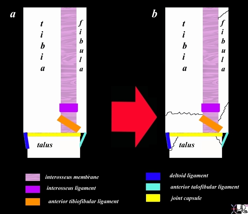
The diagram (a) shows the major anchoring mechanisms that bind the tibia, fibula and talus including the interosseous membrane (light pink), the interosseus ligament (purple) anterior tibiofibular ligament (orange) the deltoid ligament (royal blue) anterior talofibular ligament (teal blue). With a sudden force (red arrow b) on the medial malleolus for example, any number of fractures can result since the force will push on tibia and the will be imparted through ligaments either with push or pull on the distal fibula, proximal fibula, medial or lateral talus. As a result a single excessive force may be transmitted via ligamentous and muscular attachments to any number of bones and a combination of fractures may result. CODE shaft diaphysis bone tibia, fibula and talus interosseous membrane (light pink), interosseus ligament (purple) anterior tibiofibular ligament (orange) deltoid ligament (royal blue) anterior talofibular ligament (teal blue).
Courtesy Ashley Davidoff Copyright 2011 103498f05L.8s
|
Push and Pull on Connected Structures
|
|
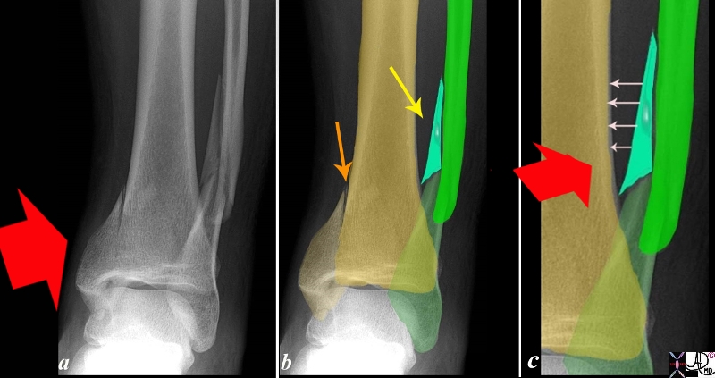
This X-ray on the antero-posterior (A-P) projection shows a vertical fracture of the distal tibia and a comminuted spiral fracture of the distal shaft of the fibula in a 26 year old male. Image a shows the impaction force (red arrow) with a anterolateral vector. Image b shows the tibial fracture (yellow arrow) and the spiral fibula fracture (yellow arrow) composed of three fragments with the lightest turquoise green overlaying the triangular or butterfly fragment. The third image (c) shows the impaction pushing force (red arrow), and the pulling force it creates on the interosseus membrane, interosseus ligament and anterior tibio-fibular ligament (white arrows) resulting in a combination of avulsion fracture (butterfly fragment turquoise green) and impaction oblique/spiral fracture (lime green and dark green). Since the mortise is intact it is likely that the anterior talofibilar ligament and deltoid ligament are not ruptured but this would also require clinical evaluation by placing stress on these ligaments.
Courtesy Ashley Davidoff Copyright 2011 101075b04L.85.8s
|
Time Zero + a Nanosecond
|
|
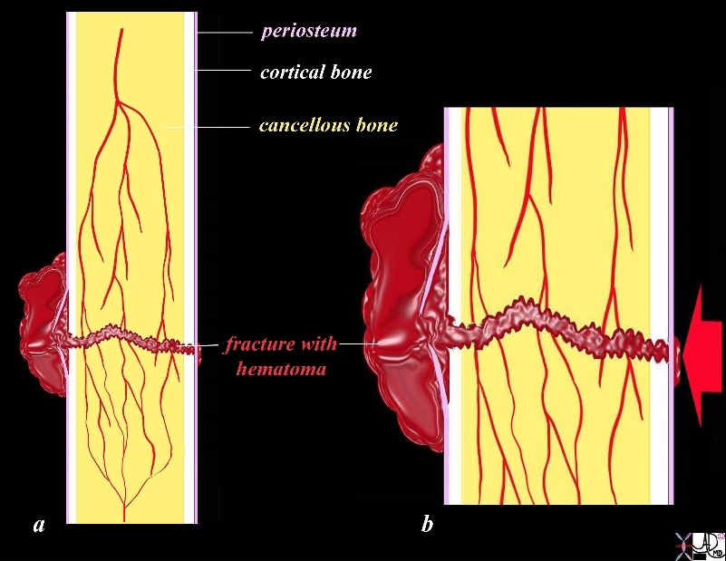
The diagram shows the macroscopic/histologic anatomy of the bone after the fracture (a) and a magnified view of the acute injury (b) revealing the periosteum (pink) cortical bone (white) and the cancellous bone embedded in the bone marrow (yellow) and hemorrhage (maroon). After the injury there is usually disruption of the periosteum on the opposite side of the force (red arrow), break of the cortical bone on one side in an incomplete fracture or both sides in a complete fracture, disruption of the softer cancellous bone and blood vessels. The rupture of the blood vessels leads to bleeding. Blood clot fills the fracture site extends and lifts the damaged periosteum off the cortical bone and spills over into the surrounding soft tissues. The disruption of the vessels at the fracture line leads to ischemia.
Courtesy Ashley Davidoff Copyright 2011 103498i11.8s
|
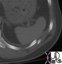
Fracture of a Rib Causing Delayed Rupturte of the Spleen |
|
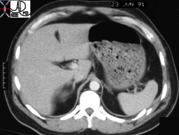
This patient was discharged from the hospital with a normal spleen but with a rib fracture in close proximity to the spleen . He re-presented to an outide institution with splenic rupture probably caused by penetration of the spleen by the fracture fragment. he required splenectomy for uncontrolled bleeding.
Courtesy Ashley Davidoff MD 20803
|
DOMElement Object
(
    [schemaTypeInfo] =>
    [tagName] => table
    [firstElementChild] => (object value omitted)
    [lastElementChild] => (object value omitted)
    [childElementCount] => 1
    [previousElementSibling] => (object value omitted)
    [nextElementSibling] =>
    [nodeName] => table
    [nodeValue] =>


Fracture of a Rib Causing Delayed Rupturte of the Spleen


This patient was discharged from the hospital with a normal spleen but with a rib fracture in close proximity to the spleen . He re-presented to an outide institution with splenic rupture probably caused by penetration of the spleen by the fracture fragment.   he required splenectomy for uncontrolled bleeding.
Courtesy Ashley Davidoff MD 20803


    [nodeType] => 1
    [parentNode] => (object value omitted)
    [childNodes] => (object value omitted)
    [firstChild] => (object value omitted)
    [lastChild] => (object value omitted)
    [previousSibling] => (object value omitted)
    [nextSibling] => (object value omitted)
    [attributes] => (object value omitted)
    [ownerDocument] => (object value omitted)
    [namespaceURI] =>
    [prefix] =>
    [localName] => table
    [baseURI] =>
    [textContent] =>


Fracture of a Rib Causing Delayed Rupturte of the Spleen


This patient was discharged from the hospital with a normal spleen but with a rib fracture in close proximity to the spleen . He re-presented to an outide institution with splenic rupture probably caused by penetration of the spleen by the fracture fragment.   he required splenectomy for uncontrolled bleeding.
Courtesy Ashley Davidoff MD 20803


)
DOMElement Object
(
    [schemaTypeInfo] =>
    [tagName] => td
    [firstElementChild] => (object value omitted)
    [lastElementChild] => (object value omitted)
    [childElementCount] => 2
    [previousElementSibling] =>
    [nextElementSibling] =>
    [nodeName] => td
    [nodeValue] =>
This patient was discharged from the hospital with a normal spleen but with a rib fracture in close proximity to the spleen . He re-presented to an outide institution with splenic rupture probably caused by penetration of the spleen by the fracture fragment.   he required splenectomy for uncontrolled bleeding.
Courtesy Ashley Davidoff MD 20803

    [nodeType] => 1
    [parentNode] => (object value omitted)
    [childNodes] => (object value omitted)
    [firstChild] => (object value omitted)
    [lastChild] => (object value omitted)
    [previousSibling] => (object value omitted)
    [nextSibling] => (object value omitted)
    [attributes] => (object value omitted)
    [ownerDocument] => (object value omitted)
    [namespaceURI] =>
    [prefix] =>
    [localName] => td
    [baseURI] =>
    [textContent] =>
This patient was discharged from the hospital with a normal spleen but with a rib fracture in close proximity to the spleen . He re-presented to an outide institution with splenic rupture probably caused by penetration of the spleen by the fracture fragment.   he required splenectomy for uncontrolled bleeding.
Courtesy Ashley Davidoff MD 20803

)
DOMElement Object
(
    [schemaTypeInfo] =>
    [tagName] => td
    [firstElementChild] => (object value omitted)
    [lastElementChild] => (object value omitted)
    [childElementCount] => 3
    [previousElementSibling] =>
    [nextElementSibling] =>
    [nodeName] => td
    [nodeValue] =>

Fracture of a Rib Causing Delayed Rupturte of the Spleen
    [nodeType] => 1
    [parentNode] => (object value omitted)
    [childNodes] => (object value omitted)
    [firstChild] => (object value omitted)
    [lastChild] => (object value omitted)
    [previousSibling] => (object value omitted)
    [nextSibling] => (object value omitted)
    [attributes] => (object value omitted)
    [ownerDocument] => (object value omitted)
    [namespaceURI] =>
    [prefix] =>
    [localName] => td
    [baseURI] =>
    [textContent] =>

Fracture of a Rib Causing Delayed Rupturte of the Spleen
)
https://beta.thecommonvein.net/wp-content/uploads/2023/05/20803.jpg
http://www.thecommonvein.net/media/20803.JPG http://www.thecommonvein.net/media/20805.JPG
DOMElement Object
(
    [schemaTypeInfo] =>
    [tagName] => table
    [firstElementChild] => (object value omitted)
    [lastElementChild] => (object value omitted)
    [childElementCount] => 1
    [previousElementSibling] => (object value omitted)
    [nextElementSibling] => (object value omitted)
    [nodeName] => table
    [nodeValue] =>


Time Zero + a Nanosecond


The diagram shows the macroscopic/histologic anatomy of the bone after the fracture (a) and a magnified view of the acute injury (b) revealing the periosteum (pink) cortical bone (white) and the cancellous bone embedded in the bone marrow (yellow) and hemorrhage (maroon). After the injury there is usually disruption of the periosteum on the opposite side of the force (red arrow), break of the cortical bone on one side in an incomplete fracture or both sides in a complete fracture, disruption of the softer cancellous bone and blood vessels. The rupture of the blood vessels leads to bleeding. Blood clot fills the fracture site extends and lifts the damaged periosteum off the cortical bone and spills over into the surrounding soft tissues. The disruption of the vessels at the fracture line leads to ischemia.
Courtesy Ashley Davidoff Copyright 2011 103498i11.8s


    [nodeType] => 1
    [parentNode] => (object value omitted)
    [childNodes] => (object value omitted)
    [firstChild] => (object value omitted)
    [lastChild] => (object value omitted)
    [previousSibling] => (object value omitted)
    [nextSibling] => (object value omitted)
    [attributes] => (object value omitted)
    [ownerDocument] => (object value omitted)
    [namespaceURI] =>
    [prefix] =>
    [localName] => table
    [baseURI] =>
    [textContent] =>


Time Zero + a Nanosecond


The diagram shows the macroscopic/histologic anatomy of the bone after the fracture (a) and a magnified view of the acute injury (b) revealing the periosteum (pink) cortical bone (white) and the cancellous bone embedded in the bone marrow (yellow) and hemorrhage (maroon). After the injury there is usually disruption of the periosteum on the opposite side of the force (red arrow), break of the cortical bone on one side in an incomplete fracture or both sides in a complete fracture, disruption of the softer cancellous bone and blood vessels. The rupture of the blood vessels leads to bleeding. Blood clot fills the fracture site extends and lifts the damaged periosteum off the cortical bone and spills over into the surrounding soft tissues. The disruption of the vessels at the fracture line leads to ischemia.
Courtesy Ashley Davidoff Copyright 2011 103498i11.8s


)
DOMElement Object
(
    [schemaTypeInfo] =>
    [tagName] => td
    [firstElementChild] => (object value omitted)
    [lastElementChild] => (object value omitted)
    [childElementCount] => 2
    [previousElementSibling] =>
    [nextElementSibling] =>
    [nodeName] => td
    [nodeValue] =>
The diagram shows the macroscopic/histologic anatomy of the bone after the fracture (a) and a magnified view of the acute injury (b) revealing the periosteum (pink) cortical bone (white) and the cancellous bone embedded in the bone marrow (yellow) and hemorrhage (maroon). After the injury there is usually disruption of the periosteum on the opposite side of the force (red arrow), break of the cortical bone on one side in an incomplete fracture or both sides in a complete fracture, disruption of the softer cancellous bone and blood vessels. The rupture of the blood vessels leads to bleeding. Blood clot fills the fracture site extends and lifts the damaged periosteum off the cortical bone and spills over into the surrounding soft tissues. The disruption of the vessels at the fracture line leads to ischemia.
Courtesy Ashley Davidoff Copyright 2011 103498i11.8s

    [nodeType] => 1
    [parentNode] => (object value omitted)
    [childNodes] => (object value omitted)
    [firstChild] => (object value omitted)
    [lastChild] => (object value omitted)
    [previousSibling] => (object value omitted)
    [nextSibling] => (object value omitted)
    [attributes] => (object value omitted)
    [ownerDocument] => (object value omitted)
    [namespaceURI] =>
    [prefix] =>
    [localName] => td
    [baseURI] =>
    [textContent] =>
The diagram shows the macroscopic/histologic anatomy of the bone after the fracture (a) and a magnified view of the acute injury (b) revealing the periosteum (pink) cortical bone (white) and the cancellous bone embedded in the bone marrow (yellow) and hemorrhage (maroon). After the injury there is usually disruption of the periosteum on the opposite side of the force (red arrow), break of the cortical bone on one side in an incomplete fracture or both sides in a complete fracture, disruption of the softer cancellous bone and blood vessels. The rupture of the blood vessels leads to bleeding. Blood clot fills the fracture site extends and lifts the damaged periosteum off the cortical bone and spills over into the surrounding soft tissues. The disruption of the vessels at the fracture line leads to ischemia.
Courtesy Ashley Davidoff Copyright 2011 103498i11.8s

)
DOMElement Object
(
    [schemaTypeInfo] =>
    [tagName] => td
    [firstElementChild] => (object value omitted)
    [lastElementChild] => (object value omitted)
    [childElementCount] => 2
    [previousElementSibling] =>
    [nextElementSibling] =>
    [nodeName] => td
    [nodeValue] =>
Time Zero + a Nanosecond

    [nodeType] => 1
    [parentNode] => (object value omitted)
    [childNodes] => (object value omitted)
    [firstChild] => (object value omitted)
    [lastChild] => (object value omitted)
    [previousSibling] => (object value omitted)
    [nextSibling] => (object value omitted)
    [attributes] => (object value omitted)
    [ownerDocument] => (object value omitted)
    [namespaceURI] =>
    [prefix] =>
    [localName] => td
    [baseURI] =>
    [textContent] =>
Time Zero + a Nanosecond

)
DOMElement Object
(
    [schemaTypeInfo] =>
    [tagName] => table
    [firstElementChild] => (object value omitted)
    [lastElementChild] => (object value omitted)
    [childElementCount] => 1
    [previousElementSibling] => (object value omitted)
    [nextElementSibling] => (object value omitted)
    [nodeName] => table
    [nodeValue] =>


Push and Pull on Connected Structures


This X-ray on the antero-posterior (A-P) projection shows a vertical fracture of the distal tibia and a comminuted spiral fracture of the distal shaft of the fibula in a 26 year old male. Image a shows the impaction force (red arrow) with a anterolateral vector. Image b shows the tibial fracture (yellow arrow) and the spiral fibula fracture (yellow arrow) composed of three fragments with the lightest turquoise green overlaying the triangular or butterfly fragment. The third image (c) shows the impaction pushing force (red arrow), and the pulling force it creates on the interosseus membrane, interosseus ligament and anterior tibio-fibular ligament (white arrows) resulting in a combination of avulsion fracture (butterfly fragment turquoise green) and impaction oblique/spiral fracture (lime green and dark green). Since the mortise is intact it is likely that the anterior talofibilar ligament and deltoid ligament are not ruptured but this would also require clinical evaluation by placing stress on these ligaments.
Courtesy Ashley Davidoff Copyright 2011 101075b04L.85.8s


    [nodeType] => 1
    [parentNode] => (object value omitted)
    [childNodes] => (object value omitted)
    [firstChild] => (object value omitted)
    [lastChild] => (object value omitted)
    [previousSibling] => (object value omitted)
    [nextSibling] => (object value omitted)
    [attributes] => (object value omitted)
    [ownerDocument] => (object value omitted)
    [namespaceURI] =>
    [prefix] =>
    [localName] => table
    [baseURI] =>
    [textContent] =>


Push and Pull on Connected Structures


This X-ray on the antero-posterior (A-P) projection shows a vertical fracture of the distal tibia and a comminuted spiral fracture of the distal shaft of the fibula in a 26 year old male. Image a shows the impaction force (red arrow) with a anterolateral vector. Image b shows the tibial fracture (yellow arrow) and the spiral fibula fracture (yellow arrow) composed of three fragments with the lightest turquoise green overlaying the triangular or butterfly fragment. The third image (c) shows the impaction pushing force (red arrow), and the pulling force it creates on the interosseus membrane, interosseus ligament and anterior tibio-fibular ligament (white arrows) resulting in a combination of avulsion fracture (butterfly fragment turquoise green) and impaction oblique/spiral fracture (lime green and dark green). Since the mortise is intact it is likely that the anterior talofibilar ligament and deltoid ligament are not ruptured but this would also require clinical evaluation by placing stress on these ligaments.
Courtesy Ashley Davidoff Copyright 2011 101075b04L.85.8s


)
DOMElement Object
(
    [schemaTypeInfo] =>
    [tagName] => td
    [firstElementChild] => (object value omitted)
    [lastElementChild] => (object value omitted)
    [childElementCount] => 2
    [previousElementSibling] =>
    [nextElementSibling] =>
    [nodeName] => td
    [nodeValue] =>
This X-ray on the antero-posterior (A-P) projection shows a vertical fracture of the distal tibia and a comminuted spiral fracture of the distal shaft of the fibula in a 26 year old male. Image a shows the impaction force (red arrow) with a anterolateral vector. Image b shows the tibial fracture (yellow arrow) and the spiral fibula fracture (yellow arrow) composed of three fragments with the lightest turquoise green overlaying the triangular or butterfly fragment. The third image (c) shows the impaction pushing force (red arrow), and the pulling force it creates on the interosseus membrane, interosseus ligament and anterior tibio-fibular ligament (white arrows) resulting in a combination of avulsion fracture (butterfly fragment turquoise green) and impaction oblique/spiral fracture (lime green and dark green). Since the mortise is intact it is likely that the anterior talofibilar ligament and deltoid ligament are not ruptured but this would also require clinical evaluation by placing stress on these ligaments.
Courtesy Ashley Davidoff Copyright 2011 101075b04L.85.8s

    [nodeType] => 1
    [parentNode] => (object value omitted)
    [childNodes] => (object value omitted)
    [firstChild] => (object value omitted)
    [lastChild] => (object value omitted)
    [previousSibling] => (object value omitted)
    [nextSibling] => (object value omitted)
    [attributes] => (object value omitted)
    [ownerDocument] => (object value omitted)
    [namespaceURI] =>
    [prefix] =>
    [localName] => td
    [baseURI] =>
    [textContent] =>
This X-ray on the antero-posterior (A-P) projection shows a vertical fracture of the distal tibia and a comminuted spiral fracture of the distal shaft of the fibula in a 26 year old male. Image a shows the impaction force (red arrow) with a anterolateral vector. Image b shows the tibial fracture (yellow arrow) and the spiral fibula fracture (yellow arrow) composed of three fragments with the lightest turquoise green overlaying the triangular or butterfly fragment. The third image (c) shows the impaction pushing force (red arrow), and the pulling force it creates on the interosseus membrane, interosseus ligament and anterior tibio-fibular ligament (white arrows) resulting in a combination of avulsion fracture (butterfly fragment turquoise green) and impaction oblique/spiral fracture (lime green and dark green). Since the mortise is intact it is likely that the anterior talofibilar ligament and deltoid ligament are not ruptured but this would also require clinical evaluation by placing stress on these ligaments.
Courtesy Ashley Davidoff Copyright 2011 101075b04L.85.8s

)
DOMElement Object
(
    [schemaTypeInfo] =>
    [tagName] => td
    [firstElementChild] => (object value omitted)
    [lastElementChild] => (object value omitted)
    [childElementCount] => 2
    [previousElementSibling] =>
    [nextElementSibling] =>
    [nodeName] => td
    [nodeValue] =>
Push and Pull on Connected Structures

    [nodeType] => 1
    [parentNode] => (object value omitted)
    [childNodes] => (object value omitted)
    [firstChild] => (object value omitted)
    [lastChild] => (object value omitted)
    [previousSibling] => (object value omitted)
    [nextSibling] => (object value omitted)
    [attributes] => (object value omitted)
    [ownerDocument] => (object value omitted)
    [namespaceURI] =>
    [prefix] =>
    [localName] => td
    [baseURI] =>
    [textContent] =>
Push and Pull on Connected Structures

)
DOMElement Object
(
    [schemaTypeInfo] =>
    [tagName] => table
    [firstElementChild] => (object value omitted)
    [lastElementChild] => (object value omitted)
    [childElementCount] => 1
    [previousElementSibling] => (object value omitted)
    [nextElementSibling] => (object value omitted)
    [nodeName] => table
    [nodeValue] =>


More Complex Issues
The Effects of Ligamnets and Muscle


The diagram (a) shows the major anchoring mechanisms that bind the tibia, fibula and talus including the interosseous membrane (light pink), the interosseus ligament (purple) anterior tibiofibular ligament (orange) the deltoid ligament (royal blue) anterior talofibular ligament (teal blue). With a sudden force (red arrow b) on the medial malleolus for example, any number of fractures can result since the force will push on tibia and the will be imparted through ligaments either with push or pull on the distal fibula, proximal fibula, medial or lateral talus. As a result a single excessive force may be transmitted via ligamentous and muscular attachments to any number of bones and a combination of fractures may result. CODE shaft diaphysis bone tibia, fibula and talus interosseous membrane (light pink), interosseus ligament (purple) anterior tibiofibular ligament (orange) deltoid ligament (royal blue) anterior talofibular ligament (teal blue).
Courtesy Ashley Davidoff Copyright 2011 103498f05L.8s


    [nodeType] => 1
    [parentNode] => (object value omitted)
    [childNodes] => (object value omitted)
    [firstChild] => (object value omitted)
    [lastChild] => (object value omitted)
    [previousSibling] => (object value omitted)
    [nextSibling] => (object value omitted)
    [attributes] => (object value omitted)
    [ownerDocument] => (object value omitted)
    [namespaceURI] =>
    [prefix] =>
    [localName] => table
    [baseURI] =>
    [textContent] =>


More Complex Issues
The Effects of Ligamnets and Muscle


The diagram (a) shows the major anchoring mechanisms that bind the tibia, fibula and talus including the interosseous membrane (light pink), the interosseus ligament (purple) anterior tibiofibular ligament (orange) the deltoid ligament (royal blue) anterior talofibular ligament (teal blue). With a sudden force (red arrow b) on the medial malleolus for example, any number of fractures can result since the force will push on tibia and the will be imparted through ligaments either with push or pull on the distal fibula, proximal fibula, medial or lateral talus. As a result a single excessive force may be transmitted via ligamentous and muscular attachments to any number of bones and a combination of fractures may result. CODE shaft diaphysis bone tibia, fibula and talus interosseous membrane (light pink), interosseus ligament (purple) anterior tibiofibular ligament (orange) deltoid ligament (royal blue) anterior talofibular ligament (teal blue).
Courtesy Ashley Davidoff Copyright 2011 103498f05L.8s


)
DOMElement Object
(
    [schemaTypeInfo] =>
    [tagName] => td
    [firstElementChild] => (object value omitted)
    [lastElementChild] => (object value omitted)
    [childElementCount] => 2
    [previousElementSibling] =>
    [nextElementSibling] =>
    [nodeName] => td
    [nodeValue] =>
The diagram (a) shows the major anchoring mechanisms that bind the tibia, fibula and talus including the interosseous membrane (light pink), the interosseus ligament (purple) anterior tibiofibular ligament (orange) the deltoid ligament (royal blue) anterior talofibular ligament (teal blue). With a sudden force (red arrow b) on the medial malleolus for example, any number of fractures can result since the force will push on tibia and the will be imparted through ligaments either with push or pull on the distal fibula, proximal fibula, medial or lateral talus. As a result a single excessive force may be transmitted via ligamentous and muscular attachments to any number of bones and a combination of fractures may result. CODE shaft diaphysis bone tibia, fibula and talus interosseous membrane (light pink), interosseus ligament (purple) anterior tibiofibular ligament (orange) deltoid ligament (royal blue) anterior talofibular ligament (teal blue).
Courtesy Ashley Davidoff Copyright 2011 103498f05L.8s

    [nodeType] => 1
    [parentNode] => (object value omitted)
    [childNodes] => (object value omitted)
    [firstChild] => (object value omitted)
    [lastChild] => (object value omitted)
    [previousSibling] => (object value omitted)
    [nextSibling] => (object value omitted)
    [attributes] => (object value omitted)
    [ownerDocument] => (object value omitted)
    [namespaceURI] =>
    [prefix] =>
    [localName] => td
    [baseURI] =>
    [textContent] =>
The diagram (a) shows the major anchoring mechanisms that bind the tibia, fibula and talus including the interosseous membrane (light pink), the interosseus ligament (purple) anterior tibiofibular ligament (orange) the deltoid ligament (royal blue) anterior talofibular ligament (teal blue). With a sudden force (red arrow b) on the medial malleolus for example, any number of fractures can result since the force will push on tibia and the will be imparted through ligaments either with push or pull on the distal fibula, proximal fibula, medial or lateral talus. As a result a single excessive force may be transmitted via ligamentous and muscular attachments to any number of bones and a combination of fractures may result. CODE shaft diaphysis bone tibia, fibula and talus interosseous membrane (light pink), interosseus ligament (purple) anterior tibiofibular ligament (orange) deltoid ligament (royal blue) anterior talofibular ligament (teal blue).
Courtesy Ashley Davidoff Copyright 2011 103498f05L.8s

)
DOMElement Object
(
    [schemaTypeInfo] =>
    [tagName] => td
    [firstElementChild] => (object value omitted)
    [lastElementChild] => (object value omitted)
    [childElementCount] => 3
    [previousElementSibling] =>
    [nextElementSibling] =>
    [nodeName] => td
    [nodeValue] =>
More Complex Issues
The Effects of Ligamnets and Muscle

    [nodeType] => 1
    [parentNode] => (object value omitted)
    [childNodes] => (object value omitted)
    [firstChild] => (object value omitted)
    [lastChild] => (object value omitted)
    [previousSibling] => (object value omitted)
    [nextSibling] => (object value omitted)
    [attributes] => (object value omitted)
    [ownerDocument] => (object value omitted)
    [namespaceURI] =>
    [prefix] =>
    [localName] => td
    [baseURI] =>
    [textContent] =>
More Complex Issues
The Effects of Ligamnets and Muscle

)


