The Common Vein Copyright 2010
Introduction
DOMElement Object
(
    [schemaTypeInfo] =>
    [tagName] => table
    [firstElementChild] => (object value omitted)
    [lastElementChild] => (object value omitted)
    [childElementCount] => 1
    [previousElementSibling] => (object value omitted)
    [nextElementSibling] =>
    [nodeName] => table
    [nodeValue] =>


Multinodular Gland Abnormal Texture on STIR Sequence


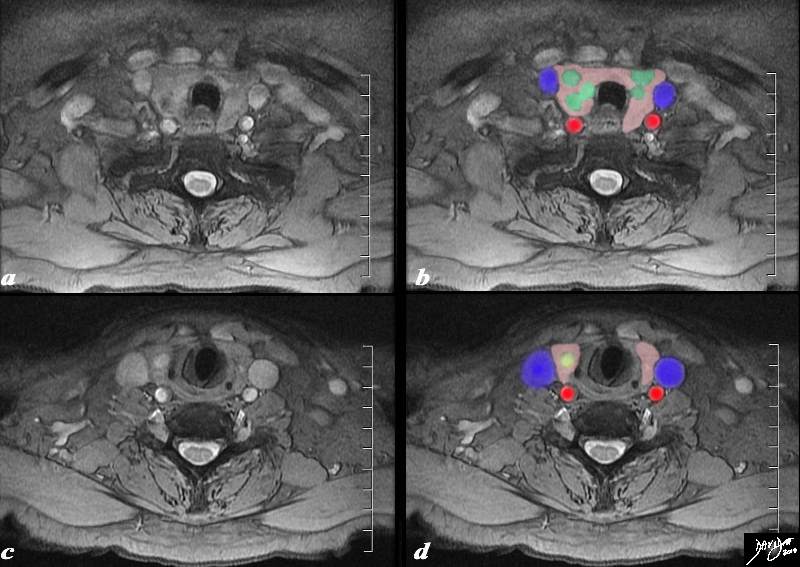
The MRI is from an 80 year old male presents with an asymptomatic multinodular goiter, consisting many nodules of varying size as identified by previous ultrasound.. The MRI using a STIR sequence shows a normal size and shape to the gland but multiple non border forming nodules (green nodules) are identified within the matrix of the gland (pink).One of the nodules (c,d) overlaid in yellow is cystic in nature. It is bright on the STIR sequence (c). The internal jugular veins (blue) form a lateral neighbor and deform the relatively soft gland. The common carotid arteries (red) form a posterolateral relation to the gland. The trachea (black) is surrounded anteriorly and laterally by the gland. These findings are consistent with a non toxic multinodular thyroid gland, – not truly a goiter since the gland is not enlarged.
Courtesy Ashley Davidoff MD Copyright 2010  94790c03.8s94790c03.8s


    [nodeType] => 1
    [parentNode] => (object value omitted)
    [childNodes] => (object value omitted)
    [firstChild] => (object value omitted)
    [lastChild] => (object value omitted)
    [previousSibling] => (object value omitted)
    [nextSibling] => (object value omitted)
    [attributes] => (object value omitted)
    [ownerDocument] => (object value omitted)
    [namespaceURI] =>
    [prefix] =>
    [localName] => table
    [baseURI] =>
    [textContent] =>


Multinodular Gland Abnormal Texture on STIR Sequence


The MRI is from an 80 year old male presents with an asymptomatic multinodular goiter, consisting many nodules of varying size as identified by previous ultrasound.. The MRI using a STIR sequence shows a normal size and shape to the gland but multiple non border forming nodules (green nodules) are identified within the matrix of the gland (pink).One of the nodules (c,d) overlaid in yellow is cystic in nature. It is bright on the STIR sequence (c). The internal jugular veins (blue) form a lateral neighbor and deform the relatively soft gland. The common carotid arteries (red) form a posterolateral relation to the gland. The trachea (black) is surrounded anteriorly and laterally by the gland. These findings are consistent with a non toxic multinodular thyroid gland, – not truly a goiter since the gland is not enlarged.
Courtesy Ashley Davidoff MD Copyright 2010  94790c03.8s94790c03.8s


)
DOMElement Object
(
    [schemaTypeInfo] =>
    [tagName] => td
    [firstElementChild] => (object value omitted)
    [lastElementChild] => (object value omitted)
    [childElementCount] => 2
    [previousElementSibling] =>
    [nextElementSibling] =>
    [nodeName] => td
    [nodeValue] =>
The MRI is from an 80 year old male presents with an asymptomatic multinodular goiter, consisting many nodules of varying size as identified by previous ultrasound.. The MRI using a STIR sequence shows a normal size and shape to the gland but multiple non border forming nodules (green nodules) are identified within the matrix of the gland (pink).One of the nodules (c,d) overlaid in yellow is cystic in nature. It is bright on the STIR sequence (c). The internal jugular veins (blue) form a lateral neighbor and deform the relatively soft gland. The common carotid arteries (red) form a posterolateral relation to the gland. The trachea (black) is surrounded anteriorly and laterally by the gland. These findings are consistent with a non toxic multinodular thyroid gland, – not truly a goiter since the gland is not enlarged.
Courtesy Ashley Davidoff MD Copyright 2010  94790c03.8s94790c03.8s

    [nodeType] => 1
    [parentNode] => (object value omitted)
    [childNodes] => (object value omitted)
    [firstChild] => (object value omitted)
    [lastChild] => (object value omitted)
    [previousSibling] => (object value omitted)
    [nextSibling] => (object value omitted)
    [attributes] => (object value omitted)
    [ownerDocument] => (object value omitted)
    [namespaceURI] =>
    [prefix] =>
    [localName] => td
    [baseURI] =>
    [textContent] =>
The MRI is from an 80 year old male presents with an asymptomatic multinodular goiter, consisting many nodules of varying size as identified by previous ultrasound.. The MRI using a STIR sequence shows a normal size and shape to the gland but multiple non border forming nodules (green nodules) are identified within the matrix of the gland (pink).One of the nodules (c,d) overlaid in yellow is cystic in nature. It is bright on the STIR sequence (c). The internal jugular veins (blue) form a lateral neighbor and deform the relatively soft gland. The common carotid arteries (red) form a posterolateral relation to the gland. The trachea (black) is surrounded anteriorly and laterally by the gland. These findings are consistent with a non toxic multinodular thyroid gland, – not truly a goiter since the gland is not enlarged.
Courtesy Ashley Davidoff MD Copyright 2010  94790c03.8s94790c03.8s

)
DOMElement Object
(
    [schemaTypeInfo] =>
    [tagName] => td
    [firstElementChild] => (object value omitted)
    [lastElementChild] => (object value omitted)
    [childElementCount] => 2
    [previousElementSibling] =>
    [nextElementSibling] =>
    [nodeName] => td
    [nodeValue] =>
Multinodular Gland Abnormal Texture on STIR Sequence
    [nodeType] => 1
    [parentNode] => (object value omitted)
    [childNodes] => (object value omitted)
    [firstChild] => (object value omitted)
    [lastChild] => (object value omitted)
    [previousSibling] => (object value omitted)
    [nextSibling] => (object value omitted)
    [attributes] => (object value omitted)
    [ownerDocument] => (object value omitted)
    [namespaceURI] =>
    [prefix] =>
    [localName] => td
    [baseURI] =>
    [textContent] =>
Multinodular Gland Abnormal Texture on STIR Sequence
)
DOMElement Object
(
    [schemaTypeInfo] =>
    [tagName] => table
    [firstElementChild] => (object value omitted)
    [lastElementChild] => (object value omitted)
    [childElementCount] => 1
    [previousElementSibling] => (object value omitted)
    [nextElementSibling] => (object value omitted)
    [nodeName] => table
    [nodeValue] =>


Thyroglossal Duct Cyst T2 Weighted Sequence


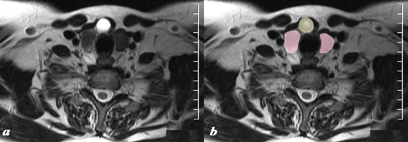
The MRI T2 weighted image through the inferior aspect of the thyroid gland (pink) shows a cystic structure (yellow), intensely T2 bright (a), in the region of the isthmus in the ventral and midline position of the gland. Findings are consistent with a thyroglossal duct cyst
Courtesy Ashley Davidoff MD Copyright 2010 97310cL.8


    [nodeType] => 1
    [parentNode] => (object value omitted)
    [childNodes] => (object value omitted)
    [firstChild] => (object value omitted)
    [lastChild] => (object value omitted)
    [previousSibling] => (object value omitted)
    [nextSibling] => (object value omitted)
    [attributes] => (object value omitted)
    [ownerDocument] => (object value omitted)
    [namespaceURI] =>
    [prefix] =>
    [localName] => table
    [baseURI] =>
    [textContent] =>


Thyroglossal Duct Cyst T2 Weighted Sequence


The MRI T2 weighted image through the inferior aspect of the thyroid gland (pink) shows a cystic structure (yellow), intensely T2 bright (a), in the region of the isthmus in the ventral and midline position of the gland. Findings are consistent with a thyroglossal duct cyst
Courtesy Ashley Davidoff MD Copyright 2010 97310cL.8


)
DOMElement Object
(
    [schemaTypeInfo] =>
    [tagName] => td
    [firstElementChild] => (object value omitted)
    [lastElementChild] => (object value omitted)
    [childElementCount] => 2
    [previousElementSibling] =>
    [nextElementSibling] =>
    [nodeName] => td
    [nodeValue] =>
The MRI T2 weighted image through the inferior aspect of the thyroid gland (pink) shows a cystic structure (yellow), intensely T2 bright (a), in the region of the isthmus in the ventral and midline position of the gland. Findings are consistent with a thyroglossal duct cyst
Courtesy Ashley Davidoff MD Copyright 2010 97310cL.8

    [nodeType] => 1
    [parentNode] => (object value omitted)
    [childNodes] => (object value omitted)
    [firstChild] => (object value omitted)
    [lastChild] => (object value omitted)
    [previousSibling] => (object value omitted)
    [nextSibling] => (object value omitted)
    [attributes] => (object value omitted)
    [ownerDocument] => (object value omitted)
    [namespaceURI] =>
    [prefix] =>
    [localName] => td
    [baseURI] =>
    [textContent] =>
The MRI T2 weighted image through the inferior aspect of the thyroid gland (pink) shows a cystic structure (yellow), intensely T2 bright (a), in the region of the isthmus in the ventral and midline position of the gland. Findings are consistent with a thyroglossal duct cyst
Courtesy Ashley Davidoff MD Copyright 2010 97310cL.8

)
DOMElement Object
(
    [schemaTypeInfo] =>
    [tagName] => td
    [firstElementChild] => (object value omitted)
    [lastElementChild] => (object value omitted)
    [childElementCount] => 2
    [previousElementSibling] =>
    [nextElementSibling] =>
    [nodeName] => td
    [nodeValue] =>
Thyroglossal Duct Cyst T2 Weighted Sequence
    [nodeType] => 1
    [parentNode] => (object value omitted)
    [childNodes] => (object value omitted)
    [firstChild] => (object value omitted)
    [lastChild] => (object value omitted)
    [previousSibling] => (object value omitted)
    [nextSibling] => (object value omitted)
    [attributes] => (object value omitted)
    [ownerDocument] => (object value omitted)
    [namespaceURI] =>
    [prefix] =>
    [localName] => td
    [baseURI] =>
    [textContent] =>
Thyroglossal Duct Cyst T2 Weighted Sequence
)
DOMElement Object
(
    [schemaTypeInfo] =>
    [tagName] => table
    [firstElementChild] => (object value omitted)
    [lastElementChild] => (object value omitted)
    [childElementCount] => 1
    [previousElementSibling] => (object value omitted)
    [nextElementSibling] => (object value omitted)
    [nodeName] => table
    [nodeValue] =>


Normal MRI


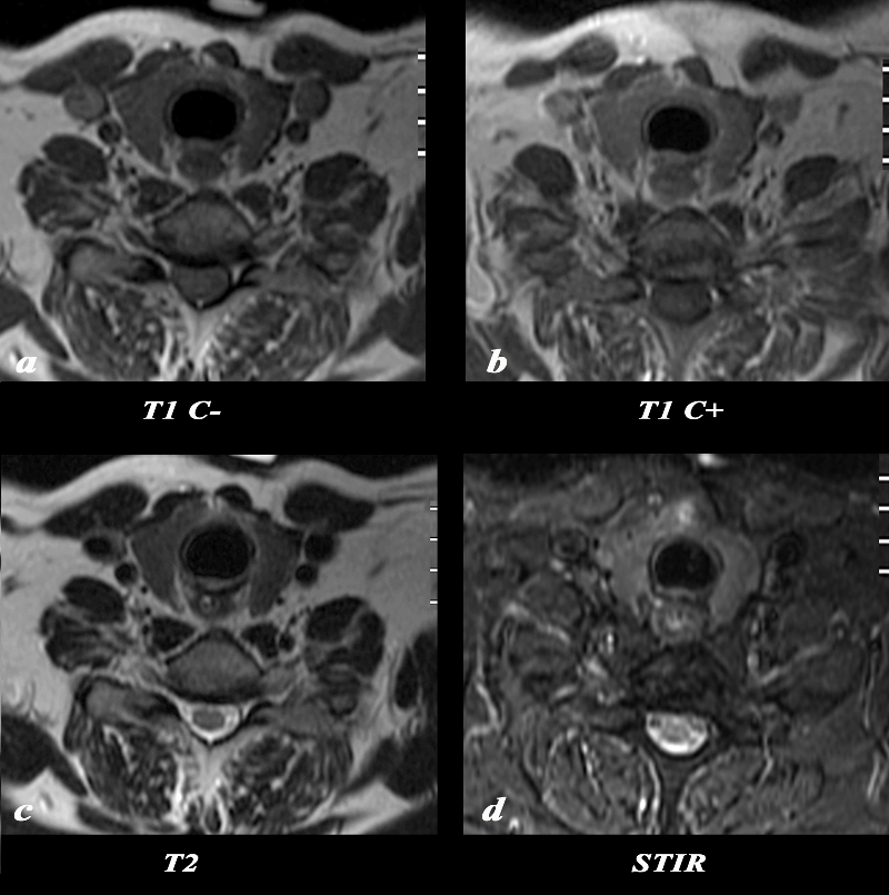
The MRI of the thyroid reveals the appearance of the thyroid on T1 weighted sequence without contrast (a), T1 with contrast (b), T2 weighted image (C), and a STIR image. The normal gland appears remarkably similar on all 4 phases showing mild enhancement and mild STIR hyperintensity.
Courtesy Ashley Davidoff MD Copyright 2010 97332c02L.8


    [nodeType] => 1
    [parentNode] => (object value omitted)
    [childNodes] => (object value omitted)
    [firstChild] => (object value omitted)
    [lastChild] => (object value omitted)
    [previousSibling] => (object value omitted)
    [nextSibling] => (object value omitted)
    [attributes] => (object value omitted)
    [ownerDocument] => (object value omitted)
    [namespaceURI] =>
    [prefix] =>
    [localName] => table
    [baseURI] =>
    [textContent] =>


Normal MRI


The MRI of the thyroid reveals the appearance of the thyroid on T1 weighted sequence without contrast (a), T1 with contrast (b), T2 weighted image (C), and a STIR image. The normal gland appears remarkably similar on all 4 phases showing mild enhancement and mild STIR hyperintensity.
Courtesy Ashley Davidoff MD Copyright 2010 97332c02L.8


)
DOMElement Object
(
    [schemaTypeInfo] =>
    [tagName] => td
    [firstElementChild] => (object value omitted)
    [lastElementChild] => (object value omitted)
    [childElementCount] => 2
    [previousElementSibling] =>
    [nextElementSibling] =>
    [nodeName] => td
    [nodeValue] =>
The MRI of the thyroid reveals the appearance of the thyroid on T1 weighted sequence without contrast (a), T1 with contrast (b), T2 weighted image (C), and a STIR image. The normal gland appears remarkably similar on all 4 phases showing mild enhancement and mild STIR hyperintensity.
Courtesy Ashley Davidoff MD Copyright 2010 97332c02L.8

    [nodeType] => 1
    [parentNode] => (object value omitted)
    [childNodes] => (object value omitted)
    [firstChild] => (object value omitted)
    [lastChild] => (object value omitted)
    [previousSibling] => (object value omitted)
    [nextSibling] => (object value omitted)
    [attributes] => (object value omitted)
    [ownerDocument] => (object value omitted)
    [namespaceURI] =>
    [prefix] =>
    [localName] => td
    [baseURI] =>
    [textContent] =>
The MRI of the thyroid reveals the appearance of the thyroid on T1 weighted sequence without contrast (a), T1 with contrast (b), T2 weighted image (C), and a STIR image. The normal gland appears remarkably similar on all 4 phases showing mild enhancement and mild STIR hyperintensity.
Courtesy Ashley Davidoff MD Copyright 2010 97332c02L.8

)
DOMElement Object
(
    [schemaTypeInfo] =>
    [tagName] => td
    [firstElementChild] => (object value omitted)
    [lastElementChild] => (object value omitted)
    [childElementCount] => 2
    [previousElementSibling] =>
    [nextElementSibling] =>
    [nodeName] => td
    [nodeValue] =>
Normal MRI
    [nodeType] => 1
    [parentNode] => (object value omitted)
    [childNodes] => (object value omitted)
    [firstChild] => (object value omitted)
    [lastChild] => (object value omitted)
    [previousSibling] => (object value omitted)
    [nextSibling] => (object value omitted)
    [attributes] => (object value omitted)
    [ownerDocument] => (object value omitted)
    [namespaceURI] =>
    [prefix] =>
    [localName] => td
    [baseURI] =>
    [textContent] =>
Normal MRI
)


